| Weight | 1 lbs |
|---|---|
| Dimensions | 9 × 5 × 2 in |
| host | rabbit |
| isotype | IgG |
| clonality | polyclonal |
| concentration | 1 mg/mL |
| applications | ICC/IF, WB |
| reactivity | p53DINP1 |
| available sizes | 100 µg |


rabbit anti-p53DINP1 polyclonal antibody 5900
$445.00
Antibody summary
- Rabbit polyclonal to p53DINP1
- Suitable for: ELISA,WB,IHC-P,IF
- Isotype: Whole IgG
- 100 µg
rabbit anti-p53DINP1 polyclonal antibody 5900
| antibody |
|---|
| Tested applications WB,IHC,IHC,ICC/IF,ELISA |
| Recommended dilutions Immunoblotting: use at 0.5-1ug/mL IHC: use at 2ug/mL. Detection of p53DINP1 in mouse liver with #56223 at 2ug/mL. Immunofluorescence: use at 5-20ugml. Detection of p53DINP1 in human lung tissue lysate with #56223 at (A) 0.5ug/mL and (B) 1ug/mL. Detection of p53DINP1 in human liver w |
| Immunogen Peptide corresponding to aa 1-14 of human p53DINP1. |
| Size and concentration 100µg and 1 mg/mL |
| Form liquid |
| Storage Instructions This antibody is stable for at least one (1) year at -20°C. Avoid multiple freeze-thaw cycles. |
| Storage buffer PBS, pH 7.4, 0.02% NaN3. |
| Purity peptide affinity purification |
| Clonality polyclonal |
| Isotype IgG |
| Compatible secondaries goat anti-rabbit IgG, H&L chain specific, peroxidase conjugated, conjugated polyclonal antibody 9512 goat anti-rabbit IgG, H&L chain specific, biotin conjugated polyclonal antibody 2079 goat anti-rabbit IgG, H&L chain specific, FITC conjugated polyclonal antibody 7863 goat anti-rabbit IgG, H&L chain specific, Cross Absorbed polyclonal antibody 2371 goat anti-rabbit IgG, H&L chain specific, biotin conjugated polyclonal antibody, crossabsorbed 1715 goat anti-rabbit IgG, H&L chain specific, FITC conjugated polyclonal antibody, crossabsorbed 1720 |
| Isotype control Rabbit polyclonal - Isotype Control |
| target relevance |
|---|
| Homo sapiens TP53INP1 Tumor protein p53-inducible nuclear protein 1 |
| Protein names Tumor protein p53-inducible nuclear protein 1 |
| Alternative names Stress-induced protein, p53-dependent damage-inducible nuclear protein 1 |
| Gene names TP53INP1 |
| Function Antiproliferative and proapoptotic protein involved in cell stress response which acts as a dual regulator of transcription and autophagy. Acts as a positive regulator of autophagy. In response to cellular stress or activation of autophagy, relocates to autophagosomes where it interacts with autophagosome-associated proteins GABARAP, GABARAPL1/L2, MAP1LC3A/B/C and regulates autophagy. Acts as an antioxidant and plays a major role in p53/TP53-driven oxidative stress response. Possesses both a p53/TP53-independent intracellular reactive oxygen species (ROS) regulatory function and a p53/TP53-dependent transcription regulatory function. Positively regulates p53/TP53 and p73/TP73 and stimulates their capacity to induce apoptosis and regulate cell cycle. In response to double-strand DNA breaks, promotes p53/TP53 phosphorylation on 'Ser-46' and subsequent apoptosis. Acts as a tumor suppressor by inducing cell death by an autophagy and caspase-dependent mechanism. Can reduce cell migration by regulating the expression of SPARC |
| Subcellular location Cytoplasm, cytosol, Nucleus, Nucleus, PML body, Cytoplasmic vesicle, autophagosome |
| Structure Interacts with p53/TP53 and HIPK2. Interacts with PRKCG, GABARAP, GABARAPL1, GABARAPL2, MAP1LC3A, MAP1LC3B and MAP1LC3C |
| Keywords Activator, Alternative splicing, Antioxidant, Apoptosis, Autophagy, Cytoplasm, Cytoplasmic vesicle, Nucleus, Proteomics identification, Reference proteome, Transcription, Transcription regulation, Tumor suppressor |
| Sequence MFQRLNKMFVGEVSSSSNQEPEFNEKEDDEWILVDFIDTCTGFSAEEEEEEEDISEESPT EHPSVFSCLPASLECLADTSDSCFLQFESCPMEESWFITPPPCFTAGGLTTIKVETSPME NLLIEHPSMSVYAVHNSCPGLSEATRGTDELHSPSSPRVEAQNEMGQHIHCYVAALAAHT TFLEQPKSFRPSQWIKEHSERQPLNRNSLRRQNLTRDCHPRQVKHNGWVVHQPCPRQYNY |
| UniProt accession: Q96A56 |
Data
 |
| Western blot analysis of p53DINP1 expression in human lung tissue lysate with p53DINP1 antibody at (A) 0.5 and (B) 1 µg/mL. |
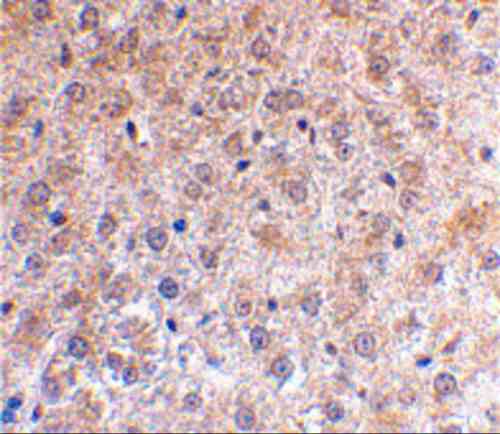 |
| Immunohistochemical staining of mouse liver using p53DINP1 antibody at 2 µg/mL. |
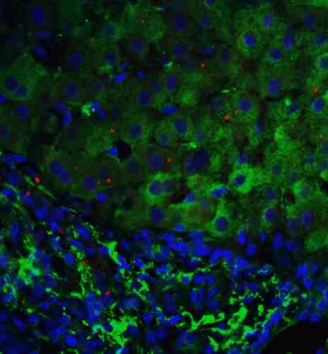 |
| Immunofluorescence of p53DINP1 in human liver tissue with p53DINP1 antibody at 5 µg/mL.
Green: p53DINP1 antibody (5900) Red: Phylloidin staining Blue: DAPI staining |
FAQ & Publications
Frequently Asked Questions
What applications has the rabbit anti-p53DINP1 polyclonal antibody 5900 been validated for?
This antibody has been tested and is suitable for use in ELISA, Western blotting (WB), immunohistochemistry on paraffin-embedded tissue (IHC-P), and immunofluorescence (IF) applications.
How should the rabbit anti-p53DINP1 polyclonal antibody 5900 be stored to maintain stability?
The antibody should be stored at -20°C and is stable for at least one year under these conditions. It is recommended to avoid multiple freeze-thaw cycles to preserve antibody integrity.
Publications
| pmid | title | authors | citation |
|---|---|---|---|
| We haven't added any publications to our database yet. | |||
Published literature highly relevant to the biological target of this product and referencing this antibody or clone are retrieved from the PubMed database provided by the United States National Library of Medicine at the National Institutes of Health.
Protocols
| relevant to this product |
|---|
| Western blot IHC ICC |
Documents
| Batch Number | QC File | SDS |
|---|---|---|
| To view batch-specific Safety Datasheets and Quality Certificates associated with your account, please Log In. | ||
Only logged in customers who have purchased this product may leave a review.


Reviews
There are no reviews yet.