| Weight | 1 lbs |
|---|---|
| Dimensions | 9 × 5 × 2 in |
| host | rabbit |
| isotype | IgG |
| clonality | polyclonal |
| concentration | 1 mg/mL |
| applications | ICC/IF, WB |
| reactivity | NAK/TBK1 (CT) |
| available sizes | 100 µg |

rabbit anti-NAK/TBK1 (CT) polyclonal antibody 4716
$445.00
Antibody summary
- Rabbit polyclonal to NAK/TBK1 (CT)
- Suitable for: ELISA,WB,ICC
- Isotype: Whole IgG
- 100 µg
rabbit anti-NAK/TBK1 (CT) polyclonal antibody 4716
| antibody |
|---|
| Tested applications WB,ICC/IF,ELISA |
| Recommended dilutions Immunoblotting: use at 0.5-1 ug/mL. In immunoblots, a band of 84 kD is detected. Positive control: MOLT4 cell lysate. |
| Immunogen Peptide corresponding to aa 712-727 of human NAK/TBK1. This sequence is identical to that of mouse NAK/TBK1. |
| Size and concentration 100µg and lot specific |
| Form liquid |
| Storage Instructions This antibody is stable for at least one (1) year at -20°C. Avoid multiple freeze- thaw cycles. |
| Storage buffer PBS, pH 7.4. |
| Purity peptide affinity purification |
| Clonality polyclonal |
| Isotype IgG |
| Compatible secondaries goat anti-rabbit IgG, H&L chain specific, peroxidase conjugated, conjugated polyclonal antibody 9512 goat anti-rabbit IgG, H&L chain specific, biotin conjugated polyclonal antibody 2079 goat anti-rabbit IgG, H&L chain specific, FITC conjugated polyclonal antibody 7863 goat anti-rabbit IgG, H&L chain specific, Cross Absorbed polyclonal antibody 2371 goat anti-rabbit IgG, H&L chain specific, biotin conjugated polyclonal antibody, crossabsorbed 1715 goat anti-rabbit IgG, H&L chain specific, FITC conjugated polyclonal antibody, crossabsorbed 1720 |
| Isotype control Rabbit polyclonal - Isotype Control |
| target relevance |
|---|
| Homo sapiens TBK1 Serine/threonine-protein kinase TBK1 |
| Protein names Serine/threonine-protein kinase TBK1 |
| Alternative names NF-kappa-B-activating kinase, T2K, TANK-binding kinase 1 |
| Gene names TBK1 |
| Protein family Belongs to the protein kinase superfamily. Ser/Thr protein kinase family. I-kappa-B kinase subfamily |
| Function Serine/threonine kinase that plays an essential role in regulating inflammatory responses to foreign agents (PubMed:10581243, PubMed:11839743, PubMed:12692549, PubMed:12702806, PubMed:14703513, PubMed:15367631, PubMed:15485837, PubMed:18583960, PubMed:21138416, PubMed:23453971, PubMed:23453972, PubMed:23746807, PubMed:25636800, PubMed:26611359, PubMed:32404352, PubMed:34363755, PubMed:32298923). Following activation of toll-like receptors by viral or bacterial components, associates with TRAF3 and TANK and phosphorylates interferon regulatory factors (IRFs) IRF3 and IRF7 as well as DDX3X (PubMed:12692549, PubMed:12702806, PubMed:14703513, PubMed:15367631, PubMed:18583960, PubMed:25636800). This activity allows subsequent homodimerization and nuclear translocation of the IRFs leading to transcriptional activation of pro-inflammatory and antiviral genes including IFNA and IFNB (PubMed:12702806, PubMed:15367631, PubMed:25636800, PubMed:32972995). In order to establish such an antiviral state, TBK1 form several different complexes whose composition depends on the type of cell and cellular stimuli (PubMed:23453971, PubMed:23453972, PubMed:23746807). Plays a key role in IRF3 activation: acts by first phosphorylating innate adapter proteins MAVS, STING1 and TICAM1 on their pLxIS motif, leading to recruitment of IRF3, thereby licensing IRF3 for phosphorylation by TBK1 (PubMed:25636800, PubMed:30842653, PubMed:37926288). Phosphorylated IRF3 dissociates from the adapter proteins, dimerizes, and then enters the nucleus to induce expression of interferons (PubMed:25636800). Thus, several scaffolding molecules including FADD, TRADD, MAVS, AZI2, TANK or TBKBP1/SINTBAD can be recruited to the TBK1-containing-complexes (PubMed:21931631). Under particular conditions, functions as a NF-kappa-B effector by phosphorylating NF-kappa-B inhibitor alpha/NFKBIA, IKBKB or RELA to translocate NF-Kappa-B to the nucleus (PubMed:10783893, PubMed:15489227). Restricts bacterial proliferation by phosphorylating the autophagy receptor OPTN/Optineurin on 'Ser-177', thus enhancing LC3 binding affinity and antibacterial autophagy (PubMed:21617041). Phosphorylates SMCR8 component of the C9orf72-SMCR8 complex, promoting autophagosome maturation (PubMed:27103069). Phosphorylates ATG8 proteins MAP1LC3C and GABARAPL2, thereby preventing their delipidation and premature removal from nascent autophagosomes (PubMed:31709703). Seems to play a role in energy balance regulation by sustaining a state of chronic, low-grade inflammation in obesity, which leads to a negative impact on insulin sensitivity (By similarity). Attenuates retroviral budding by phosphorylating the endosomal sorting complex required for transport-I (ESCRT-I) subunit VPS37C (PubMed:21270402). Phosphorylates Borna disease virus (BDV) P protein (PubMed:16155125). Plays an essential role in the TLR3- and IFN-dependent control of herpes virus HSV-1 and HSV-2 infections in the central nervous system (PubMed:22851595). Acts both as a positive and negative regulator of the mTORC1 complex, depending on the context: activates mTORC1 in response to growth factors by catalyzing phosphorylation of MTOR, while it limits the mTORC1 complex by promoting phosphorylation of RPTOR (PubMed:29150432, PubMed:31530866). Acts as a positive regulator of the mTORC2 complex by mediating phosphorylation of MTOR, leading to increased phosphorylation and activation of AKT1 (By similarity). Phosphorylates and activates AKT1 (PubMed:21464307). Involved in the regulation of TNF-induced RIPK1-mediated cell death, probably acting via CYLD phosphorylation that in turn controls RIPK1 ubiquitination status (PubMed:34363755). Also participates in the differentiation of T follicular regulatory cells together with the receptor ICOS (PubMed:27135603) |
| Catalytic activity L-seryl-[protein] + ATP = O-phospho-L-seryl-[protein] + ADP + H(+) L-threonyl-[protein] + ATP = O-phospho-L-threonyl-[protein] + ADP + H(+) |
| Subcellular location Cytoplasm |
| Structure (Microbial infection) Interacts (via N-terminus) with Severe fever with thrombocytopenia virus (SFTSV) NSs; this interaction antagonizes TBK1 phosphorylation and sequesters TBK1 in NSs-induced cytoplasmic inclusion bodies thereby inhibiting the IFN responses |
| Post-translational modification Autophosphorylation at Ser-172 activates the kinase, and is an essential step for virus-triggered signaling. Phosphorylated by IKBKB/IKKB at Ser-172. Phosphorylation requires homodimerization and ubiquitination at Lys-30 and Lys-401. Dephosphorylated at Ser-172 by PPM1B and this negatively regulates its role in mediating antiviral response 'Lys-63'-linked polyubiquitination by MIB1 after RNA virus infection, or by NRDP1 after LPS stimulation at Lys-30 and Lys-401, participates in kinase activation. 'Lys-48'-linked polyubiquitination at Lys-670 by DTX4 leads to proteasomal degradation. 'Lys-48'-linked polyubiquitination by TRAIP also leads to proteasomal degradation. 'Lys-48'-linked polyubiquitination by TRAF7; leading to proteasomal degradation (PubMed:37086853). 'Lys-63'-linked polyubiquitination by RNF128 at Lys-30 and Lys-401 leads to the activation of antiviral responses. 'Lys-48'-linked polyubiquitination after 'lys-33'-linked deubiquitination by USP38 promotes TBK1 degradation (PubMed:27692986) (Microbial infection) Interaction with SARS-CoV-2 M protein induces 'Lys-48'-linked ubiquitination which leads to proteasomal degradation (Microbial infection) Deubiquitinated by Epstein-Barr virus BPLF1 on both 'Lys-48' and 'Lys-63'-linked ubiquitin chains; leading to inhibition of type I interfewron production Monomethylation at Lys-607 by SETD4 maximizes TBK1 activation and promotes efficient interferon signaling |
| Involvement in disease Glaucoma 1, open angle, P A form of primary open angle glaucoma (POAG). POAG is characterized by a specific pattern of optic nerve and visual field defects. The angle of the anterior chamber of the eye is open, and usually the intraocular pressure is increased. However, glaucoma can occur at any intraocular pressure. The disease is generally asymptomatic until the late stages, by which time significant and irreversible optic nerve damage has already taken place. GLC1P is characterized by early onset, thin central corneas and low intraocular pressure. Frontotemporal dementia and/or amyotrophic lateral sclerosis 4 A neurodegenerative disorder characterized by frontotemporal dementia and/or amyotrophic lateral sclerosis in affected individuals. There is high intrafamilial variation. Frontotemporal dementia is characterized by frontal and temporal lobe atrophy associated with neuronal loss, gliosis, and dementia. Patients exhibit progressive changes in social, behavioral, and/or language function. Amyotrophic lateral sclerosis is characterized by the death of motor neurons in the brain, brainstem, and spinal cord, resulting in fatal paralysis. Encephalopathy, acute, infection-induced, 8, herpes-specific A rare, often fatal complication of herpes simplex infection, caused by virus spreading in the central nervous system. Disease manifestations include low-grade fever, severe headache, nausea, vomiting, and lethargy. Neurological features include confusion, acute memory disturbances, disorientation, behavioral changes, hemiparesis and seizures. Autoinflammation with arthritis and vasculitis An autosomal recessive disorder characterized by onset of chronic and systemic autoinflammation in infancy or early childhood. Affected individuals have recurrent fever, erythematous skin rashes, vasculitis, oral aphthous lesions, and polyarthritis. Additional variable features are poor overall growth, microcytic anemia, mild intellectual disability, and seizures. |
| Keywords 3D-structure, Amyotrophic lateral sclerosis, Antiviral defense, ATP-binding, Coiled coil, Cytoplasm, Disease variant, Glaucoma, Host-virus interaction, Immunity, Innate immunity, Isopeptide bond, Kinase, Methylation, Neurodegeneration, Nucleotide-binding, Phosphoprotein, Proteomics identification, Reference proteome, Serine/threonine-protein kinase, Transferase, Ubl conjugation |
| Sequence MQSTSNHLWLLSDILGQGATANVFRGRHKKTGDLFAIKVFNNISFLRPVDVQMREFEVLK KLNHKNIVKLFAIEEETTTRHKVLIMEFCPCGSLYTVLEEPSNAYGLPESEFLIVLRDVV GGMNHLRENGIVHRDIKPGNIMRVIGEDGQSVYKLTDFGAARELEDDEQFVSLYGTEEYL HPDMYERAVLRKDHQKKYGATVDLWSIGVTFYHAATGSLPFRPFEGPRRNKEVMYKIITG KPSGAISGVQKAENGPIDWSGDMPVSCSLSRGLQVLLTPVLANILEADQEKCWGFDQFFA ETSDILHRMVIHVFSLQQMTAHKIYIHSYNTATIFHELVYKQTKIISSNQELIYEGRRLV LEPGRLAQHFPKTTEENPIFVVSREPLNTIGLIYEKISLPKVHPRYDLDGDASMAKAITG VVCYACRIASTLLLYQELMRKGIRWLIELIKDDYNETVHKKTEVVITLDFCIRNIEKTVK VYEKLMKINLEAAELGEISDIHTKLLRLSSSQGTIETSLQDIDSRLSPGGSLADAWAHQE GTHPKDRNVEKLQVLLNCMTEIYYQFKKDKAERRLAYNEEQIHKFDKQKLYYHATKAMTH FTDECVKKYEAFLNKSEEWIRKMLHLRKQLLSLTNQCFDIEEEVSKYQEYTNELQETLPQ KMFTASSGIKHTMTPIYPSSNTLVEMTLGMKKLKEEMEGVVKELAENNHILERFGSLTMD GGLRNVDCL |
| UniProt accession: Q9UHD2 |
Data
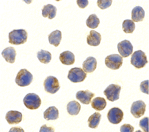 |
| Immunocytochemistry of NAK in MOLT4 cells with NAK antibody at 10 µg/mL. |
FAQ & Publications
Frequently Asked Questions
What applications is the rabbit anti-NAK/TBK1 (CT) polyclonal antibody 4716 validated for?
This antibody is validated for use in ELISA, Western blot (WB), and immunocytochemistry/immunofluorescence (ICC/IF) applications.
How should the rabbit anti-NAK/TBK1 (CT) antibody 4716 be stored to ensure stability?
The antibody is stable for at least one year when stored at -20°C. It is recommended to avoid multiple freeze-thaw cycles to maintain antibody integrity.
What species does the immunogen peptide for this antibody correspond to, and is there cross-reactivity?
The immunogen is a peptide corresponding to amino acids 712-727 of human NAK/TBK1, which is identical to the mouse sequence, indicating reactivity with both human and mouse NAK/TBK1.
What is the recommended dilution for using this antibody in immunoblotting, and what size band is expected?
For immunoblotting, the antibody should be used at 0.5-1 µg/mL. It detects a band at approximately 84 kDa, with MOLT4 cell lysate serving as a positive control.
What type of antibody is the rabbit anti-NAK/TBK1 (CT) 4716 in terms of host and clonality?
This product is a rabbit-derived polyclonal antibody of the IgG isotype.
Publications
| pmid | title | authors | citation |
|---|---|---|---|
| We haven't added any publications to our database yet. | |||
Published literature highly relevant to the biological target of this product and referencing this antibody or clone are retrieved from the PubMed database provided by the United States National Library of Medicine at the National Institutes of Health.
Protocols
| relevant to this product |
|---|
| Western blot IHC ICC |
Documents
| Batch Number | QC File | SDS |
|---|---|---|
| To view batch-specific Safety Datasheets and Quality Certificates associated with your account, please Log In. | ||
Only logged in customers who have purchased this product may leave a review.


Reviews
There are no reviews yet.