| Weight | 1 lbs |
|---|---|
| Dimensions | 9 × 5 × 2 in |
| host | rabbit |
| isotype | IgG |
| clonality | polyclonal |
| concentration | 1 mg/mL |
| applications | ICC/IF, WB |
| reactivity | MyD88 (IN) |
| available sizes | 100 µg |


rabbit anti-MYD88 (IN) polyclonal antibody 5407
$445.00
Antibody summary
- Rabbit polyclonal to MYD88 (IN)
- Suitable for: ELISA,WB,IHC-P,IF,IP
- Isotype: IgG
- 100 µg
rabbit anti-MYD88 (IN) polyclonal antibody 5407
| antibody |
|---|
| Tested applications WB,IHC,IHC,ICC/IF,ELISA |
| Recommended dilutions Immunoblotting: use at 1:500-1:1,000 dilution. Positive control: Whole cell lysate from Jurkat cells. |
| Immunogen Peptide corresponding to aa 233-248 of human MyD88. The sequence differs from mouse MyD88 by two amino acids. |
| Size and concentration 100µg and lot specific |
| Form liquid |
| Storage Instructions This antibody is stable for at least one (1) year at -20°C. Avoid multiple freeze- thaw cycles. |
| Storage buffer PBS, pH 7.4. |
| Purity peptide affinity purification |
| Clonality polyclonal |
| Isotype IgG |
| Compatible secondaries goat anti-rabbit IgG, H&L chain specific, peroxidase conjugated, conjugated polyclonal antibody 9512 goat anti-rabbit IgG, H&L chain specific, biotin conjugated polyclonal antibody 2079 goat anti-rabbit IgG, H&L chain specific, FITC conjugated polyclonal antibody 7863 goat anti-rabbit IgG, H&L chain specific, Cross Absorbed polyclonal antibody 2371 goat anti-rabbit IgG, H&L chain specific, biotin conjugated polyclonal antibody, crossabsorbed 1715 goat anti-rabbit IgG, H&L chain specific, FITC conjugated polyclonal antibody, crossabsorbed 1720 |
| Isotype control Rabbit polyclonal - Isotype Control |
| target relevance |
|---|
| Homo sapiens MYD88 Myeloid differentiation primary response protein MyD88 |
| Protein names Myeloid differentiation primary response protein MyD88 |
| Gene names MYD88 |
| Function Adapter protein involved in the Toll-like receptor and IL-1 receptor signaling pathway in the innate immune response (PubMed:15361868, PubMed:18292575, PubMed:33718825, PubMed:37971847). Acts via IRAK1, IRAK2, IRF7 and TRAF6, leading to NF-kappa-B activation, cytokine secretion and the inflammatory response (PubMed:15361868, PubMed:19506249, PubMed:24316379, PubMed:40638072). Increases IL-8 transcription (PubMed:9013863). Involved in IL-18-mediated signaling pathway. Activates IRF1 resulting in its rapid migration into the nucleus to mediate an efficient induction of IFN-beta, NOS2/INOS, and IL12A genes. Upon TLR8 activation by GU-rich single-stranded RNA (GU-rich RNA) derived from viruses such as SARS-CoV-2, SARS-CoV and HIV-1, induces IL1B release through NLRP3 inflammasome activation (PubMed:33718825). MyD88-mediated signaling in intestinal epithelial cells is crucial for maintenance of gut homeostasis and controls the expression of the antimicrobial lectin REG3G in the small intestine (By similarity) |
| Subcellular location Cytoplasm, Nucleus |
| Structure (Microbial infection) Interacts with human cytomegalovirus protein UL88; this interaction degrades MYD88 and reduces innate immune activation |
| Post-translational modification Ubiquitinated; undergoes 'Lys-63'-linked polyubiquitination. OTUD4 specifically hydrolyzes 'Lys-63'-linked polyubiquitinated MYD88 (PubMed:29395066). Deubiquitinated by USP3 that cleaves 'Lys-63'-linked ubiquitin chains leading to inhibition of MYD88-induced NF-kappa-B signaling (PubMed:37971847) (Microbial infection) Ubiquitinated by human herpesvirus 8 (KSHV) protein RTA/ORF50, leading to proteasomal degradation and suppression of TLR4 signaling pathway |
| Involvement in disease Immunodeficiency 68 An autosomal recessive primary immunodeficiency characterized by life-threatening, often recurrent, pyogenic bacterial infections, including invasive pneumococcal disease, beginning in infancy or early childhood. Macroglobulinemia, Waldenstrom, 1 A malignant B-cell neoplasm characterized by lymphoplasmacytic infiltration of the bone marrow and hypersecretion of monoclonal immunoglobulin M (IgM) protein. Clinical features are variable and include anemia, thrombocytopenia, hepatosplenomegaly, and lymphadenopathy. Many patients have asymptomatic or indolent disease. |
| Keywords 3D-structure, Alternative splicing, Antiviral defense, Cytoplasm, Disease variant, Immunity, Inflammatory response, Innate immunity, Nucleus, Phosphoprotein, Proteomics identification, Reference proteome, Ubl conjugation |
| Sequence MAAGGPGAGSAAPVSSTSSLPLAALNMRVRRRLSLFLNVRTQVAADWTALAEEMDFEYLE IRQLETQADPTGRLLDAWQGRPGASVGRLLELLTKLGRDDVLLELGPSIEEDCQKYILKQ QQEEAEKPLQVAAVDSSVPRTAELAGITTLDDPLGHMPERFDAFICYCPSDIQFVQEMIR QLEQTNYRLKLCVSDRDVLPGTCVWSIASELIEKRCRRMVVVVSDDYLQSKECDFQTKFA LSLSPGAHQKRLIPIKYKAMKKEFPSILRFITVCDYTNPCTKSWFWTRLAKALSLP |
| UniProt accession: Q99836 |
Data
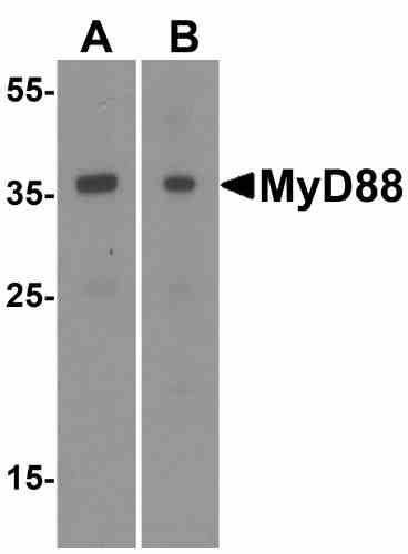 |
| Western Blot Validation of MyD88 in HeLa (A) and Jurket (B) Cells Loading: 15 µg of lysates per lane. Antibodies: 5407 (1 µg/mL) 1 h incubation at RT in 5% NFDM/TBST.Secondary: Goat anti-rabbit IgG HRP conjugate at 1:10000 dilution. |
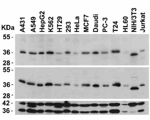 |
| Independent Antibody Validation (IAV) via Protein Expression Profile in Cell Lines Loading: 15 µg of lysates per lane. Antibodies: MyD88 5407 (2 µg/mL), MyD88 2127 (2 µg/mL), beta-actin (1 µg/mL), and GAPDH (0.02 µg/mL), 1 h incubation at RT in 5% NFDM/TBST.Secondary: Goat anti-rabbit IgG HRP conjugate at 1:10000 dilution. |
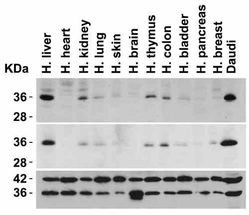 |
| Independent Antibody Validation (IAV) via Protein Expression Profile in Human Tissues Loading: 15 µg of lysates per lane. Antibodies: MyD88 5407 (2 µg/mL), MyD88 2127 (2 µg/mL), beta-actin (1 µg/mL), and GAPDH (0.02 µg/mL), 1 h incubation at RT in 5% NFDM/TBST.Secondary: Goat anti-rabbit IgG HRP conjugate at 1:10000 dilution. |
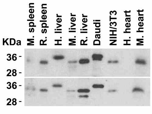 |
| Animal Species Reactivity Loading: Lysates/proteins at 15 µg per lane. Antibodies: 5407 (2 µg/mL) or 2127 (2 µg/mL). 1 h incubation at RT in 5% NFDM/TBST.Secondary: Goat anti-rabbit IgG HRP conjugate at 1:10000 dilution. |
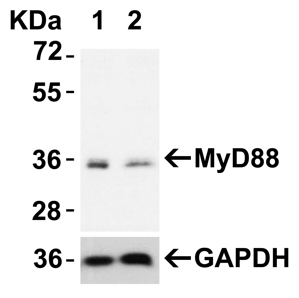 |
| Validation with MyD88 siRNA Knockdown in HeLa Cells HeLa cells were transfected with control siRNAs (lane 1) or MyD88 siRNAs (lane 2) Loading: 10 µg of HeLa whole cell lysates per lane. Antibodies: 5407 (2 µg/mL), 1 h incubation at RT in 5% NFDM/TBST.Secondary: Goat anti-rabbit IgG HRP conjugate at 1:10000 dilution. |
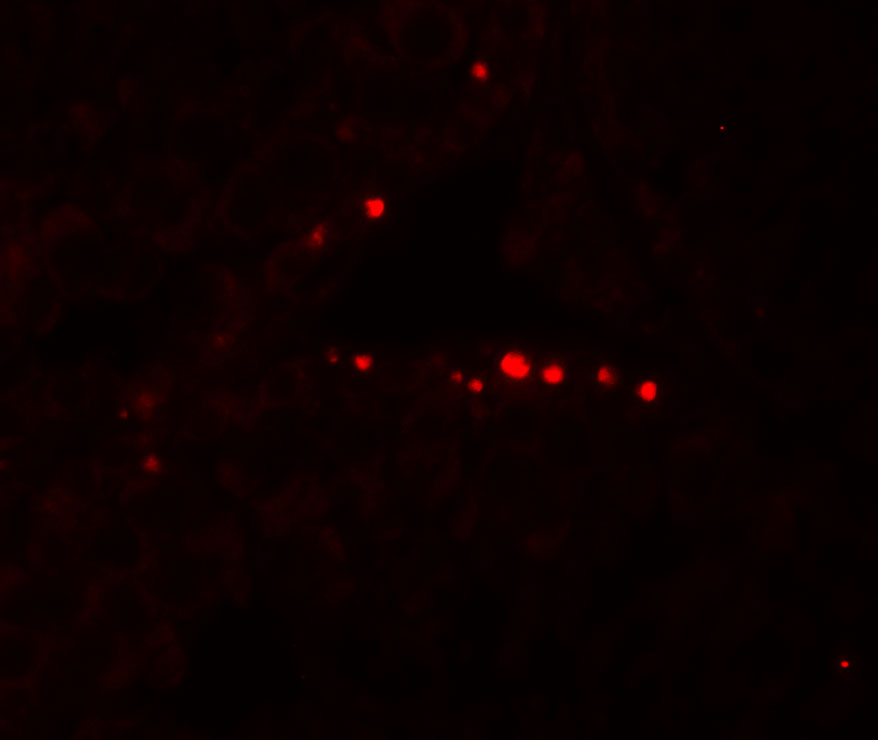 |
| Immunofluorescence Validation of MyD88 n Human Testis Immunofluorescent analysis of 4% paraformaldehyde-fixed human testis tissue labeling MyD88 with 5407 at 20 µg/mL, followed by goat anti-rabbit IgG secondary antibody at 1/500 dilution (red). Image showing nucleus staining on human testis cells. |
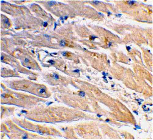 |
| Immunohistochemistry Validation of MyD88 in Human Heart Immunohistochemical analysis of paraffin-embedded human heart tissue using anti-MyD88 antibody (5407) at 2 µg/mL. Tissue was fixed with formaldehyde and blocked with 10% serum for 1 h at RT; antigen retrieval was by heat mediation with a citrate buffer (pH6). Samples were incubated with primary antibody overnight at 4C. A goat anti-rabbit IgG H&L (HRP) at 1/250 was used as secondary. Counter stained with Hematoxylin. |
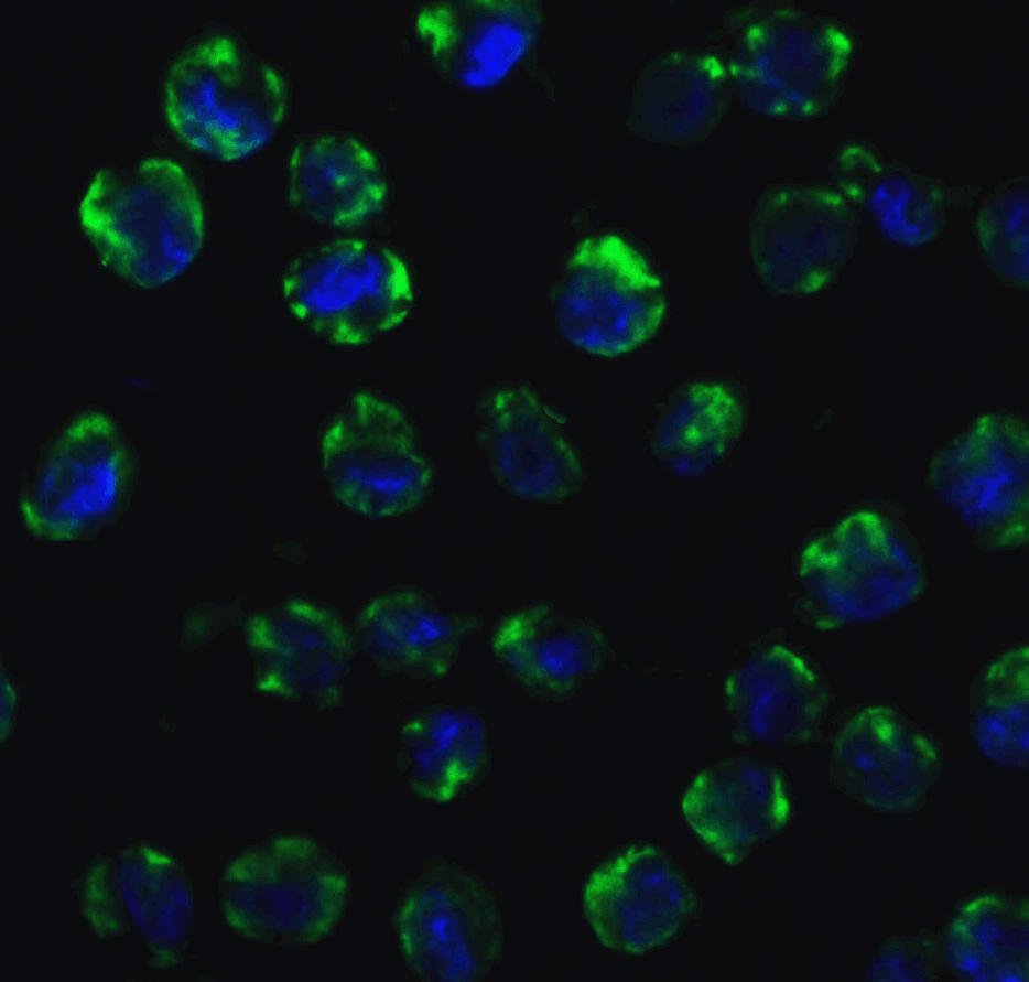 |
| Immunofluorescence Validation of MyD88 in K562 Cells Immunofluorescent analysis of 4% paraformaldehyde-fixed K562 cells labeling MyD88 with 5407 at 10 µg/mL, followed by Goat anti-rabbit IgG secondary antibody at 1/500 dilution (green) and DAPI staining (blue). |
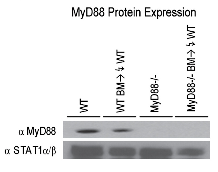 |
| KO Validation in Mouse Macrophages (Miller et al., 2006) Bone marrow-derived macrophages from wild type (WT) mice and MyD88 knockout mice were assessed for MyD88 protein expression by anti-MyD88 antibodies. MyD88 expression was detected in WT mice, but not in MyD88 knockout mice. |
FAQ & Publications
Frequently Asked Questions
What applications has the rabbit anti-MYD88 (IN) polyclonal antibody 5407 been validated for?
The rabbit anti-MYD88 (IN) polyclonal antibody 5407 is suitable and tested for applications including Western Blot (WB), Immunohistochemistry (IHC-P), Immunofluorescence (IF/ICC), Immunoprecipitation (IP), and ELISA.
How should the rabbit anti-MYD88 (IN) polyclonal antibody 5407 be stored to maintain stability?
This antibody should be stored at -20°C and is stable for at least one year under these conditions. It is important to avoid multiple freeze-thaw cycles to maintain antibody integrity.
Publications
| pmid | title | authors | citation |
|---|---|---|---|
| We haven't added any publications to our database yet. | |||
Published literature highly relevant to the biological target of this product and referencing this antibody or clone are retrieved from the PubMed database provided by the United States National Library of Medicine at the National Institutes of Health.
Protocols
| relevant to this product |
|---|
| Western blot IHC ICC |
Documents
| Batch Number | QC File | SDS |
|---|---|---|
| To view batch-specific Safety Datasheets and Quality Certificates associated with your account, please Log In. | ||
Only logged in customers who have purchased this product may leave a review.


Reviews
There are no reviews yet.