| Weight | 1 lbs |
|---|---|
| Dimensions | 9 × 5 × 2 in |
| host | rabbit |
| isotype | IgG |
| clonality | polyclonal |
| concentration | 1 mg/mL |
| applications | ICC/IF, WB |
| reactivity | Livin |
| available sizes | 100 µg |


rabbit anti-Livin polyclonal antibody 6637
$445.00
Antibody summary
- Rabbit polyclonal to Livin
- Suitable for: ELISA,WB,IHC-P,IF
- Isotype: IgG
- 100 µg
rabbit anti-Livin polyclonal antibody 6637
| antibody |
|---|
| Tested applications WB,IHC,IHC,ICC/IF,ELISA |
| Recommended dilutions Immunoblotting: use at 0.5-1.0ug/mL. Positive control: Raji cell lysate. Immunohistochemistry: use at 5ug/mL. |
| Immunogen Peptide corresponding to aa 264- 280 of the short form and aa 281-298 of the long form of human Livin (accession no. |
| Size and concentration 100µg and lot specific |
| Form liquid |
| Storage Instructions This antibody is stable for at least one (1) year at -20°C. Avoid multiple freeze-thaw cycles. |
| Storage buffer PBS, pH 7.4. |
| Purity peptide affinity purification |
| Clonality polyclonal |
| Isotype IgG |
| Compatible secondaries goat anti-rabbit IgG, H&L chain specific, peroxidase conjugated, conjugated polyclonal antibody 9512 goat anti-rabbit IgG, H&L chain specific, biotin conjugated polyclonal antibody 2079 goat anti-rabbit IgG, H&L chain specific, FITC conjugated polyclonal antibody 7863 goat anti-rabbit IgG, H&L chain specific, Cross Absorbed polyclonal antibody 2371 goat anti-rabbit IgG, H&L chain specific, biotin conjugated polyclonal antibody, crossabsorbed 1715 goat anti-rabbit IgG, H&L chain specific, FITC conjugated polyclonal antibody, crossabsorbed 1720 |
| Isotype control Rabbit polyclonal - Isotype Control |
| target relevance |
|---|
| Homo sapiens BIRC7 Baculoviral IAP repeat-containing protein 7 |
| Protein names Baculoviral IAP repeat-containing protein 7 |
| Alternative names Kidney inhibitor of apoptosis protein, Livin, Melanoma inhibitor of apoptosis protein, RING finger protein 50, RING-type E3 ubiquitin transferase BIRC7 |
| Gene names BIRC7 |
| Protein family Belongs to the IAP family |
| Function Apoptotic regulator capable of exerting proapoptotic and anti-apoptotic activities and plays crucial roles in apoptosis, cell proliferation, and cell cycle control (PubMed:11024045, PubMed:11084335, PubMed:11162435, PubMed:16729033, PubMed:17294084). Its anti-apoptotic activity is mediated through the inhibition of CASP3, CASP7 and CASP9, as well as by its E3 ubiquitin-protein ligase activity (PubMed:11024045, PubMed:16729033). As it is a weak caspase inhibitor, its anti-apoptotic activity is thought to be due to its ability to ubiquitinate DIABLO/SMAC targeting it for degradation thereby promoting cell survival (PubMed:16729033). May contribute to caspase inhibition, by blocking the ability of DIABLO/SMAC to disrupt XIAP/BIRC4-caspase interactions (PubMed:16729033). Protects against apoptosis induced by TNF or by chemical agents such as adriamycin, etoposide or staurosporine (PubMed:11084335, PubMed:11162435, PubMed:11865055). Suppression of apoptosis is mediated by activation of MAPK8/JNK1, and possibly also of MAPK9/JNK2 (PubMed:11865055). This activation depends on TAB1 and MAP3K7/TAK1 (PubMed:11865055). In vitro, inhibits CASP3 and proteolytic activation of pro-CASP9 (PubMed:11024045) |
| Catalytic activity S-ubiquitinyl-[E2 ubiquitin-conjugating enzyme]-L-cysteine + [acceptor protein]-L-lysine = [E2 ubiquitin-conjugating enzyme]-L-cysteine + N(6)-ubiquitinyl-[acceptor protein]-L-lysine. |
| Subcellular location Nucleus, Cytoplasm, Golgi apparatus |
| Structure Binds to CASP9. Interaction with DIABLO/SMAC via the BIR domain disrupts binding to CASP9 and apoptotic suppressor activity. Interacts with TAB1. In vitro, interacts with CASP3 and CASP7 via its BIR domain |
| Post-translational modification Autoubiquitinated and undergoes proteasome-mediated degradation The truncated protein (tLivin) not only loses its anti-apoptotic effect but also acquires a pro-apoptotic effect |
| Keywords 3D-structure, Alternative splicing, Apoptosis, Cytoplasm, Golgi apparatus, Metal-binding, Nucleus, Protease inhibitor, Proteomics identification, Reference proteome, Thiol protease inhibitor, Transferase, Ubl conjugation, Ubl conjugation pathway, Zinc, Zinc-finger |
| Sequence MGPKDSAKCLHRGPQPSHWAAGDGPTQERCGPRSLGSPVLGLDTCRAWDHVDGQILGQLR PLTEEEEEEGAGATLSRGPAFPGMGSEELRLASFYDWPLTAEVPPELLAAAGFFHTGHQD KVRCFFCYGGLQSWKRGDDPWTEHAKWFPSCQFLLRSKGRDFVHSVQETHSQLLGSWDPW EEPEDAAPVAPSVPASGYPELPTPRREVQSESAQEPGGVSPAEAQRAWWVLEPPGARDVE AQLRRLQEERTCKVCLDRAVSIVFVPCGHLVCAECAPGLQLCPICRAPVRSRVRTFLS |
| UniProt accession: Q96CA5 |
Data
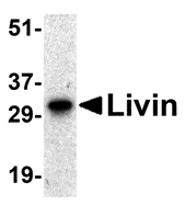 |
| Western blot analysis of Livin expression in human Raji cell lysate with Livin antibody at 0.5 µg/mL. |
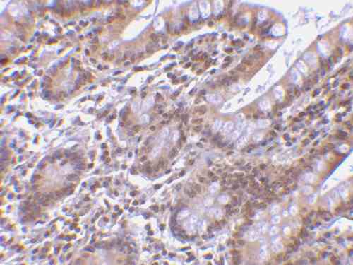 |
| Immunohistochemistry of Livin in human small intestine tissue with Livin antibody at 5 µg/mL. |
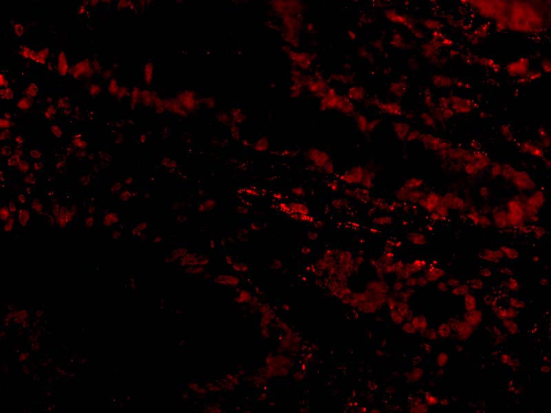 |
| Immunofluorescence of Livin in Human Small Intestine cells with Livin antibody at 20 µg/mL. |
FAQ & Publications
Frequently Asked Questions
What applications is the rabbit anti-Livin polyclonal antibody 6637 validated for?
This antibody is suitable for use in ELISA, Western blotting (WB), immunohistochemistry on paraffin-embedded tissues (IHC-P), and immunofluorescence (IF). Recommended dilutions are 0.5-1.0 µg/mL for immunoblotting and 5 µg/mL for immunohistochemistry.
How should the rabbit anti-Livin polyclonal antibody 6637 be stored to maintain stability?
The antibody should be stored at -20°C and is stable for at least one year under these conditions. It is important to avoid multiple freeze-thaw cycles to preserve antibody integrity.
What is the immunogen used to generate the rabbit anti-Livin polyclonal antibody 6637?
The immunogen corresponds to a peptide spanning amino acids 264-280 of the short form and amino acids 281-298 of the long form of human Livin protein (accession number provided in product details).
Publications
| pmid | title | authors | citation |
|---|---|---|---|
| We haven't added any publications to our database yet. | |||
Published literature highly relevant to the biological target of this product and referencing this antibody or clone are retrieved from the PubMed database provided by the United States National Library of Medicine at the National Institutes of Health.
Protocols
| relevant to this product |
|---|
| Western blot IHC ICC |
Documents
| Batch Number | QC File | SDS |
|---|---|---|
| To view batch-specific Safety Datasheets and Quality Certificates associated with your account, please Log In. | ||
Only logged in customers who have purchased this product may leave a review.


Reviews
There are no reviews yet.