| Weight | 1 lbs |
|---|---|
| Dimensions | 9 × 5 × 2 in |
| host | rabbit |
| isotype | IgG |
| clonality | polyclonal |
| concentration | 1 mg/mL |
| applications | ICC/IF, WB |
| reactivity | IRAK-M |
| available sizes | 100 µg |


rabbit anti-IRAK-M BP polyclonal antibody 2359
$445.00
Antibody summary
- Rabbit polyclonal to IRAK-M BP
- Suitable for: ELISA,WB,IHC-P,IF
- Isotype: IgG
- 100 µg
rabbit anti-IRAK-M BP polyclonal antibody 2359
| antibody |
|---|
| Tested applications WB,IHC,IHC,ICC/IF,ELISA |
| Recommended dilutions Immunoblotting: use at 1:500-1:1,000 dilution. Positive control: Tissue lysate from mouse spleen or rat kidney. |
| Immunogen Peptide corresponding to aa 581-596 of human IRAK-M. |
| Size and concentration 100µg and lot specific |
| Form liquid |
| Storage Instructions This antibody is stable for at least one (1) year at -20°C. Avoid multiple freeze- thaw cycles. |
| Storage buffer PBS, pH 7.4. |
| Purity peptide affinity purification |
| Clonality polyclonal |
| Isotype IgG |
| Compatible secondaries goat anti-rabbit IgG, H&L chain specific, peroxidase conjugated, conjugated polyclonal antibody 9512 goat anti-rabbit IgG, H&L chain specific, biotin conjugated polyclonal antibody 2079 goat anti-rabbit IgG, H&L chain specific, FITC conjugated polyclonal antibody 7863 goat anti-rabbit IgG, H&L chain specific, Cross Absorbed polyclonal antibody 2371 goat anti-rabbit IgG, H&L chain specific, biotin conjugated polyclonal antibody, crossabsorbed 1715 goat anti-rabbit IgG, H&L chain specific, FITC conjugated polyclonal antibody, crossabsorbed 1720 |
| Isotype control Rabbit polyclonal - Isotype Control |
| target relevance |
|---|
| Homo sapiens IRAK3 Interleukin-1 receptor-associated kinase 3 |
| Protein names Interleukin-1 receptor-associated kinase 3 |
| Alternative names IL-1 receptor-associated kinase M, Inactive IL-1 receptor-associated kinase 3 |
| Gene names IRAK3 |
| Protein family Belongs to the protein kinase superfamily. TKL Ser/Thr protein kinase family. Pelle subfamily |
| Function Putative inactive protein kinase which regulates signaling downstream of immune receptors including IL1R and Toll-like receptors (PubMed:10383454, PubMed:29686383). Inhibits dissociation of IRAK1 and IRAK4 from the Toll-like receptor signaling complex by either inhibiting the phosphorylation of IRAK1 and IRAK4 or stabilizing the receptor complex (By similarity). Upon IL33-induced lung inflammation, positively regulates expression of IL6, CSF3, CXCL2 and CCL5 mRNAs in dendritic cells (PubMed:29686383) |
| Subcellular location Cytoplasm, Nucleus |
| Structure Monomer (PubMed:33238146). Homodimer; disulfide-linked (PubMed:33238146). May interact with IRAK4 (when phosphorylated) (PubMed:33238146). Interacts (when phosphorylated at Ser-110) with PIN1 (via WW domain) in response to IL33-mediated (but not TLR4 ligand LPS) dendritic cell stimulation (PubMed:29686383) |
| Involvement in disease Asthma-related traits 5 Asthma-related traits include clinical symptoms of asthma, such as coughing, wheezing, dyspnea, bronchial hyperresponsiveness as assessed by methacholine challenge test, serum IgE levels, atopy and atopic dermatitis. |
| Keywords 3D-structure, Alternative splicing, Asthma, ATP-binding, Cytoplasm, Disulfide bond, Nucleotide-binding, Nucleus, Phosphoprotein, Proteomics identification, Reference proteome |
| Sequence MAGNCGARGALSAHTLLFDLPPALLGELCAVLDSCDGALGWRGLAERLSSSWLDVRHIEK YVDQGKSGTRELLWSWAQKNKTIGDLLQVLQEMGHRRAIHLITNYGAVLSPSEKSYQEGG FPNILFKETANVTVDNVLIPEHNEKGILLKSSISFQNIIEGTRNFHKDFLIGEGEIFEVY RVEIQNLTYAVKLFKQEKKMQCKKHWKRFLSELEVLLLFHHPNILELAAYFTETEKFCLI YPYMRNGTLFDRLQCVGDTAPLPWHIRIGILIGISKAIHYLHNVQPCSVICGSISSANIL LDDQFQPKLTDFAMAHFRSHLEHQSCTINMTSSSSKHLWYMPEEYIRQGKLSIKTDVYSF GIVIMEVLTGCRVVLDDPKHIQLRDLLRELMEKRGLDSCLSFLDKKVPPCPRNFSAKLFC LAGRCAATRAKLRPSMDEVLNTLESTQASLYFAEDPPTSLKSFRCPSPLFLENVPSIPVE DDESQNNNLLPSDEGLRIDRMTQKTPFECSQSEVMFLSLDKKPESKRNEEACNMPSSSCE ESWFPKYIVPSQDLRPYKVNIDPSSEAPGHSCRSRPVESSCSSKFSWDEYEQYKKE |
| UniProt accession: Q9Y616 |
Data
 |
| Western blot analysis of IRAK-M in (M) mouse spleen and (R) rat liver tissue lysates with IRAK-M antibody at 1 µg/mL. |
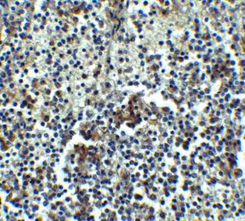 |
| Immunohistochemistry of IRAK-M in human spleen tissue with IRAK-M antibody at 5 µg/mL. |
 |
| Immunofluorescence of IRAK-M in Rat Liver tissue with IRAK-M antibody at 10 µg/mL. |
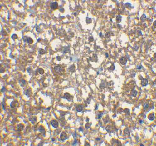 |
| Immunohistochemical staining of rat liver tissue using IRAK-M antibody at 2 µg/mL. |
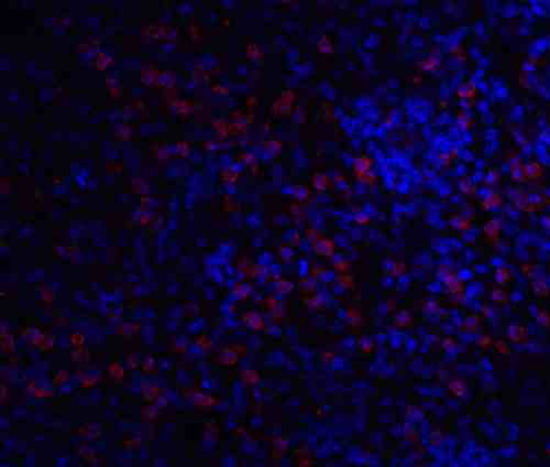 |
| Immunofluorescence of IRAK2 in A20 cells with IRAK2 antibody at 20 µg/mL.
Red: IRAK-M Antibody (2359) Blue: DAPI staining |
FAQ & Publications
Frequently Asked Questions
What applications has the rabbit anti-IRAK-M BP polyclonal antibody 2359 been validated for?
This antibody has been tested and validated for use in Western blotting (WB), immunohistochemistry (IHC-P), immunofluorescence (IF/ICC), and ELISA assays.
How should the rabbit anti-IRAK-M BP polyclonal antibody 2359 be stored to maintain its stability?
The antibody should be stored at -20°C and is stable for at least one year under these conditions. It is important to avoid multiple freeze-thaw cycles to preserve antibody integrity.
Publications
| pmid | title | authors | citation |
|---|---|---|---|
| We haven't added any publications to our database yet. | |||
Published literature highly relevant to the biological target of this product and referencing this antibody or clone are retrieved from the PubMed database provided by the United States National Library of Medicine at the National Institutes of Health.
Protocols
| relevant to this product |
|---|
| Western blot IHC ICC |
Documents
| Batch Number | QC File | SDS |
|---|---|---|
| To view batch-specific Safety Datasheets and Quality Certificates associated with your account, please Log In. | ||
Only logged in customers who have purchased this product may leave a review.


Reviews
There are no reviews yet.