| Weight | 1 lbs |
|---|---|
| Dimensions | 9 × 5 × 2 in |
| host | rabbit |
| isotype | IgG |
| clonality | polyclonal |
| concentration | 1 mg/mL |
| applications | IP |
| reactivity | human |
| available sizes | 10 µL, 100 µL |

rabbit anti-HO1 polyclonal antibody 1028
Price range: $100.00 through $2,600.00
Antibody summary
- Rabbit polyclonal to HO-1 (Heme oxygenase-1)
- Suitable for: IP
- Reacts with: Hu
- Isotype: IgG
- 100 µL, 10 µl
rabbit anti- ho1 polyclonal antibody 1028
| target relevance |
|---|
| Protein names Heme oxygenase 1 (HO-1) (EC 1.14.14.18) [Cleaved into: Heme oxygenase 1 soluble form] |
| Gene names HMOX1,HMOX1 HO HO1 |
| Protein family Heme oxygenase family |
| Mass 32819Da |
| Function FUNCTION: [Heme oxygenase 1]: Catalyzes the oxidative cleavage of heme at the alpha-methene bridge carbon, released as carbon monoxide (CO), to generate biliverdin IXalpha, while releasing the central heme iron chelate as ferrous iron (PubMed:11121422, PubMed:19556236, PubMed:7703255). Affords protection against programmed cell death and this cytoprotective effect relies on its ability to catabolize free heme and prevent it from sensitizing cells to undergo apoptosis (PubMed:20055707). {ECO:0000269|PubMed:11121422, ECO:0000269|PubMed:19556236, ECO:0000269|PubMed:7703255, ECO:0000303|PubMed:20055707}.; FUNCTION: [Heme oxygenase 1]: (Microbial infection) During SARS-COV-2 infection, promotes SARS-CoV-2 ORF3A-mediated autophagy but is unlikely to be required for ORF3A-mediated induction of reticulophagy. {ECO:0000269|PubMed:35239449}.; FUNCTION: [Heme oxygenase 1 soluble form]: Catalyzes the oxidative cleavage of heme at the alpha-methene bridge carbon, released as carbon monoxide (CO), to generate biliverdin IXalpha, while releasing the central heme iron chelate as ferrous iron. {ECO:0000269|PubMed:7703255}. |
| Catalytic activity CATALYTIC ACTIVITY: Reaction=heme b + 3 reduced [NADPH--hemoprotein reductase] + 3 O2 = biliverdin IXalpha + CO + Fe(2+) + 3 oxidized [NADPH--hemoprotein reductase] + 3 H2O + H(+); Xref=Rhea:RHEA:21764, Rhea:RHEA-COMP:11964, Rhea:RHEA-COMP:11965, ChEBI:CHEBI:15377, ChEBI:CHEBI:15378, ChEBI:CHEBI:15379, ChEBI:CHEBI:17245, ChEBI:CHEBI:29033, ChEBI:CHEBI:57618, ChEBI:CHEBI:57991, ChEBI:CHEBI:58210, ChEBI:CHEBI:60344; EC=1.14.14.18; Evidence={ECO:0000269|PubMed:11121422, ECO:0000269|PubMed:19556236, ECO:0000269|PubMed:7703255}; PhysiologicalDirection=left-to-right; Xref=Rhea:RHEA:21765; Evidence={ECO:0000305|PubMed:7703255}; |
| Subellular location SUBCELLULAR LOCATION: Endoplasmic reticulum membrane {ECO:0000269|PubMed:19556236, ECO:0000269|PubMed:22419571, ECO:0000269|PubMed:27184847}; Single-pass type IV membrane protein {ECO:0000255}; Cytoplasmic side {ECO:0000269|PubMed:22419571}. |
| Tissues TISSUE SPECIFICITY: Expressed at higher levels in renal cancer tissue than in normal tissue (at protein level). {ECO:0000269|PubMed:20855888}. |
| Structure SUBUNIT: (Microbial infection) Interacts with SARS-CoV-2 ORF3A protein; the interaction promotes ORF3A-induced autophagy but is unlikely to be involved in ORF3A-mediated induction of reticulophagy. {ECO:0000269|PubMed:35239449}.; SUBUNIT: Homodimer and higher order homooligomer (PubMed:19556236). Oligomerization is crucial for its stability and function in the endoplasmic reticulum (PubMed:19556236). Interacts with FLVCR2; this interaction is potentiated in the presence of heme. {ECO:0000250|UniProtKB:P14901, ECO:0000269|PubMed:19556236}. |
| Post-translational modification PTM: A soluble form arises by proteolytic removal of the membrane anchor. {ECO:0000305|PubMed:7703255}. |
| Domain DOMAIN: The transmembrane domain is necessary for its oligomerization. {ECO:0000269|PubMed:19556236}. |
| Involvement in disease DISEASE: Heme oxygenase 1 deficiency (HMOX1D) [MIM:614034]: A disease characterized by impaired stress hematopoiesis, resulting in marked erythrocyte fragmentation and intravascular hemolysis, coagulation abnormalities, endothelial damage, and iron deposition in renal and hepatic tissues. Clinical features include persistent hemolytic anemia, asplenia, nephritis, generalized erythematous rash, growth retardation and hepatomegaly. {ECO:0000269|PubMed:9884342}. Note=The disease is caused by variants affecting the gene represented in this entry. |
| Target Relevance information above includes information from UniProt accession: P09601 |
| The UniProt Consortium |
Data
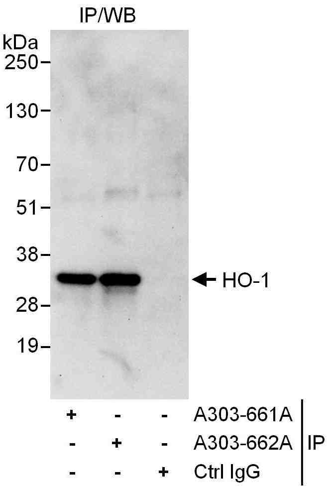 |
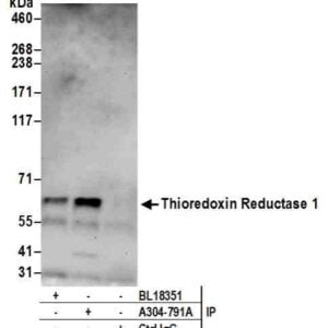
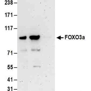
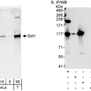
| Detection of human HO-1 by western blot of immunoprecipitates. Samples: Whole cell lysate (1 mg for IP, 20% of IP loaded) from HeLa cells. Antibodies: Affinity purified rabbit anti-HO-1 antibody #1028 used for IP at 6 µg/mg lysate. HO-1 was also immunoprecipitated by rabbit anti-HO-1 antibody, which recognizes a downstream epitope. For blotting immunoprecipitated HO-1 was used at 1 µg/ml. Detection: Chemiluminescence with an exposure time of 30 seconds. |
Publications
| pmid | title | authors | citation |
|---|---|---|---|
| We haven't added any publications to our database yet. | |||
Protocols
| relevant to this product |
|---|
| Western blot |
Documents
| # | SDS | Certificate | |
|---|---|---|---|
| Please enter your product and batch number here to retrieve product datasheet, SDS, and QC information. | |||
Only logged in customers who have purchased this product may leave a review.

Reviews
There are no reviews yet.