| Weight | 1 lbs |
|---|---|
| Dimensions | 9 × 5 × 2 in |
| host | rabbit |
| isotype | IgG |
| clonality | polyclonal |
| concentration | 1 mg/mL |
| applications | ICC/IF, WB |
| reactivity | CXCR4 (NT) |
| available sizes | 100 µg |


rabbit anti-HIV Co-Receptor CXCR4 (NT) polyclonal antibody 6344
$445.00
Antibody summary
- Rabbit polyclonal to HIV Co-Receptor CXCR4 (NT)
- Suitable for: ELISA,WB,ICC,IP,IF,FC,IHC-P
- Isotype: IgG
- 100 µg
rabbit anti-HIV Co-Receptor CXCR4 (NT) polyclonal antibody 6344
| antibody |
|---|
| Tested applications WB,IHC,IHC,ICC/IF,ELISA,IP |
| Recommended dilutions Immunoblotting: use at 1:1,000-1:2,000 dilution. Immunoprecipitation: use at 1:1,000- 1:2,000 dilution. Immunocytochemistry: use at 1:1,000- 1:1,2000 dilution. Positive control: Whole cell lysate from HeLa cells. |
| Immunogen Peptide corresponding to the N- terminus of human CXCR4. |
| Size and concentration 100µg and lot specific |
| Form liquid |
| Storage Instructions This antibody is stable for at least one (1) year at -20°C. Avoid multiple freeze-thaw cycles. |
| Storage buffer PBS, pH 7.4. |
| Purity peptide affinity purification |
| Clonality polyclonal |
| Isotype IgG |
| Compatible secondaries goat anti-rabbit IgG, H&L chain specific, peroxidase conjugated, conjugated polyclonal antibody 9512 goat anti-rabbit IgG, H&L chain specific, biotin conjugated polyclonal antibody 2079 goat anti-rabbit IgG, H&L chain specific, FITC conjugated polyclonal antibody 7863 goat anti-rabbit IgG, H&L chain specific, Cross Absorbed polyclonal antibody 2371 goat anti-rabbit IgG, H&L chain specific, biotin conjugated polyclonal antibody, crossabsorbed 1715 goat anti-rabbit IgG, H&L chain specific, FITC conjugated polyclonal antibody, crossabsorbed 1720 |
| Isotype control Rabbit polyclonal - Isotype Control |
| target relevance |
|---|
| Homo sapiens CXCR4 C-X-C chemokine receptor type 4 |
| Protein names C-X-C chemokine receptor type 4 |
| Alternative names FB22, Fusin, HM89, LCR1, Leukocyte-derived seven transmembrane domain receptor, Lipopolysaccharide-associated protein 3, NPYRL, Stromal cell-derived factor 1 receptor |
| Gene names CXCR4 |
| Protein family Belongs to the G-protein coupled receptor 1 family |
| Function Receptor for the C-X-C chemokine CXCL12/SDF-1 that transduces a signal by increasing intracellular calcium ion levels and enhancing MAPK1/MAPK3 activation (PubMed:10074102, PubMed:10452968, PubMed:10644702, PubMed:10825158, PubMed:18799424, PubMed:20048153, PubMed:20505072, PubMed:24912431, PubMed:28978524, PubMed:8752280, PubMed:8752281). Ligand binding causes a conformation change that triggers signaling via guanine nucleotide-binding proteins (G proteins) and modulates the activity of downstream effectors, such as adenylate cyclase (PubMed:16725153, PubMed:17197449, PubMed:18799424, PubMed:39093700). CXCR4 is coupled to G(i) G alpha proteins and mediates inhibition of adenylate cyclase (PubMed:17197449, PubMed:39093700). Involved in the AKT signaling cascade (PubMed:24912431). Plays a role in regulation of cell migration, e.g. during wound healing (PubMed:28978524). Also acts as a receptor for extracellular ubiquitin; leading to enhanced intracellular calcium ions and reduced cellular cAMP levels (PubMed:20228059). Binds bacterial lipopolysaccharide (LPS) et mediates LPS-induced inflammatory response, including TNF secretion by monocytes (PubMed:11276205). Involved in hematopoiesis and in cardiac ventricular septum formation (By similarity). Also plays an essential role in vascularization of the gastrointestinal tract, probably by regulating vascular branching and/or remodeling processes in endothelial cells (By similarity). Involved in cerebellar development; in the CNS, could mediate hippocampal-neuron surviva (By similarity) |
| Subcellular location Cell membrane, Cell junction, Early endosome, Late endosome, Lysosome |
| Structure (Microbial infection) Interacts with Staphylococcus aureus protein SSL10 |
| Post-translational modification Phosphorylated on agonist stimulation (PubMed:20048153, PubMed:37209686). Phosphorylation of the P-X-P-P motif promotes association with beta-arrestin ARRB1, leading to receptor desensitization and negative regulation of G-protein coupled receptor signaling (PubMed:37209686). Phosphorylation at Ser-324 and Ser-325 leads to recruitment of ITCH, ubiquitination and protein degradation (PubMed:14602072, PubMed:19116316) Ubiquitinated after ligand binding, leading to its degradation (PubMed:28978524). Ubiquitinated by ITCH at the cell membrane on agonist stimulation (PubMed:14602072, PubMed:34927784). The ubiquitin-dependent mechanism, endosomal sorting complex required for transport (ESCRT), then targets CXCR4 for lysosomal degradation. This process is dependent also on prior Ser-/Thr-phosphorylation in the C-terminal of CXCR4. Also binding of ARRB1 to STAM negatively regulates CXCR4 sorting to lysosomes though modulating ubiquitination of SFR5S Sulfation on Tyr-21 is required for efficient binding of CXCL12/SDF-1alpha and promotes its dimerization. Tyr-7 and Tyr-12 are sulfated in a sequential manner after Tyr-21 is almost fully sulfated, with the binding affinity for CXCL12/SDF-1alpha increasing with the number of sulfotyrosines present. Sulfotyrosines Tyr-7 and Tyr-12 occupy clefts on opposing CXCL12 subunits, thus bridging the CXCL12 dimer interface and promoting CXCL12 dimerization O- and N-glycosylated. Asn-11 is the principal site of N-glycosylation. There appears to be very little or no glycosylation on Asn-176. N-glycosylation masks coreceptor function in both X4 and R5 laboratory-adapted and primary HIV-1 strains through inhibiting interaction with their Env glycoproteins. The O-glycosylation chondroitin sulfate attachment does not affect interaction with CXCL12/SDF-1alpha nor its coreceptor activity |
| Involvement in disease WHIM syndrome 1 An autosomal dominant immunologic disease characterized by neutropenia, hypogammaglobulinemia and extensive human papillomavirus (HPV) infection. Despite the peripheral neutropenia, bone marrow aspirates from affected individuals contain abundant mature myeloid cells, a condition termed myelokathexis. |
| Keywords 3D-structure, Alternative splicing, Cell junction, Cell membrane, Disease variant, Disulfide bond, Endosome, G-protein coupled receptor, Glycoprotein, Host cell receptor for virus entry, Host-virus interaction, Isopeptide bond, Lysosome, Membrane, Phosphoprotein, Proteoglycan, Proteomics identification, Receptor, Reference proteome, Sulfation, Transducer, Transmembrane, Transmembrane helix, Ubl conjugation |
| Sequence MEGISIYTSDNYTEEMGSGDYDSMKEPCFREENANFNKIFLPTIYSIIFLTGIVGNGLVI LVMGYQKKLRSMTDKYRLHLSVADLLFVITLPFWAVDAVANWYFGNFLCKAVHVIYTVNL YSSVLILAFISLDRYLAIVHATNSQRPRKLLAEKVVYVGVWIPALLLTIPDFIFANVSEA DDRYICDRFYPNDLWVVVFQFQHIMVGLILPGIVILSCYCIIISKLSHSKGHQKRKALKT TVILILAFFACWLPYYIGISIDSFILLEIIKQGCEFENTVHKWISITEALAFFHCCLNPI LYAFLGAKFKTSAQHALTSVSRGSSLKILSKGKRGGHSSVSTESESSSFHSS |
| UniProt accession: P61073 |
Data
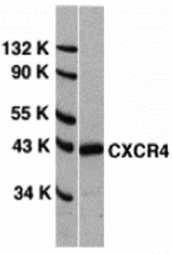 |
| Western Blot Validation of CXCR4 in HeLa Cells Loading: 15 µg of lysates per lane. Antibodies: 6344 (1 µg/mL), 1 h incubation at RT in 5% NFDM/TBST. Secondary: Goat anti-rabbit IgG HRP conjugate at 1:10000 dilution. |
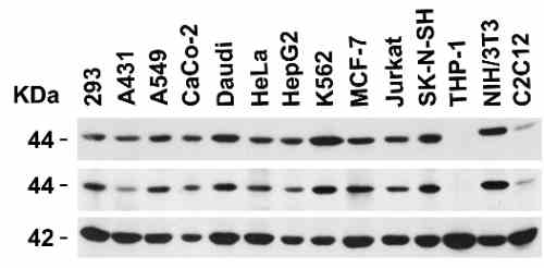 |
| Independent Antibody Validation (IAV) via Protein Expression Profile in Cell Lines Loading: 15 µg of lysates per lane. Antibodies: 6344 (1 µg/mL), 1012 (1 µg/mL), and beta-actin (1 µg/mL), 1 h incubation at RT in 5% NFDM/TBST. Secondary: Goat anti-rabbit IgG HRP conjugate at 1:10000 dilution. |
 |
| Validation with CXCR4 siRNA Knockdown in HeLa Cells HeLa cells were transfected with control siRNAs (lane 1) or CXCR4 siRNAs (lane 2) Loading: 10 µg of HeLa whole cell lysates per lane. Antibodies: 6344 (2 µg/mL), 1 h incubation at RT in 5% NFDM/TBST. Secondary: Goat anti-rabbit IgG HRP conjugate at 1:10000 dilution. |
 |
| Animal Species Reactivity Loading: Lysates/proteins at 20 µg per lane. Antibodies: 6344 (2 µg/mL) or 1012 (2 µg/mL). 1 h incubation at RT in 5% NFDM/TBST. Secondary: Goat anti-rabbit IgG HRP conjugate at 1:10000 dilution. |
 |
| Recombinant Protein Test Loading: CXCR4 partial recombinant protein (Novus Biologicals, Cat# H00007852-Q01). Lane 1: Anti-CXCR4 antibody (0.1 µg/mL) 1 h incubation at RT in 5% NFDM/TBST. Lane 2: Coomassie blue staining. Secondary: Goat anti-rabbit IgG HRP conjugate at 1:10000 dilution. |
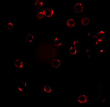 |
| Immunofluorescence Validation of CXCR4 in HeLa Cells Immunofluorescent analysis of 4% paraformaldehyde-fixed HeLa cells labeling CXCR4 with 6344 at 20 µg/mL, followed by goat anti-rabbit IgG secondary antibody at 1/500 dilution (red). Image showing both membrane and cytoplasmic staining on HeLa cells. |
 |
| Flow Cytometry Validation of CXCR4 in HeLa Cells Overlay histogram showing HeLa cells stained with 6344 (red line, 1ug/1x106 cells). 1 h incubation at 4C in 2% FBS/PBS. Followed by secondary antibody 488 goat anti-rabbit IgG (H+L) at 1/500 dilution for 1 h 4C. Isotype control antibody (Green line) was mouse IgG1 (1ug/1x106 cells) used under the same conditions. |
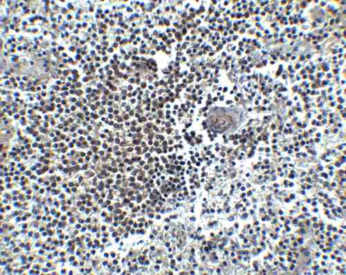 |
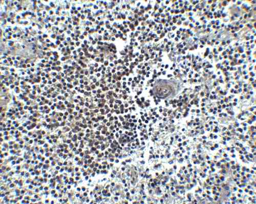
| Immunohistochemistry Validation of CXCR4 in Human Spleen Immunohistochemical analysis of paraffin-embedded human spleen tissue using anti-CXCR4 antibody (6344) at 5 µg/mL. Tissue was fixed with formaldehyde and blocked with 10% serum for 1 h at RT; antigen retrieval was by heat mediation with a citrate buffer (pH6). Samples were incubated with primary antibody overnight at 4C. A Goat anti-rabbit IgG H&L (HRP) at 1/250 was used as secondary. Counter stained with Hematoxylin. |
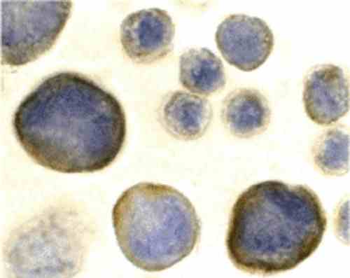 |
| Immunocytochemistry Validation of CXCR4 in HeLa Cells Immunocytochemical analysis of HeLa cells using anti-CXCR4 antibody (6344) at 2 µg/mL. Cells was fixed with formaldehyde and blocked with 10% serum for 1 h at RT; antigen retrieval was by heat mediation with a citrate buffer (pH6). Samples were incubated with primary antibody overnight at 4C. A goat anti-rabbit IgG H&L (HRP) at 1/250 was used as secondary. Counter stained with Hematoxylin. |
FAQ & Publications
Frequently Asked Questions
What applications is the rabbit anti-HIV Co-Receptor CXCR4 (NT) polyclonal antibody 6344 suitable for?
This antibody is suitable for ELISA, Western Blot (WB), Immunocytochemistry (ICC), Immunoprecipitation (IP), Immunofluorescence (IF), Flow Cytometry (FC), and Immunohistochemistry on paraffin-embedded tissues (IHC-P).
How should the rabbit anti-HIV Co-Receptor CXCR4 (NT) antibody be stored to maintain stability?
The antibody should be stored at -20°C and is stable for at least one year under these conditions. It is important to avoid multiple freeze-thaw cycles to preserve antibody integrity.
What is the immunogen used to generate this CXCR4 polyclonal antibody?
The immunogen is a peptide corresponding to the N-terminus of the human CXCR4 protein.
Is the rabbit anti-HIV Co-Receptor CXCR4 (NT) polyclonal antibody validated for use in human cell lines?
Yes, validation data includes Western blotting and immunofluorescence analyses using HeLa human cell lysates, confirming its specificity and utility in human cell models.
Publications
| pmid | title | authors | citation |
|---|---|---|---|
| We haven't added any publications to our database yet. | |||
Published literature highly relevant to the biological target of this product and referencing this antibody or clone are retrieved from the PubMed database provided by the United States National Library of Medicine at the National Institutes of Health.
Protocols
| relevant to this product |
|---|
| Western blot IHC ICC |
Documents
| Batch Number | QC File | SDS |
|---|---|---|
| To view batch-specific Safety Datasheets and Quality Certificates associated with your account, please Log In. | ||
Only logged in customers who have purchased this product may leave a review.


Reviews
There are no reviews yet.