| Weight | 1 lbs |
|---|---|
| Dimensions | 9 × 5 × 2 in |
| host | mouse |
| isotype | IgG |
| clonality | monoclonal |
| concentration | concentrate, predilute |
| applications | IHC |
| reactivity | human |
| available size | 0.1 mL, 0.5 mL, 1 mL concentrated, 7 mL prediluted |

rabbit anti-HBsAg monoclonal antibody (ZR393) 6209
Price range: $160.00 through $528.00
Antibody summary
- Rabbit monoclonal to Hepatitis B Virus (HBV) HBsAg
- Suitable for: Immunohistochemistry (formalin-fixed, paraffin-embedded tissues)
- Reacts with: Human
- Isotype:IgG
- Control: Hepatitis B infected liver
- Visualization: Cytoplasmic
- 0.1, 0.5, 1.0 mL concentrated, 7 mL prediluted
rabbit anti-HBsAg monoclonal antibody ZR393 6209
| target relevance |
|---|
| Hepatitis B virus genotype C subtype ayr (isolate Human/Japan/Okamoto/-) S Large envelope protein |
| Protein names Large envelope protein |
| Alternative names L glycoprotein, L-HBsAg, Large S protein, Large surface protein, Major surface antigen |
| Gene names S |
| Protein family Belongs to the orthohepadnavirus major surface antigen family |
| Function The large envelope protein exists in two topological conformations, one which is termed 'external' or Le-HBsAg and the other 'internal' or Li-HBsAg. In its external conformation the protein attaches the virus to cell receptors and thereby initiating infection. This interaction determines the species specificity and liver tropism. This attachment induces virion internalization predominantly through caveolin-mediated endocytosis. The large envelope protein also assures fusion between virion membrane and endosomal membrane. In its internal conformation the protein plays a role in virion morphogenesis and mediates the contact with the nucleocapsid like a matrix protein |
| Subcellular location Virion membrane |
| Structure Interacts with isoform L. Interacts with the antigens of satellite virus HDV (HDVAgs); this interaction is required for encapsidation of HDV genomic RNA |
| Post-translational modification Isoform M is N-terminally acetylated by host at a ratio of 90%, and N-glycosylated by host at the pre-S2 region Myristoylated |
| Keywords 3D-structure, Acetylation, Alternative initiation, Alternative splicing, Caveolin-mediated endocytosis of virus by host, Fusion of virus membrane with host endosomal membrane, Fusion of virus membrane with host membrane, Glycoprotein, Host-virus interaction, Lipoprotein, Membrane, Myristate, Reference proteome, Transmembrane, Transmembrane helix, Viral attachment to host cell, Viral envelope protein, Viral penetration into host cytoplasm, Virion, Virus endocytosis by host, Virus entry into host cell |
| Sequence MGGWSSKPRQGMGTNLSVPNPLGFFPDHQLDPAFGANSNNPDWDFNPNKDHWPEANQVGA GAFGPGFTPPHGGLLGWSPQAQGILTTLPAAPPPASTNRQSGRQPTPISPPLRDSHPQAM QWNSTTFHQALLDPRVRGLYFPAGGSSSGTVNPVPTTASPISSIFSRTGDPAPNMESTTS GFLGPLLVLQAGFFLLTRILTIPQSLDSWWTSLNFLGGAPTCPGQNSQSPTSNHSPTSCP PTCPGYRWMCLRRFIIFLFILLLCLIFLLVLLDYQGMLPVCPLLPGTSTTSTGPCRTCTI PAQGTSMFPSCCCTKPSDGNCTCIPIPSSWAFARFLWEWASVRFSWLSLLVPFVQWFVGL SPTVWLSAIWMMWYWGPSLYNILSPFLPLLPIFFCLWVYI |
| UniProt accession: Q76R62 |
Data
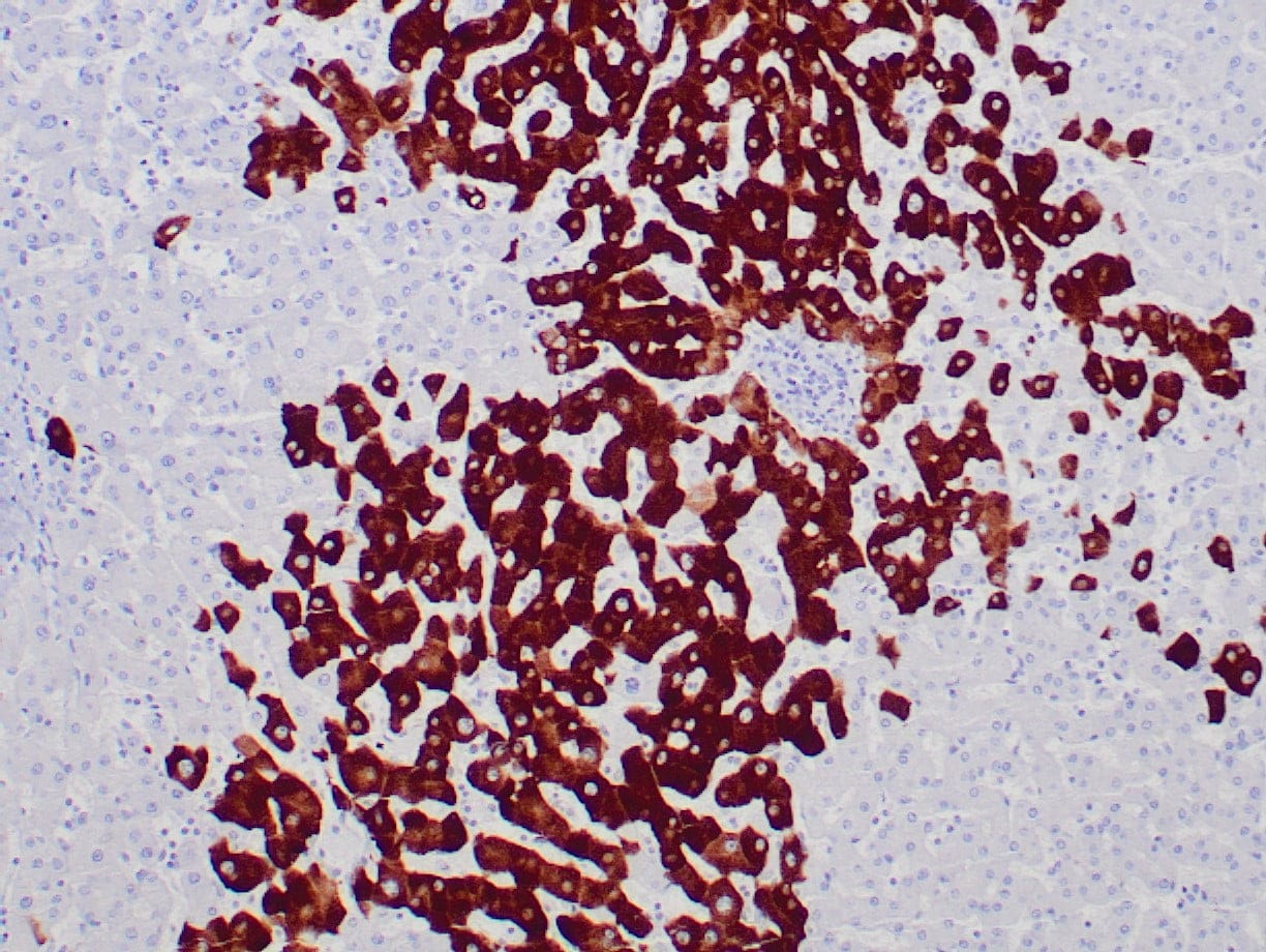 |
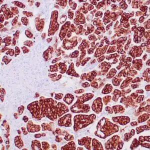
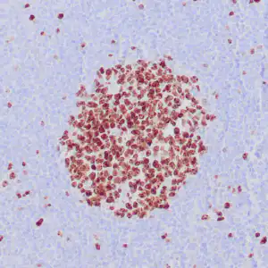
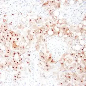
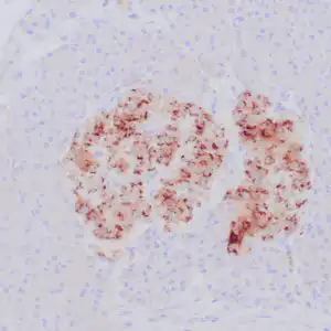
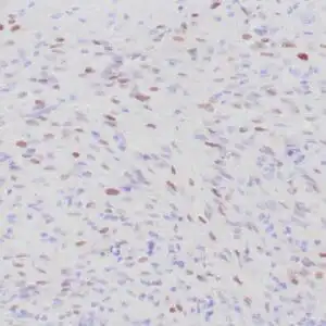
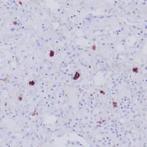
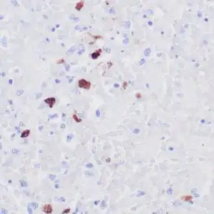
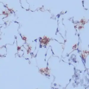
| Human liver infected by hepatitis B virus stained with anti-HBsAg antibody using peroxidase-conjugate and DAB chromogen. Note the cytoplasmic staining of infected hepatocytes. |
FAQ & Publications
Frequently Asked Questions
What is the recommended application and dilution for the rabbit anti-HBsAg monoclonal antibody (ZR393)?
This antibody is suitable for immunohistochemistry (IHC) on formalin-fixed, paraffin-embedded tissues with a recommended dilution of 1:100-200 when used in concentrated form.
How should the rabbit anti-HBsAg monoclonal antibody (ZR393) be stored to maintain its stability?
For short-term storage, keep the antibody at 2-8°C. For long-term storage, it should be kept at -20°C. Avoid repeated freeze/thaw cycles to preserve antibody integrity.
Which species does the rabbit anti-HBsAg monoclonal antibody (ZR393) react with and what is its host and clonality?
The antibody is reactive with human samples. It is a rabbit monoclonal antibody with an IgG isotype.
Publications
| pmid | title | authors | citation |
|---|---|---|---|
| We haven't added any publications to our database yet. | |||
Published literature highly relevant to the biological target of this product and referencing this antibody or clone are retrieved from the PubMed database provided by the United States National Library of Medicine at the National Institutes of Health.
Protocols
| relevant to this product |
|---|
| IHC |
Documents
| Batch Number | QC File | SDS |
|---|---|---|
| To view batch-specific Safety Datasheets and Quality Certificates associated with your account, please Log In. | ||
Only logged in customers who have purchased this product may leave a review.


Reviews
There are no reviews yet.