| Weight | 1 lbs |
|---|---|
| Dimensions | 9 × 5 × 2 in |
| host | rabbit |
| isotype | IgG |
| clonality | polyclonal |
| concentration | 1 mg/mL |
| applications | ICC/IF, WB |
| reactivity | F1Aa |
| available sizes | 100 µg |


rabbit anti-F1A α (CT) polyclonal antibody 8308
$445.00
Antibody summary
- Rabbit polyclonal to F1A α (CT)
- Suitable for: ELISA,WB,IHC-P,IF
- Isotype: IgG
- 100 µg
rabbit anti-F1A α (CT) polyclonal antibody 8308
| antibody |
|---|
| Tested applications WB,IHC,IHC,ICC/IF,ELISA |
| Recommended dilutions Immunoblotting: use at 1ug/mL. Immunohistochemistry: use at 5ug/mL. Positive control: Mouse liver tissue lysate. These are recommended concentrations. Enduser should determine optimal concentrations for their applications. |
| Immunogen Peptide corresponding to aa 609- 622 of human F1A (accession no. AAF05314). This sequence is identical to that of mouse. |
| Size and concentration 100µg and lot specific |
| Form liquid |
| Storage Instructions This antibody is stable for at least one (1) year at -20°C. Avoid multiple freeze-thaw cycles. |
| Storage buffer PBS, pH 7.4. |
| Purity peptide affinity purification |
| Clonality polyclonal |
| Isotype IgG |
| Compatible secondaries goat anti-rabbit IgG, H&L chain specific, peroxidase conjugated, conjugated polyclonal antibody 9512 goat anti-rabbit IgG, H&L chain specific, biotin conjugated polyclonal antibody 2079 goat anti-rabbit IgG, H&L chain specific, FITC conjugated polyclonal antibody 7863 goat anti-rabbit IgG, H&L chain specific, Cross Absorbed polyclonal antibody 2371 goat anti-rabbit IgG, H&L chain specific, biotin conjugated polyclonal antibody, crossabsorbed 1715 goat anti-rabbit IgG, H&L chain specific, FITC conjugated polyclonal antibody, crossabsorbed 1720 |
| Isotype control Rabbit polyclonal - Isotype Control |
| target relevance |
|---|
| Homo sapiens FEM1B Protein fem-1 homolog B |
| Protein names Protein fem-1 homolog B |
| Alternative names FEM1-beta, Fem-1-like death receptor-binding protein alpha, Fem-1-like in apoptotic pathway protein alpha |
| Gene names FEM1B |
| Protein family Belongs to the fem-1 family |
| Function Substrate-recognition component of a Cul2-RING (CRL2) E3 ubiquitin-protein ligase complex of the DesCEND (destruction via C-end degrons) pathway, which recognizes a C-degron located at the extreme C terminus of target proteins, leading to their ubiquitination and degradation (PubMed:29779948, PubMed:33398168, PubMed:33398170). The C-degron recognized by the DesCEND pathway is usually a motif of less than ten residues and can be present in full-length proteins, truncated proteins or proteolytically cleaved forms (PubMed:29779948, PubMed:33398168, PubMed:33398170). The CRL2(FEM1B) complex specifically recognizes proteins ending with -Gly-Leu-Asp-Arg, such as CDK5R1, leading to their ubiquitination and degradation (PubMed:33398168, PubMed:33398170). Also acts as a regulator of the reductive stress response by mediating ubiquitination of reduced FNIP1: in response to reductive stress, the CRL2(FEM1B) complex specifically recognizes a conserved Cys degron in FNIP1 when this degron is reduced, leading to FNIP1 degradation and subsequent activation of mitochondria to recalibrate reactive oxygen species (ROS) (By similarity). Mechanistically, recognizes and binds reduced FNIP1 through two interface zinc ions, which act as a molecular glue that recruit reduced FNIP1 to FEM1B (By similarity). Promotes ubiquitination of GLI1, suppressing GLI1 transcriptional activator activity (PubMed:24076122). Promotes ubiquitination and degradation of ANKRD37 (By similarity). Promotes ubiquitination and degradation of SLBP (PubMed:28118078). Involved in apoptosis by acting as a death receptor-associated protein that mediates apoptosis (PubMed:10542291). Also involved in glucose homeostasis in pancreatic islet (By similarity). May also act as an adapter/mediator in replication stress-induced signaling that leads to the activation of CHEK1 (PubMed:19330022) |
| Subcellular location Cytoplasm, Nucleus |
| Structure Component of a CRL2 E3 ubiquitin-protein ligase complex, also named ECS (Elongin BC-CUL2/5-SOCS-box protein) complex, composed of CUL2, Elongin BC (ELOB and ELOC), RBX1 and substrate-specific adapter FEM1B (PubMed:15601820, PubMed:29779948). Homooligomer (PubMed:10542291). Interacts with PPM1F and PHTF1 (PubMed:11559703). Interacts with the death domain of FAS/TNFRSF6 and TNFRSF1A (PubMed:10542291). Interacts with CHEK1 (PubMed:19330022). Interacts with NKX3-1 (By similarity) |
| Involvement in disease Neurodevelopmental disorder with behavioral, ear, and skeletal abnormalities An autosomal dominant neurodevelopmental disorder characterized by global developmental delay, impaired intellectual development, and behavioral abnormalities. Additional variable features include brain anomalies, clubfeet, skeletal abnormalities, facial dysmorphism, dysplastic ears, and hearing loss. |
| Keywords 3D-structure, ANK repeat, Apoptosis, Cytoplasm, Deafness, Disease variant, Intellectual disability, Metal-binding, Nucleus, Proteomics identification, Reference proteome, Repeat, TPR repeat, Ubl conjugation pathway, Zinc |
| Sequence MEGLAGYVYKAASEGKVLTLAALLLNRSESDIRYLLGYVSQQGGQRSTPLIIAARNGHAK VVRLLLEHYRVQTQQTGTVRFDGYVIDGATALWCAAGAGHFEVVKLLVSHGANVNHTTVT NSTPLRAACFDGRLDIVKYLVENNANISIANKYDNTCLMIAAYKGHTDVVRYLLEQRADP NAKAHCGATALHFAAEAGHIDIVKELIKWRAAIVVNGHGMTPLKVAAESCKADVVELLLS HADCDRRSRIEALELLGASFANDRENYDIIKTYHYLYLAMLERFQDGDNILEKEVLPPIH AYGNRTECRNPQELESIRQDRDALHMEGLIVRERILGADNIDVSHPIIYRGAVYADNMEF EQCIKLWLHALHLRQKGNRNTHKDLLRFAQVFSQMIHLNETVKAPDIECVLRCSVLEIEQ SMNRVKNISDADVHNAMDNYECNLYTFLYLVCISTKTQCSEEDQCKINKQIYNLIHLDPR TREGFTLLHLAVNSNTPVDDFHTNDVCSFPNALVTKLLLDCGAEVNAVDNEGNSALHIIV QYNRPISDFLTLHSIIISLVEAGAHTDMTNKQNKTPLDKSTTGVSEILLKTQMKMSLKCL AARAVRANDINYQDQIPRTLEEFVGFH |
| UniProt accession: Q9UK73 |
Data
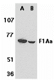 |
| Western blot analysis of F1A alpha in mouse (A) and rat (B) liver tissue lysates with F1A alpha antibody at 1 µg/mL. |
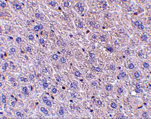 |
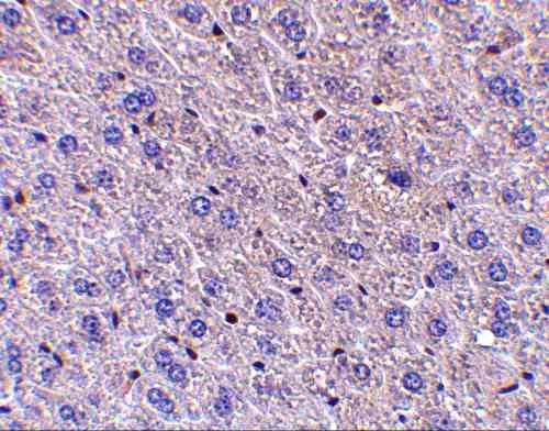
| Immunohistochemistry of F1Aα in mouse liver tissue with F1Aα antibody at 5 µg/mL. |
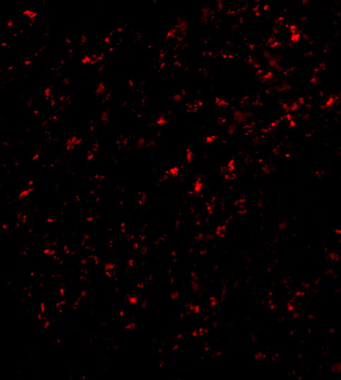 |
| Immunofluorescence of F1A alpha in Mouse Liver cells with F1A alpha antibody at 20 µg/mL. |
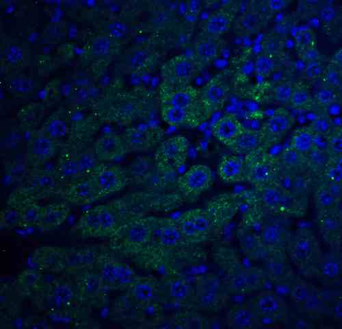 |
| Immunofluorescence of F1A alpha in mouse liver tissue with F1A alpha antibody at 20 µg/mL.
Green: F1A alpha Antibody (8308) Blue: DAPI staining |
FAQ & Publications
Frequently Asked Questions
What applications has the rabbit anti-F1A α (CT) polyclonal antibody 8308 been validated for?
This antibody is suitable and has been tested for use in Western blot (WB), immunohistochemistry (IHC-P), immunofluorescence (IF), immunocytochemistry (ICC/IF), and ELISA.
How should I store the rabbit anti-F1A α (CT) polyclonal antibody to maintain its stability?
The antibody is stable for at least one year when stored at -20°C. It is recommended to avoid multiple freeze-thaw cycles to preserve antibody integrity.
What is the immunogen used to generate the rabbit anti-F1A α (CT) polyclonal antibody 8308?
The immunogen is a peptide corresponding to amino acids 609-622 of human F1A (accession no. AAF05314), which is identical to the mouse sequence.
Publications
| pmid | title | authors | citation |
|---|---|---|---|
| We haven't added any publications to our database yet. | |||
Published literature highly relevant to the biological target of this product and referencing this antibody or clone are retrieved from the PubMed database provided by the United States National Library of Medicine at the National Institutes of Health.
Protocols
| relevant to this product |
|---|
| Western blot IHC ICC |
Documents
| Batch Number | QC File | SDS |
|---|---|---|
| To view batch-specific Safety Datasheets and Quality Certificates associated with your account, please Log In. | ||
Only logged in customers who have purchased this product may leave a review.


Reviews
There are no reviews yet.