| Weight | 1 lbs |
|---|---|
| Dimensions | 9 × 5 × 2 in |
| host | rabbit |
| isotype | IgG |
| clonality | polyclonal |
| concentration | 1 mg/mL |
| applications | ICC/IF, WB |
| reactivity | DR6 (NT) |
| available sizes | 100 µg |

rabbit anti-DR6 (NT) polyclonal antibody 5107
$445.00
Antibody summary
- Rabbit polyclonal to DR6 (NT)
- Suitable for: ELISA,WB
- Isotype: IgG
- 100 µg
rabbit anti-DR6 (NT) polyclonal antibody 5107
| antibody |
|---|
| Tested applications WB,ELISA |
| Recommended dilutions Immunoblotting : use at 2ug/mL. Positive control: Whole cell lysate from K562 or Raji cells. These are recommended concentrations. Enduser should determine optimal concentrations for their applications. |
| Immunogen Peptide corresponding to aa 42-56 of human DR6 precursor (accession no. AF068868). |
| Size and concentration 100µg and lot specific |
| Form liquid |
| Storage Instructions This antibody is stable for at least one (1) year at -20°C. Avoid multiple freeze-thaw cycles. |
| Storage buffer PBS, pH 7.4. |
| Purity peptide affinity purification |
| Clonality polyclonal |
| Isotype IgG |
| Compatible secondaries goat anti-rabbit IgG, H&L chain specific, peroxidase conjugated, conjugated polyclonal antibody 9512 goat anti-rabbit IgG, H&L chain specific, biotin conjugated polyclonal antibody 2079 goat anti-rabbit IgG, H&L chain specific, FITC conjugated polyclonal antibody 7863 goat anti-rabbit IgG, H&L chain specific, Cross Absorbed polyclonal antibody 2371 goat anti-rabbit IgG, H&L chain specific, biotin conjugated polyclonal antibody, crossabsorbed 1715 goat anti-rabbit IgG, H&L chain specific, FITC conjugated polyclonal antibody, crossabsorbed 1720 |
| Isotype control Rabbit polyclonal - Isotype Control |
| target relevance |
|---|
| Homo sapiens TNFRSF21 Tumor necrosis factor receptor superfamily member 21 |
| Protein names Tumor necrosis factor receptor superfamily member 21 |
| Alternative names Death receptor 6 |
| Gene names TNFRSF21 |
| Function Promotes apoptosis, possibly via a pathway that involves the activation of NF-kappa-B. Can also promote apoptosis mediated by BAX and by the release of cytochrome c from the mitochondria into the cytoplasm. Trophic-factor deprivation triggers the cleavage of surface APP by beta-secretase to release sAPP-beta which is further cleaved to release an N-terminal fragment of APP (N-APP). Negatively regulates oligodendrocyte survival, maturation and myelination. Plays a role in signaling cascades triggered by stimulation of T-cell receptors, in the adaptive immune response and in the regulation of T-cell differentiation and proliferation. Negatively regulates T-cell responses and the release of cytokines such as IL4, IL5, IL10, IL13 and IFNG by Th2 cells. Negatively regulates the production of IgG, IgM and IgM in response to antigens. May inhibit the activation of JNK in response to T-cell stimulation. Also acts as a regulator of pyroptosis: recruits CASP8 in response to reactive oxygen species (ROS) and subsequent oxidation, leading to activation of GSDMC (PubMed:34012073) |
| Subcellular location Cell membrane |
| Structure (Microbial infection) Interacts with hepatitis C virus (HCV) non-structural protein 5A; this interaction allows the modulation by the virus of JNK, p38 MAPK, STAT3, and Akt signaling pathways in a DR6-dependent manner |
| Post-translational modification Oxidized in response to reactive oxygen species (ROS), leading to endocytosis |
| Keywords 3D-structure, Adaptive immunity, Apoptosis, Cell membrane, Disulfide bond, Glycoprotein, Host-virus interaction, Immunity, Lipoprotein, Membrane, Oxidation, Palmitate, Proteomics identification, Receptor, Reference proteome, Repeat, Signal, Transmembrane, Transmembrane helix |
| Sequence MGTSPSSSTALASCSRIARRATATMIAGSLLLLGFLSTTTAQPEQKASNLIGTYRHVDRA TGQVLTCDKCPAGTYVSEHCTNTSLRVCSSCPVGTFTRHENGIEKCHDCSQPCPWPMIEK LPCAALTDRECTCPPGMFQSNATCAPHTVCPVGWGVRKKGTETEDVRCKQCARGTFSDVP SSVMKCKAYTDCLSQNLVVIKPGTKETDNVCGTLPSFSSSTSPSPGTAIFPRPEHMETHE VPSSTYVPKGMNSTESNSSASVRPKVLSSIQEGTVPDNTSSARGKEDVNKTLPNLQVVNH QQGPHHRHILKLLPSMEATGGEKSSTPIKGPKRGHPRQNLHKHFDINEHLPWMIVLFLLL VLVVIVVCSIRKSSRTLKKGPRQDPSAIVEKAGLKKSMTPTQNREKWIYYCNGHGIDILK LVAAQVGSQWKDIYQFLCNASEREVAAFSNGYTADHERAYAALQHWTIRGPEASLAQLIS ALRQHRRNDVVEKIRGLMEDTTQLETDKLALPMSPSPLSPSPIPSPNAKLENSALLTVEP SPQDKNKGFFVDESEPLLRCDSTSSGSSALSRNGSFITKEKKDTVLRQVRLDPCDLQPIF DDMLHFLNPEELRVIEEIPQAEDKLDRLFEIIGVKSQEASQTLLDSVYSHLPDLL |
| UniProt accession: O75509 |
Data
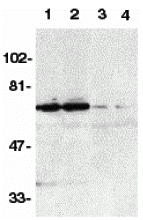 |
| Western blot analysis of DR6 in K562 (1,3) and Raji (2,4) whole cell lysate in the absence (1,2) or presence (3,4) of blocking peptide (Catalog no. 5107P) with DR6 antibody at 1:500 dilution. |
FAQ & Publications
Frequently Asked Questions
What applications is the Rabbit anti-DR6 (NT) Polyclonal Antibody 5107 validated for?
This antibody is tested and suitable for use in Western blot (WB) and ELISA assays.
What is the recommended concentration for immunoblotting using this antibody?
For immunoblotting, it is recommended to use the antibody at a concentration of 2 µg/mL. However, end users should optimize the concentration for their specific applications.
How should the Rabbit anti-DR6 (NT) Polyclonal Antibody 5107 be stored to maintain stability?
The antibody should be stored at -20°C and is stable for at least one year under these conditions. It is important to avoid multiple freeze-thaw cycles to preserve antibody integrity.
What is the immunogen used to generate this Rabbit anti-DR6 (NT) Polyclonal Antibody?
The immunogen is a peptide corresponding to amino acids 42 to 56 of the human DR6 precursor protein (accession number AF068868).
Which secondary antibodies are compatible with this Rabbit anti-DR6 (NT) Polyclonal Antibody?
Compatible secondary antibodies include goat anti-rabbit IgG antibodies that are H&L chain specific, available conjugated to peroxidase, biotin, or FITC, including cross-absorbed variants for reduced background.
Publications
| pmid | title | authors | citation |
|---|---|---|---|
| We haven't added any publications to our database yet. | |||
Published literature highly relevant to the biological target of this product and referencing this antibody or clone are retrieved from the PubMed database provided by the United States National Library of Medicine at the National Institutes of Health.
Protocols
| relevant to this product |
|---|
| Western blot IHC ICC |
Documents
| Batch Number | QC File | SDS |
|---|---|---|
| To view batch-specific Safety Datasheets and Quality Certificates associated with your account, please Log In. | ||
Only logged in customers who have purchased this product may leave a review.


Reviews
There are no reviews yet.