| Weight | 1 lbs |
|---|---|
| Dimensions | 9 × 5 × 2 in |
| host | rabbit |
| isotype | IgG |
| clonality | polyclonal |
| concentration | 1 mg/mL |
| applications | ICC/IF, WB |
| reactivity | DR5 |
| available sizes | 100 µg |


rabbit anti-DR5 polyclonal antibody 9289
$445.00
Antibody summary
- Rabbit polyclonal to DR5
- Suitable for: ELISA,WB,IHC-P,IF
- Isotype: IgG
- 100 µg
rabbit anti-DR5 polyclonal antibody 9289
| antibody |
|---|
| Tested applications WB,IHC,IHC,ICC/IF,ELISA |
| Recommended dilutions Immunoblotting : use at 2ug/mL. Immunocytochemistry: use at 5ug/mL. Positive control: Whole cell lysate from HeLa cells or K562 cells. These are recommended concentrations. Enduser should determine optimal concentrations for their applications. |
| Immunogen Peptide corresponding to aa 388- 407 of human DR5 precursor (accession no. AF012535). |
| Size and concentration 100µg and lot specific |
| Form liquid |
| Storage Instructions This antibody is stable for at least one (1) year at -20°C. Avoid multiple freeze-thaw cycles. |
| Storage buffer PBS, pH 7.4. |
| Purity peptide affinity purification |
| Clonality polyclonal |
| Isotype IgG |
| Compatible secondaries goat anti-rabbit IgG, H&L chain specific, peroxidase conjugated, conjugated polyclonal antibody 9512 goat anti-rabbit IgG, H&L chain specific, biotin conjugated polyclonal antibody 2079 goat anti-rabbit IgG, H&L chain specific, FITC conjugated polyclonal antibody 7863 goat anti-rabbit IgG, H&L chain specific, Cross Absorbed polyclonal antibody 2371 goat anti-rabbit IgG, H&L chain specific, biotin conjugated polyclonal antibody, crossabsorbed 1715 goat anti-rabbit IgG, H&L chain specific, FITC conjugated polyclonal antibody, crossabsorbed 1720 |
| Isotype control Rabbit polyclonal - Isotype Control |
| target relevance |
|---|
| Homo sapiens TNFRSF10B Tumor necrosis factor receptor superfamily member 10B |
| Protein names Tumor necrosis factor receptor superfamily member 10B |
| Alternative names Death receptor 5, TNF-related apoptosis-inducing ligand receptor 2 |
| Gene names TNFRSF10B |
| Function Receptor for the cytotoxic ligand TNFSF10/TRAIL (PubMed:10549288). The adapter molecule FADD recruits caspase-8 to the activated receptor. The resulting death-inducing signaling complex (DISC) performs caspase-8 proteolytic activation which initiates the subsequent cascade of caspases (aspartate-specific cysteine proteases) mediating apoptosis. Promotes the activation of NF-kappa-B. Essential for ER stress-induced apoptosis |
| Subcellular location Membrane |
| Structure Monomer (PubMed:10549288). Can interact with TRADD and RIPK1. Interacts with HCMV protein UL141; this interaction prevents TNFRSF10B cell surface expression. Two TNFRSF10B monomers interact with a UL141 homodimer. Three TNFRSF10B molecules interact with TNFSF10 homotrimer (PubMed:10549288). In the absence of stimulation, interacts with BIRC2, DDX3X and GSK3B. The interaction with BIRC2 and DDX3X is further enhanced upon receptor stimulation and accompanied by DDX3X and BIRC2 cleavage (PubMed:18846110) |
| Post-translational modification (Microbial infection) Glycosylated on Arg residue by S.typhimurium protein Ssek3 |
| Involvement in disease Squamous cell carcinoma of the head and neck A non-melanoma skin cancer affecting the head and neck. The hallmark of cutaneous SCC is malignant transformation of normal epidermal keratinocytes. |
| Keywords 3D-structure, Alternative splicing, Apoptosis, Direct protein sequencing, Disulfide bond, Glycoprotein, Membrane, Proteomics identification, Receptor, Reference proteome, Repeat, Signal, Transmembrane, Transmembrane helix |
| Sequence MEQRGQNAPAASGARKRHGPGPREARGARPGPRVPKTLVLVVAAVLLLVSAESALITQQD LAPQQRAAPQQKRSSPSEGLCPPGHHISEDGRDCISCKYGQDYSTHWNDLLFCLRCTRCD SGEVELSPCTTTRNTVCQCEEGTFREEDSPEMCRKCRTGCPRGMVKVGDCTPWSDIECVH KESGTKHSGEVPAVEETVTSSPGTPASPCSLSGIIIGVTVAAVVLIVAVFVCKSLLWKKV LPYLKGICSGGGGDPERVDRSSQRPGAEDNVLNEIVSILQPTQVPEQEMEVQEPAEPTGV NMLSPGESEHLLEPAEAERSQRRRLLVPANEGDPTETLRQCFDDFADLVPFDSWEPLMRK LGLMDNEIKVAKAEAAGHRDTLYTMLIKWVNKTGRDASVHTLLDALETLGERLAKQKIED HLLSSGKFMYLEGNADSAMS |
| UniProt accession: O14763 |
Data
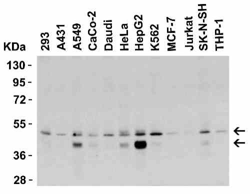 |
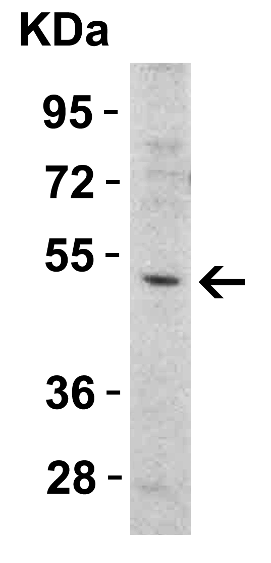
| Western Blot Validation in Human Cell Lines Loading: 15 µg of lysates per lane. Antibodies: DR5 9289, (0.5 µg/mL), 1h incubation at RT in 5% NFDM/TBST.Secondary: Goat anti-rabbit IgG HRP conjugate at 1:10000 dilution. |
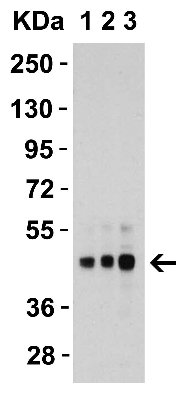 |
| Western Blot Validation in Human HepG2 Cells Loading: 15 µg of lysates per lane. Antibodies: DR5 9289, 1h incubation at RT in 5% NFDM/TBST.Secondary: Goat anti-rabbit IgG HRP conjugate at 1:10000 dilution.Lane 1: 1 µg/mLLane 2: 2 µg/mLLane 3: 4 µg/mL |
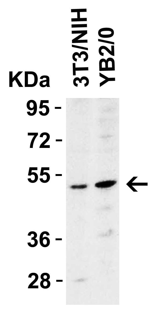 |
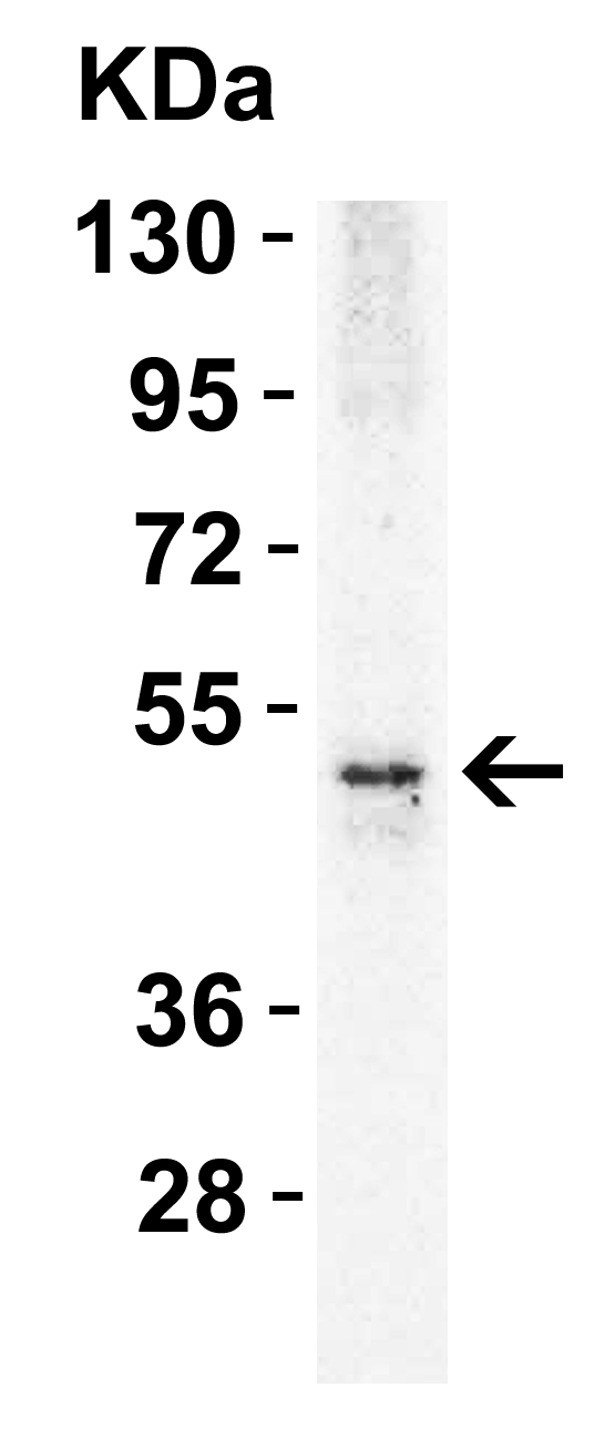
| Western Blot Validation in Mouse and Rat Cell Lines Loading: 15 µg of lysates per lane. Antibodies: DR5 9289, (2 µg/mL), 1h incubation at RT in 5% NFDM/TBST.Secondary: Goat anti-rabbit IgG HRP |
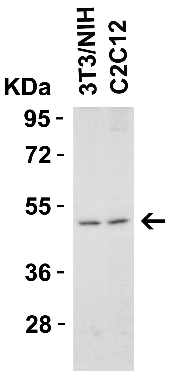 |
| Western Blot Validation in Mouse Cell Lines Loading: 15 µg of lysates per lane. Antibodies: DR5 9289, (1 µg/mL), 1h incubation at RT in 5% NFDM/TBST.Secondary: Goat anti-rabbit IgG HRP conjugate at 1:10000 dilution. |
 |
| Western Blot Validation in Mouse Heart Loading: 15 µg of lysatesper lane. Antibodies: DR5 9289, (1 µg/mL), 1h incubation at RT in 5% NFDM/TBST.Secondary: Goat anti-rabbit IgG HRP conjugate at 1:10000 dilution. |
 |
| Western Blot Validation in Rat Skeletal Muscle Loading: 15 µg of lysate per lane. Antibodies: DR5 9289, (1 µg/mL), 1h incubation at RT in 5% NFDM/TBST.Secondary: Goat anti-rabbit IgG HRP conjugate at 1:10000 dilution. |
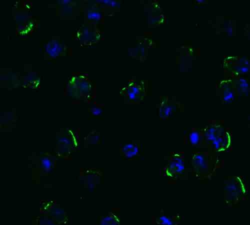 |
| Immunofluorescence Validation of DR5 in Human HepG2 Cells Immunofluorescent analysis of 4% paraformaldehyde-fixed human HepG2 cells labeling DR5 with 9289 at 5 µg/mL, followed by goat anti-rabbit IgG secondary antibody at 1/500 dilution (green) and DAPI (blue). |
 |
| Immunofluorescence Validation of DR5 in Human Testis Immunofluorescent analysis of 4% paraformaldehyde-fixed human testis tissue labeling DR5 with 9289 at 10 µg/mL, followed by goat anti-rabbit IgG secondary antibody at 1/500 dilution (green) and DAPI (blue). |
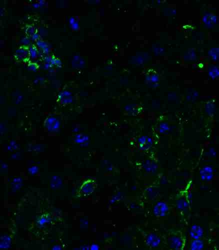 |
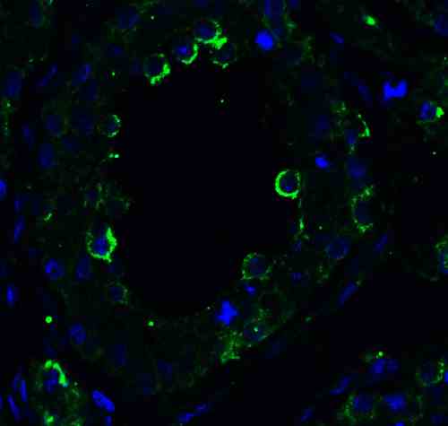
| Immunofluorescence Validation of DR5 in Mouse Pancreas Immunofluorescent analysis of 4% paraformaldehyde-fixed mouse pancreas tissue labeling DR5 with 9289 at 10 µg/mL, followed by goat anti-rabbit IgG secondary antibody at 1/500 dilution (green) and DAPI (blue). |
FAQ & Publications
Frequently Asked Questions
What applications is the rabbit anti-DR5 polyclonal antibody suitable for?
This antibody is suitable for ELISA, Western Blot (WB), Immunohistochemistry on paraffin-embedded tissues (IHC-P), and Immunofluorescence (IF). It has also been validated for Immunocytochemistry (ICC).
How should I store the rabbit anti-DR5 polyclonal antibody to maintain its stability?
The antibody should be stored at -20°C and is stable for at least one year under these conditions. It is important to avoid multiple freeze-thaw cycles to preserve antibody integrity.
What is the recommended dilution for using this antibody in Western Blot and Immunocytochemistry?
For Western Blot, use the antibody at 2 µg/mL. For Immunocytochemistry, a concentration of 5 µg/mL is recommended. These are suggested starting concentrations and optimal dilutions may need to be determined empirically for specific applications.
What is the immunogen used to generate the rabbit anti-DR5 polyclonal antibody?
The immunogen is a peptide corresponding to amino acids 388-407 of the human DR5 precursor protein, based on accession number AF012535.
Can this rabbit anti-DR5 polyclonal antibody detect DR5 in multiple species?
Yes, the antibody has been validated for Western Blot in human, mouse, and rat cell lines and tissues, indicating cross-reactivity with DR5 from these species.
Publications
| pmid | title | authors | citation |
|---|---|---|---|
| We haven't added any publications to our database yet. | |||
Published literature highly relevant to the biological target of this product and referencing this antibody or clone are retrieved from the PubMed database provided by the United States National Library of Medicine at the National Institutes of Health.
Protocols
| relevant to this product |
|---|
| Western blot IHC ICC |
Documents
| Batch Number | QC File | SDS |
|---|---|---|
| To view batch-specific Safety Datasheets and Quality Certificates associated with your account, please Log In. | ||
Only logged in customers who have purchased this product may leave a review.


Reviews
There are no reviews yet.