| Weight | 1 lbs |
|---|---|
| Dimensions | 9 × 5 × 2 in |
| host | rabbit |
| isotype | IgG |
| clonality | polyclonal |
| concentration | 1 mg/mL |
| applications | ICC/IF, WB |
| reactivity | DR4 (NT) |
| available sizes | 100 µg |


rabbit anti-DR4 (NT) polyclonal antibody 6001
$445.00
Antibody summary
- Rabbit polyclonal to DR4 (NT)
- Suitable for: ELISA,WB,IF,ICC
- Isotype: IgG
- 100 µg
rabbit anti-DR4 (NT) polyclonal antibody 6001
| antibody |
|---|
| Tested applications WB,IHC,IHC,ICC/IF,ELISA |
| Recommended dilutions Immunoblotting : use at 2ug/mL. Positive control: Whole cell lysate from HeLa, K562, or Jurkat cells. These are recommended concentrations. Enduser should determine optimal concentration for their applications. |
| Immunogen Peptide corresponding to aa 1-20 of human DR4 mature protein (accession no. ACC51226). |
| Size and concentration 100µg and lot specific |
| Form liquid |
| Storage Instructions This antibody is stable for at least one (1) year at -20°C. Avoid multiple freeze-thaw cycles. |
| Storage buffer PBS, pH 7.4. |
| Purity peptide affinity purification |
| Clonality polyclonal |
| Isotype IgG |
| Compatible secondaries goat anti-rabbit IgG, H&L chain specific, peroxidase conjugated, conjugated polyclonal antibody 9512 goat anti-rabbit IgG, H&L chain specific, biotin conjugated polyclonal antibody 2079 goat anti-rabbit IgG, H&L chain specific, FITC conjugated polyclonal antibody 7863 goat anti-rabbit IgG, H&L chain specific, Cross Absorbed polyclonal antibody 2371 goat anti-rabbit IgG, H&L chain specific, biotin conjugated polyclonal antibody, crossabsorbed 1715 goat anti-rabbit IgG, H&L chain specific, FITC conjugated polyclonal antibody, crossabsorbed 1720 |
| Isotype control Rabbit polyclonal - Isotype Control |
| target relevance |
|---|
| Homo sapiens TNFRSF10A Tumor necrosis factor receptor superfamily member 10A |
| Protein names Tumor necrosis factor receptor superfamily member 10A |
| Alternative names Death receptor 4, TNF-related apoptosis-inducing ligand receptor 1 |
| Gene names TNFRSF10A |
| Function Receptor for the cytotoxic ligand TNFSF10/TRAIL (PubMed:26457518, PubMed:38532423). The adapter molecule FADD recruits caspase-8 to the activated receptor. The resulting death-inducing signaling complex (DISC) performs caspase-8 proteolytic activation which initiates the subsequent cascade of caspases (aspartate-specific cysteine proteases) mediating apoptosis (PubMed:19090789). Promotes the activation of NF-kappa-B (PubMed:9430227) |
| Subcellular location Cell membrane, Membrane raft, Cytoplasm, cytosol |
| Structure (Microbial infection) Interacts with HCMV protein UL141; this interaction prevents TNFRSF10A cell surface expression |
| Post-translational modification Palmitoylated (PubMed:19090789). Palmitoylation of TNFRSF10A is required for its association with lipid rafts, oligomerization and function in TRAIL-induced cell death (PubMed:19090789). Palmitoylated by ZDHHC3 (Probable) |
| Keywords 3D-structure, Apoptosis, Cell membrane, Cytoplasm, Disulfide bond, Glycoprotein, Membrane, Methylation, Phosphoprotein, Proteomics identification, Receptor, Reference proteome, Repeat, Signal, Transmembrane, Transmembrane helix |
| Sequence MAPPPARVHLGAFLAVTPNPGSAASGTEAAAATPSKVWGSSAGRIEPRGGGRGALPTSMG QHGPSARARAGRAPGPRPAREASPRLRVHKTFKFVVVGVLLQVVPSSAATIKLHDQSIGT QQWEHSPLGELCPPGSHRSEHPGACNRCTEGVGYTNASNNLFACLPCTACKSDEEERSPC TTTRNTACQCKPGTFRNDNSAEMCRKCSRGCPRGMVKVKDCTPWSDIECVHKESGNGHNI WVILVVTLVVPLLLVAVLIVCCCIGSGCGGDPKCMDRVCFWRLGLLRGPGAEDNAHNEIL SNADSLSTFVSEQQMESQEPADLTGVTVQSPGEAQCLLGPAEAEGSQRRRLLVPANGADP TETLMLFFDKFANIVPFDSWDQLMRQLDLTKNEIDVVRAGTAGPGDALYAMLMKWVNKTG RNASIHTLLDALERMEERHAREKIQDLLVDSGKFIYLEDGTGSAVSLE |
| UniProt accession: O00220 |
Data
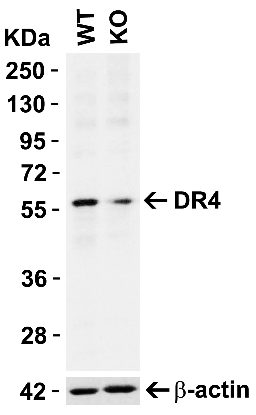 |
| KO Validation in HeLa Cells Loading: 10 µg of HeLa WT cell lysates or DR4 KO cell lysates. Antibodies: DR4 6001 (1 µg/mL) and beta-actin 3779 (1 µg/mL), 1 h incubation at RT in 5% NFDM/TBST.Secondary: Goat Anti-Rabbit IgG HRP conjugate at 1:10000 dilution. |
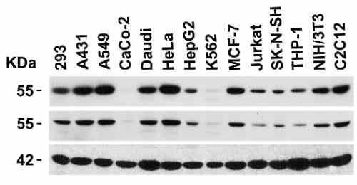 |
| Independent Antibody Validation (IAV) via Protein Expression Profile in Cell Lines Loading: 15 µg of lysates per lane. Antibodies: DR4 2404 (1 µg/mL), DR4 6001 (4 µg/mL), beta-actin (1 µg/mL), and GAPDH (0.02 µg/mL), 1h incubation at RT in 5% NFDM/TBST.Secondary: Goat anti-rabbit IgG HRP conjugate at 1:10000 dilution. |
 |
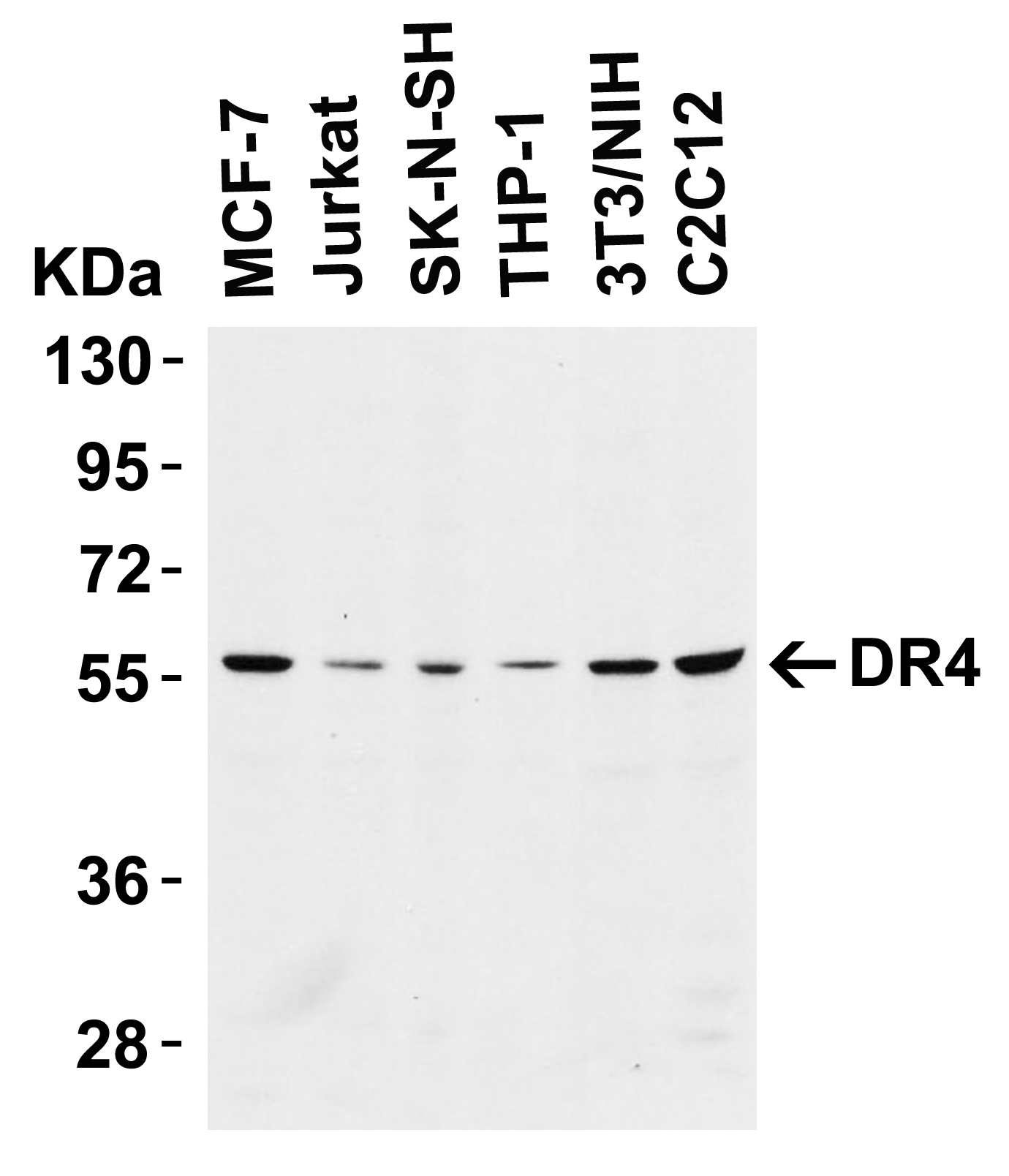
| Western Blot Validation in Cell Lines Loading: 15 µg of cell lysates per lane. Antibodies: DR4 6001 (4ug/mL), 1h incubation at RT in 5% NFDM/TBST.Secondary: Goat anti-rabbit IgG HRP conjugate at 1:10000 dilution. |
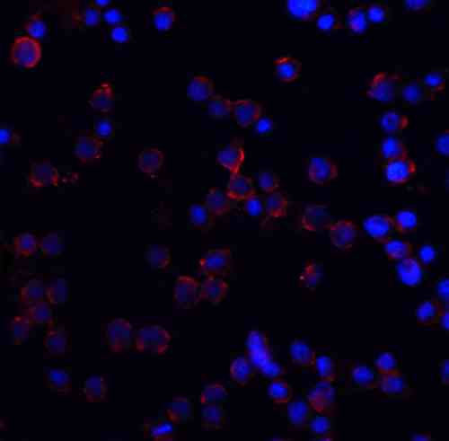 |
| Immunofluorescence Validation of DR4 in HeLa Cells Immunofluorescent analysis of 4% paraformaldehyde-fixed HeLa cells labeling DR4 with 6001 at 5 µg/mL, followed by goat anti-rabbit IgG secondary antibody at 1/500 dilution (red) and DAPI staining (blue). Image showing membrane staining on HeLa cells. |
 |
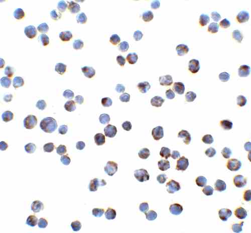
| Immunocytochemistry Validation of DR4 in HeLa Cells Immunocytochemical analysis of HeLa cells using anti-DR4 antibody (6001) at 2 µg/mL. Cells was fixed with formaldehyde and blocked with 10% serum for 1 h at RT; antigen retrieval was by heat mediation with a citrate buffer (pH6). Samples were incubated with primary antibody overnight at 4C. A goat anti-rabbit IgG H&L (HRP) at 1/250 was used as secondary. Counter stained with Hematoxylin. |
 |
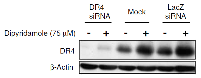
| KD Validation in SW480 Cells (Goda et al., 2008) The expression of DR4 was knocked down via DR4 siRNA, 24 h latercells were treated with dipyridamole for 24 h. DR4 protein expression detected by anti-DR4 antibodies was disrupted. Dipyridamole up-regulated the expression of DR4. |
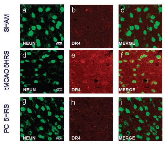 |
| Immunofluorescence Validation of DR4 in Rat Brain (Cantarella et al., 2014) DR4 protein expression detected by anti-DR4 antibodies was increased after transient brain ischemia (tMCAO) and decreased after pre-conditioning stimulus. Confocal microscopic images displaying NeuN (a,d, g) (green), DR4 (b, e, h) (red), and Merge (c, f, i) (yellow) in the brain peri-ischemic region of rats after 5 h. |
 |
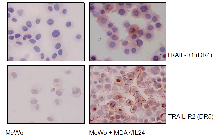
| Immunocytochemistry Validation of DR4 in Human Melanoma Cells (Ekmekcioglu et al., 2008) MeWo melanoma cells were exposed to affinity-purified MDA7/IL-24. After 48 h of treatment, cells were collected and cytospins prepared for cytochemical assessment of their TRAIL receptor (R1 and R2) expression (anti-DR4 or anti-DR5, AEC, hematoxylin). Both DR4 and DR5 expression were upregulated in MeWo cells after treatment. |
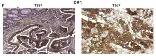 |
| Immunohistochemistry Validation of DR4 in Human Colon Tumors (Devetzi et al., 2016) DR4 expression in human colon tumors detected by anti-DR4 antibodies. Strong immunoreactivity is shown for DR4 in T167. Moderate immunoreactivity is shown for DR4 in T187. |
FAQ & Publications
Frequently Asked Questions
What applications has the rabbit anti-DR4 (NT) polyclonal antibody 6001 been validated for?
This antibody has been tested and validated for use in Western blot (WB), immunohistochemistry (IHC), immunocytochemistry/immunofluorescence (ICC/IF), and ELISA applications.
How should the rabbit anti-DR4 (NT) polyclonal antibody 6001 be stored to maintain stability?
The antibody should be stored at -20°C and is stable for at least one year under these conditions. It is important to avoid multiple freeze-thaw cycles to preserve antibody integrity.
What is the immunogen used to generate the rabbit anti-DR4 (NT) polyclonal antibody 6001?
The immunogen is a peptide corresponding to amino acids 1-20 of the human DR4 mature protein, based on accession number ACC51226.
Which secondary antibodies are compatible with the rabbit anti-DR4 (NT) polyclonal antibody 6001 for detection?
Compatible secondary antibodies include goat anti-rabbit IgG, H&L chain specific, with various conjugations such as peroxidase, biotin, and FITC, including cross-absorbed versions suitable for different detection methods.
Publications
| pmid | title | authors | citation |
|---|---|---|---|
| We haven't added any publications to our database yet. | |||
Published literature highly relevant to the biological target of this product and referencing this antibody or clone are retrieved from the PubMed database provided by the United States National Library of Medicine at the National Institutes of Health.
Protocols
| relevant to this product |
|---|
| Western blot IHC ICC |
Documents
| Batch Number | QC File | SDS |
|---|---|---|
| To view batch-specific Safety Datasheets and Quality Certificates associated with your account, please Log In. | ||
Only logged in customers who have purchased this product may leave a review.


Reviews
There are no reviews yet.