| Weight | 1 lbs |
|---|---|
| Dimensions | 9 × 5 × 2 in |
| host | rabbit |
| isotype | IgG |
| clonality | polyclonal |
| concentration | 1 mg/mL |
| applications | ICC/IF, WB |
| reactivity | DR3 (CT) |
| available sizes | 100 µg |


rabbit anti-DR3 (CT) polyclonal antibody 5310
$445.00
Antibody summary
- Rabbit polyclonal to DR3 (CT)
- Suitable for: ELISA,WB,IF
- Isotype: IgG
- 100 µg
rabbit anti-DR3 (CT) polyclonal antibody 5310
| antibody |
|---|
| Tested applications WB,ICC/IF,ELISA |
| Recommended dilutions Immunoblotting: use at 2ug/mL. Positive control: Whole cell lysate from Jurkat cells. These are recommended concentrations. Enduser should determine optimal concentrations for their applications. |
| Immunogen Peptide corresponding to aa 398-417 of human DR3 (accession no. AAQ88676). |
| Size and concentration 100µg and lot specific |
| Form liquid |
| Storage Instructions This antibody is stable for at least one (1) year at -20°C. Avoid multiple freeze-thaw cycles. |
| Storage buffer PBS, pH 7.4. |
| Purity peptide affinity purification |
| Clonality polyclonal |
| Isotype IgG |
| Compatible secondaries goat anti-rabbit IgG, H&L chain specific, peroxidase conjugated, conjugated polyclonal antibody 9512 goat anti-rabbit IgG, H&L chain specific, biotin conjugated polyclonal antibody 2079 goat anti-rabbit IgG, H&L chain specific, FITC conjugated polyclonal antibody 7863 goat anti-rabbit IgG, H&L chain specific, Cross Absorbed polyclonal antibody 2371 goat anti-rabbit IgG, H&L chain specific, biotin conjugated polyclonal antibody, crossabsorbed 1715 goat anti-rabbit IgG, H&L chain specific, FITC conjugated polyclonal antibody, crossabsorbed 1720 |
| Isotype control Rabbit polyclonal - Isotype Control |
| target relevance |
|---|
| Homo sapiens TNFRSF25 Tumor necrosis factor receptor superfamily member 25 |
| Protein names Tumor necrosis factor receptor superfamily member 25 |
| Alternative names Apo-3, Apoptosis-inducing receptor AIR, Apoptosis-mediating receptor DR3, Apoptosis-mediating receptor TRAMP, Death receptor 3, Lymphocyte-associated receptor of death, Protein WSL, Protein WSL-1 |
| Gene names TNFRSF25 |
| Function Receptor for TNFSF12/APO3L/TWEAK. Interacts directly with the adapter TRADD. Mediates activation of NF-kappa-B and induces apoptosis. May play a role in regulating lymphocyte homeostasis |
| Subcellular location Secreted |
| Structure Homodimer. Interacts strongly via the death domains with TNFRSF1 and TRADD to activate at least two distinct signaling cascades, apoptosis and NF-kappa-B signaling. Interacts with BAG4 |
| Post-translational modification (Microbial infection) Glycosylated at Arg-352 by enteropathogenic E.coli protein NleB1 Glycosylated |
| Keywords 3D-structure, Alternative splicing, Apoptosis, Cell membrane, Disulfide bond, Glycoprotein, Membrane, Proteomics identification, Receptor, Reference proteome, Repeat, Secreted, Signal, Transmembrane, Transmembrane helix |
| Sequence MEQRPRGCAAVAAALLLVLLGARAQGGTRSPRCDCAGDFHKKIGLFCCRGCPAGHYLKAP CTEPCGNSTCLVCPQDTFLAWENHHNSECARCQACDEQASQVALENCSAVADTRCGCKPG WFVECQVSQCVSSSPFYCQPCLDCGALHRHTRLLCSRRDTDCGTCLPGFYEHGDGCVSCP TSTLGSCPERCAAVCGWRQMFWVQVLLAGLVVPLLLGATLTYTYRHCWPHKPLVTADEAG MEALTPPPATHLSPLDSAHTLLAPPDSSEKICTVQLVGNSWTPGYPETQEALCPQVTWSW DQLPSRALGPAAAPTLSPESPAGSPAMMLQPGPQLYDVMDAVPARRWKEFVRTLGLREAE IEAVEVEIGRFRDQQYEMLKRWRQQQPAGLGAVYAALERMGLDGCVEDLRSRLQRGP |
| UniProt accession: Q93038 |
Data
 |
| Western blot analysis of DR3 in Jurkat total cell lysate with DR3 antibody at 1:500 dilution. |
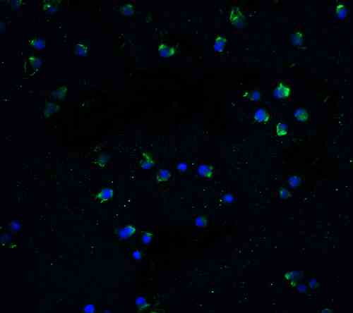 |
| Immunofluorescence of DR3 in Jurkat cells with DR3 antibody at 20 µg/mL.
Green: DR3 Antibody (5310) Blue: DAPI staining |
FAQ & Publications
Frequently Asked Questions
What applications is the rabbit anti-DR3 (CT) polyclonal antibody 5310 validated for?
This antibody is validated for use in ELISA, Western Blot (WB), and Immunofluorescence (IF) applications.
How should the rabbit anti-DR3 (CT) polyclonal antibody 5310 be stored to maintain stability?
The antibody should be stored at -20°C and is stable for at least one year under these conditions. Avoid multiple freeze-thaw cycles to preserve antibody integrity.
What is the recommended concentration and positive control for immunoblotting with this antibody?
For immunoblotting, use the antibody at 2 µg/mL. A suitable positive control is whole cell lysate from Jurkat cells.
What is the immunogen used to generate the rabbit anti-DR3 (CT) polyclonal antibody 5310?
The immunogen is a peptide corresponding to amino acids 398-417 of human DR3, based on the accession number AAQ88676.
Publications
| pmid | title | authors | citation |
|---|---|---|---|
| We haven't added any publications to our database yet. | |||
Published literature highly relevant to the biological target of this product and referencing this antibody or clone are retrieved from the PubMed database provided by the United States National Library of Medicine at the National Institutes of Health.
Protocols
| relevant to this product |
|---|
| Western blot IHC ICC |
Documents
| Batch Number | QC File | SDS |
|---|---|---|
| To view batch-specific Safety Datasheets and Quality Certificates associated with your account, please Log In. | ||
Only logged in customers who have purchased this product may leave a review.


Reviews
There are no reviews yet.