| Weight | 1 lbs |
|---|---|
| Dimensions | 9 × 5 × 2 in |
| host | rabbit |
| isotype | IgG |
| clonality | polyclonal |
| concentration | 1 mg/mL |
| applications | ICC/IF, WB |
| reactivity | p62dok |
| available sizes | 100 µg |

rabbit anti-DOK (CT) polyclonal antibody 3680
$445.00
Antibody summary
- Rabbit polyclonal to DOK (CT)
- Suitable for: ELISA,WB,ICC,IF
- Isotype: IgG
- 100 µg
rabbit anti-DOK (CT) polyclonal antibody 3680
| antibody |
|---|
| Tested applications WB,ICC/IF,ELISA |
| Recommended dilutions Immunoblotting: use at 1:1,000-1:2,000 dilution. Positive control: Whole cell lysate of Jurkat cells. |
| Immunogen Peptide corresponding to aa 425- 439 of human p62dok. |
| Size and concentration 100µg and lot specific |
| Form liquid |
| Storage Instructions This antibody is stable for at least one (1) year at -20°C. Avoid multiple freeze-thaw cycles. |
| Storage buffer PBS, pH 7.4. |
| Purity peptide affinity purification |
| Clonality polyclonal |
| Isotype IgG |
| Compatible secondaries goat anti-rabbit IgG, H&L chain specific, peroxidase conjugated, conjugated polyclonal antibody 9512 goat anti-rabbit IgG, H&L chain specific, biotin conjugated polyclonal antibody 2079 goat anti-rabbit IgG, H&L chain specific, FITC conjugated polyclonal antibody 7863 goat anti-rabbit IgG, H&L chain specific, Cross Absorbed polyclonal antibody 2371 goat anti-rabbit IgG, H&L chain specific, biotin conjugated polyclonal antibody, crossabsorbed 1715 goat anti-rabbit IgG, H&L chain specific, FITC conjugated polyclonal antibody, crossabsorbed 1720 |
| Isotype control Rabbit polyclonal - Isotype Control |
| target relevance |
|---|
| Homo sapiens DOK1 Docking protein 1 |
| Protein names Docking protein 1 |
| Alternative names Downstream of tyrosine kinase 1, p62(dok), pp62 |
| Gene names DOK1 |
| Protein family Belongs to the DOK family. Type A subfamily |
| Function DOK proteins are enzymatically inert adaptor or scaffolding proteins. They provide a docking platform for the assembly of multimolecular signaling complexes. DOK1 appears to be a negative regulator of the insulin signaling pathway. Modulates integrin activation by competing with talin for the same binding site on ITGB3 |
| Subcellular location Cytoplasm, perinuclear region |
| Structure Interacts with ABL1 (By similarity). Interacts with RasGAP and INPP5D/SHIP1. Interacts directly with phosphorylated ITGB3. Interacts with SRMS (via the SH2 and SH3 domains) |
| Post-translational modification Constitutively tyrosine-phosphorylated. Phosphorylated by TEC (By similarity). Phosphorylated by LYN (By similarity). Phosphorylated on tyrosine residues by the insulin receptor kinase. Results in the negative regulation of the insulin signaling pathway. Phosphorylated on tyrosine residues by SRMS |
| Keywords 3D-structure, Acetylation, Alternative initiation, Alternative splicing, Cytoplasm, Nucleus, Phosphoprotein, Proteomics identification, Reference proteome |
| Sequence MDGAVMEGPLFLQSQRFGTKRWRKTWAVLYPASPHGVARLEFFDHKGSSSGGGRGSSRRL DCKVIRLAECVSVAPVTVETPPEPGATAFRLDTAQRSHLLAADAPSSAAWVQTLCRNAFP KGSWTLAPTDNPPKLSALEMLENSLYSPTWEGSQFWVTVQRTEAAERCGLHGSYVLRVEA ERLTLLTVGAQSQILEPLLSWPYTLLRRYGRDKVMFSFEAGRRCPSGPGTFTFQTAQGND IFQAVETAIHRQKAQGKAGQGHDVLRADSHEGEVAEGKLPSPPGPQELLDSPPALYAEPL DSLRIAPCPSQDSLYSDPLDSTSAQAGEGVQRKKPLYWDLYEHAQQQLLKAKLTDPKEDP IYDEPEGLAPVPPQGLYDLPREPKDAWWCQARVKEEGYELPYNPATDDYAVPPPRSTKPL LAPKPQGPAFPEPGTATGSGIKSHNSALYSQVQKSGASGSWDCGLSRVGTDKTGVKSEGS T |
| UniProt accession: Q99704 |
Data
 |
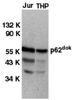
| Western blot analysis of DOK1 in Jurkat (Jur) and THP-1 (THP) cell lysates with DOK1 antibody at 1 µg/mL. |
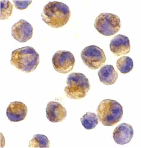 |
| Immunocytochemistry of DOK1 in K562 cells with DOK1 antibody at 2 µg/mL. |
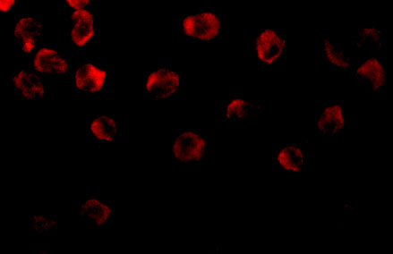 |
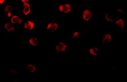
| Immunofluorescence of DOK1 in K562 cells with DOK1 antibody at 10 µg/mL. |
FAQ & Publications
Frequently Asked Questions
What applications is the rabbit anti-DOK (CT) polyclonal antibody suitable for?
This antibody is suitable for ELISA, Western blotting (WB), immunocytochemistry (ICC), and immunofluorescence (IF) applications.
How should the rabbit anti-DOK (CT) antibody be stored to maintain stability?
The antibody should be stored at -20°C and is stable for at least one year under these conditions. Avoid multiple freeze-thaw cycles to preserve antibody integrity.
What is the immunogen used to generate the rabbit anti-DOK (CT) polyclonal antibody?
The immunogen is a peptide corresponding to amino acids 425-439 of the human p62dok protein.
Which species does the rabbit anti-DOK (CT) polyclonal antibody react with?
This antibody reacts with the human p62dok protein, also known as Docking protein 1 or DOK1.
Publications
| pmid | title | authors | citation |
|---|---|---|---|
| We haven't added any publications to our database yet. | |||
Published literature highly relevant to the biological target of this product and referencing this antibody or clone are retrieved from the PubMed database provided by the United States National Library of Medicine at the National Institutes of Health.
Protocols
| relevant to this product |
|---|
| Western blot IHC ICC |
Documents
| Batch Number | QC File | SDS |
|---|---|---|
| To view batch-specific Safety Datasheets and Quality Certificates associated with your account, please Log In. | ||
Only logged in customers who have purchased this product may leave a review.


Reviews
There are no reviews yet.