| Weight | 1 lbs |
|---|---|
| Dimensions | 9 × 5 × 2 in |
| host | rabbit |
| isotype | IgG |
| clonality | polyclonal |
| concentration | 1 mg/mL |
| applications | ICC/IF, WB |
| reactivity | DEDAF |
| available sizes | 100 µg |


rabbit anti-DEDAF polyclonal antibody 3011
$445.00
Antibody summary
- Rabbit polyclonal to DEDAF
- Suitable for: ELISA,WB,IHC-P,IF
- Isotype: IgG
- 100 µg
rabbit anti-DEDAF polyclonal antibody 3011
| antibody |
|---|
| Tested applications WB,IHC,IHC,ICC/IF,ELISA |
| Recommended dilutions Immunoblotting: use at 1ug/mL. Immunohistochemistry: use at 10ug/mL. These are recommended concentrations. Enduser should determine optimal concentrations for their applications. Positive control: Whole cell lysate from A549 or HepG2 cells. |
| Immunogen Peptide corresponding to aa 215-229 of human DEDAF (accession no. AF179286). This sequence is identical to that of mouse. |
| Size and concentration 100µg and lot specific |
| Form liquid |
| Storage Instructions This antibody is stable for at least one (1) year at -20°C. Avoid multiple freeze-thaw cycles. |
| Storage buffer PBS, pH 7.4. |
| Purity peptide affinity purification |
| Clonality polyclonal |
| Isotype IgG |
| Compatible secondaries goat anti-rabbit IgG, H&L chain specific, peroxidase conjugated, conjugated polyclonal antibody 9512 goat anti-rabbit IgG, H&L chain specific, biotin conjugated polyclonal antibody 2079 goat anti-rabbit IgG, H&L chain specific, FITC conjugated polyclonal antibody 7863 goat anti-rabbit IgG, H&L chain specific, Cross Absorbed polyclonal antibody 2371 goat anti-rabbit IgG, H&L chain specific, biotin conjugated polyclonal antibody, crossabsorbed 1715 goat anti-rabbit IgG, H&L chain specific, FITC conjugated polyclonal antibody, crossabsorbed 1720 |
| Isotype control Rabbit polyclonal - Isotype Control |
| target relevance |
|---|
| Homo sapiens RYBP RING1 and YY1-binding protein |
| Protein names RING1 and YY1-binding protein |
| Alternative names Apoptin-associating protein 1, Death effector domain-associated factor, YY1 and E4TF1-associated factor 1 |
| Gene names RYBP |
| Function Component of a Polycomb group (PcG) multiprotein PRC1-like complex, a complex class required to maintain the transcriptionally repressive state of many genes, including Hox genes, throughout development. PcG PRC1-like complex acts via chromatin remodeling and modification of histones; it mediates monoubiquitination of histone H2A 'Lys-119', rendering chromatin heritably changed in its expressibility (PubMed:25519132). Component of a PRC1-like complex that mediates monoubiquitination of histone H2A 'Lys-119' on the X chromosome and is required for normal silencing of one copy of the X chromosome in XX females. May stimulate ubiquitination of histone H2A 'Lys-119' by recruiting the complex to target sites (By similarity). Inhibits ubiquitination and subsequent degradation of TP53, and thereby plays a role in regulating transcription of TP53 target genes (PubMed:19098711). May also regulate the ubiquitin-mediated proteasomal degradation of other proteins like FANK1 to regulate apoptosis (PubMed:14765135, PubMed:27060496). May be implicated in the regulation of the transcription as a repressor of the transcriptional activity of E4TF1 (PubMed:11953439). May bind to DNA (By similarity). May play a role in the repression of tumor growth and metastasis in breast cancer by down-regulating SRRM3 (PubMed:27748911) |
| Subcellular location Nucleus, Cytoplasm, Nucleus, nucleoplasm |
| Structure Monomer. Component of repressive BCOR complex containing Polycomb group subcomplex at least composed of BCOR, PCGF1, RING1 and RNF2/RING2 (PubMed:16943429). Component of PCR1-like complexes (PubMed:20696397, PubMed:26687479). Interacts with PCGF1 (PubMed:26687479). Part of a PCR1-like complex that contains AUTS2, PCGF5, RNF2, CSNK2B and RYBP (PubMed:25519132). Interacts with RNF2; the interaction is direct (PubMed:20696397). Interacts with CBX2, YAF2, RING1 and RNF2 (By similarity). Interacts with ubiquitin and ubiquitinated proteins (By similarity). Interacts with ubiquitinated histone H2A (By similarity). Interacts with apoptin, DEDD, FADD, CASP8, CASP10, YY1 and GABPB1 (PubMed:11395500, PubMed:11953439, PubMed:14765135). Together with GABPB1 and YY1, it forms a ternary complex, probably being the bridge factor between these two transcription factors (PubMed:11953439). Interacts with MDM2, and thereby inhibits ubiquitination of TP53 (PubMed:19098711). Identified in a ternary complex containing MDM2, TP53 and RYBP (PubMed:19098711). Interacts with FANK1; may prevent the ubiquitin-mediated proteasomal degradation of FANK1 (PubMed:27060496). Interacts with IFT57 (By similarity) |
| Post-translational modification Monoubiquitinated |
| Keywords 3D-structure, Apoptosis, Cytoplasm, DNA-binding, Isopeptide bond, Metal-binding, Nucleus, Phosphoprotein, Proteomics identification, Reference proteome, Repressor, Transcription, Transcription regulation, Ubl conjugation, Zinc, Zinc-finger |
| Sequence MTMGDKKSPTRPKRQAKPAADEGFWDCSVCTFRNSAEAFKCSICDVRKGTSTRKPRINSQ LVAQQVAQQYATPPPPKKEKKEKVEKQDKEKPEKDKEISPSVTKKNTNKKTKPKSDILKD PPSEANSIQSANATTKTSETNHTSRPRLKNVDRSTAQQLAVTVGNVTVIITDFKEKTRSS STSSSTVTSSAGSEQQNQSSSGSESTDKGSSRSSTPKGDMSAVNDESF |
| UniProt accession: Q8N488 |
Data
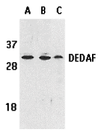 |
| Western blot analysis of DEDAF expression in human A549 (lane A), HepG2 (lane B), and mouse 3T3 (lane C) cell lysates with DEDAF antibody at 1 µg/mL. |
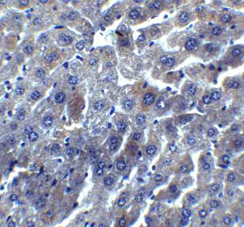 |
| Immunohistochemistry of DEDAF in mouse liver tissue with DEDAF antibody at 5 µg/mL. |
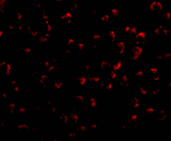 |
| Immunofluorescence of DEDAF in A549 cells with DEDAF antibody at 20 µg/mL. |
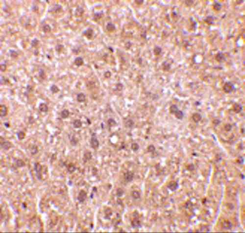 |
| Immunohistochemistry of DEDAF in mouse liver tissue with DEDAF antibody at 10 µg/mL. |
 |
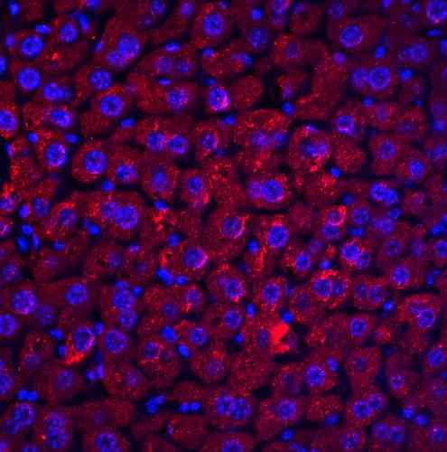
| Immunofluorescence of DEDAF in mouse liver tissue with DEDAF antibody at 20 µg/mL. |
FAQ & Publications
Frequently Asked Questions
What is the recommended dilution for using the rabbit anti-DEDAF polyclonal antibody in immunoblotting applications?
For immunoblotting (Western blot), it is recommended to use the rabbit anti-DEDAF polyclonal antibody at a concentration of 1 µg/mL. However, end users should optimize this concentration for their specific experiments.
How should the rabbit anti-DEDAF polyclonal antibody be stored to maintain its stability?
This antibody should be stored at -20°C and is stable for at least one year under these conditions. It is important to avoid multiple freeze-thaw cycles to preserve antibody integrity.
Which species does the immunogen peptide used to generate this antibody correspond to, and how does this affect cross-reactivity?
The immunogen is a peptide corresponding to amino acids 215-229 of human DEDAF (accession no. AF179286). This peptide sequence is identical to that of mouse DEDAF, suggesting the antibody is reactive with both human and mouse DEDAF proteins.
Publications
| pmid | title | authors | citation |
|---|---|---|---|
| We haven't added any publications to our database yet. | |||
Published literature highly relevant to the biological target of this product and referencing this antibody or clone are retrieved from the PubMed database provided by the United States National Library of Medicine at the National Institutes of Health.
Protocols
| relevant to this product |
|---|
| Western blot IHC ICC |
Documents
| Batch Number | QC File | SDS |
|---|---|---|
| To view batch-specific Safety Datasheets and Quality Certificates associated with your account, please Log In. | ||
Only logged in customers who have purchased this product may leave a review.


Reviews
There are no reviews yet.