| Weight | 1 lbs |
|---|---|
| Dimensions | 9 × 5 × 2 in |
| host | rabbit |
| isotype | IgG |
| clonality | polyclonal |
| concentration | 1 mg/mL |
| applications | ICC/IF, WB |
| reactivity | DC-SIGN (ED) |
| available sizes | 100 µg |

rabbit anti-DC-SIGN (ED) polyclonal antibody 9118
$445.00
Antibody summary
- Rabbit polyclonal to DC-SIGN (ED)
- Suitable for: ELISA,WB,IHC-P
- Isotype: Whole IgG
- 100 µg
rabbit anti-DC-SIGN (ED) polyclonal antibody 9118
| antibody |
|---|
| Tested applications WB,IHC,IHC,ELISA |
| Recommended dilutions Immunoblotting: use at 1-2 ug/mL. In immunoblots, a band of 44 kD is detected. Positive control: Human placenta tissue lysate. |
| Immunogen Peptide corresponding to aa 277-293 of human DC-SIGN. |
| Size and concentration 100µg and lot specific |
| Form liquid |
| Storage Instructions This antibody is stable for at least one (1) year at -20°C. Avoid multiple freeze- thaw cycles. |
| Storage buffer PBS, pH 7.4. |
| Purity peptide affinity purification |
| Clonality polyclonal |
| Isotype IgG |
| Compatible secondaries goat anti-rabbit IgG, H&L chain specific, peroxidase conjugated, conjugated polyclonal antibody 9512 goat anti-rabbit IgG, H&L chain specific, biotin conjugated polyclonal antibody 2079 goat anti-rabbit IgG, H&L chain specific, FITC conjugated polyclonal antibody 7863 goat anti-rabbit IgG, H&L chain specific, Cross Absorbed polyclonal antibody 2371 goat anti-rabbit IgG, H&L chain specific, biotin conjugated polyclonal antibody, crossabsorbed 1715 goat anti-rabbit IgG, H&L chain specific, FITC conjugated polyclonal antibody, crossabsorbed 1720 |
| Isotype control Rabbit polyclonal - Isotype Control |
| target relevance |
|---|
| Homo sapiens CD209 CD209 antigen |
| Protein names CD209 antigen |
| Alternative names C-type lectin domain family 4 member L, Dendritic cell-specific ICAM-3-grabbing non-integrin 1 |
| Gene names CD209 |
| Function Pathogen-recognition receptor expressed on the surface of immature dendritic cells (DCs) and involved in initiation of primary immune response. Thought to mediate the endocytosis of pathogens which are subsequently degraded in lysosomal compartments. The receptor returns to the cell membrane surface and the pathogen-derived antigens are presented to resting T-cells via MHC class II proteins to initiate the adaptive immune response |
| Subcellular location Secreted |
| Structure (Microbial infection) Interacts with whole M.bovis cells in a Ca(2+)-dependent and independent manner; in vitro experiments suggest it interacts with CH60.1 (groL1), DnaK, GADPH (gap) and LrpG (PubMed:21203928) |
| Keywords 3D-structure, Adaptive immunity, Alternative splicing, Calcium, Cell adhesion, Cell membrane, Disulfide bond, Endocytosis, Glycoprotein, Host cell receptor for virus entry, Host-virus interaction, Immunity, Innate immunity, Lectin, Mannose-binding, Membrane, Metal-binding, Proteomics identification, Receptor, Reference proteome, Repeat, Secreted, Signal-anchor, Transmembrane, Transmembrane helix |
| Sequence MSDSKEPRLQQLGLLEEEQLRGLGFRQTRGYKSLAGCLGHGPLVLQLLSFTLLAGLLVQV SKVPSSISQEQSRQDAIYQNLTQLKAAVGELSEKSKLQEIYQELTQLKAAVGELPEKSKL QEIYQELTRLKAAVGELPEKSKLQEIYQELTWLKAAVGELPEKSKMQEIYQELTRLKAAV GELPEKSKQQEIYQELTRLKAAVGELPEKSKQQEIYQELTRLKAAVGELPEKSKQQEIYQ ELTQLKAAVERLCHPCPWEWTFFQGNCYFMSNSQRNWHDSITACKEVGAQLVVIKSAEEQ NFLQLQSSRSNRFTWMGLSDLNQEGTWQWVDGSPLLPSFKQYWNRGEPNNVGEEDCAEFS GNGWNDDKCNLAKFWICKKSAASCSRDEEQFLSPAPATPNPPPA |
| UniProt accession: Q9NNX6 |
Data
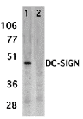 |
| Western blot analysis of DC-SIGN expression in human placenta tissue lysate in the absence (lane 1) and presence (lane 2) of blocking peptide with DC-SIGN antibody at 2 µg/mL. |
 |
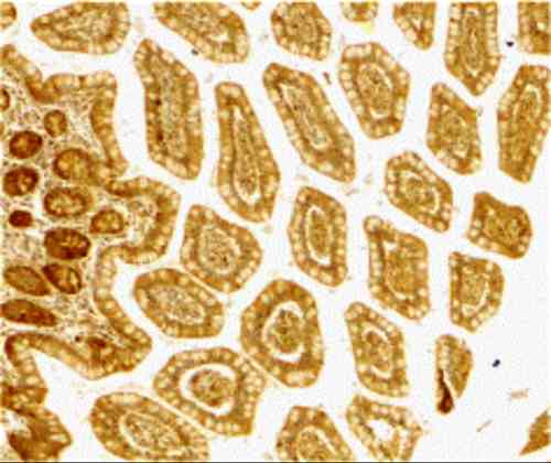
| Immunohistochemistry of DC-SIGN in human small intestine tissue with DC-SIGN antibody at 10 µg/mL. |
FAQ & Publications
Frequently Asked Questions
What applications has the rabbit anti-DC-SIGN (ED) polyclonal antibody 9118 been validated for?
This antibody is validated for use in Western blot (WB), immunohistochemistry on paraffin-embedded tissue sections (IHC-P), ELISA, and immunocytochemistry/immunofluorescence (ICC/IF) applications.
How should the rabbit anti-DC-SIGN (ED) antibody 9118 be stored to maintain stability?
The antibody should be stored at -20°C and is stable for at least one year under these conditions. It is important to avoid multiple freeze-thaw cycles to preserve antibody integrity.
What is the recommended concentration and expected band size for Western blot using this antibody?
For immunoblotting, use the antibody at 1-2 µg/mL. A specific band at approximately 44 kDa corresponding to DC-SIGN is detected in human placenta tissue lysate as a positive control.
What is the immunogen used to generate the rabbit anti-DC-SIGN (ED) polyclonal antibody 9118?
The immunogen is a peptide corresponding to amino acids 277-293 of the human DC-SIGN protein.
Which secondary antibodies are compatible with the rabbit anti-DC-SIGN (ED) polyclonal antibody 9118 for detection?
Compatible secondary antibodies include goat anti-rabbit IgG (H&L chain specific) conjugated to peroxidase, biotin, FITC, and cross-absorbed variants suitable for various detection methods.
Publications
| pmid | title | authors | citation |
|---|---|---|---|
| We haven't added any publications to our database yet. | |||
Published literature highly relevant to the biological target of this product and referencing this antibody or clone are retrieved from the PubMed database provided by the United States National Library of Medicine at the National Institutes of Health.
Protocols
| relevant to this product |
|---|
| Western blot IHC ICC |
Documents
| Batch Number | QC File | SDS |
|---|---|---|
| To view batch-specific Safety Datasheets and Quality Certificates associated with your account, please Log In. | ||
Only logged in customers who have purchased this product may leave a review.


Reviews
There are no reviews yet.