| Weight | 1 lbs |
|---|---|
| Dimensions | 9 × 5 × 2 in |
| host | rabbit |
| isotype | IgG |
| clonality | polyclonal |
| concentration | 1 mg/mL |
| applications | ICC/IF, WB |
| reactivity | CIDE-A (CT) |
| available sizes | 100 µg |


rabbit anti-CIDE-A (CT) polyclonal antibody 3080
$445.00
Antibody summary
- Rabbit polyclonal to CIDE-A (CT)
- Suitable for: ELISA,WB,IHC-P,IF
- Isotype: IgG
- 100 µg
rabbit anti-CIDE-A (CT) polyclonal antibody 3080
| antibody |
|---|
| Tested applications WB,IHC,IHC,ICC/IF,ELISA |
| Recommended dilutions Immunoblotting: use at 0.5ug/mL. Immunohistochemistry: use at 5ug/mL. These are recommended concentrations. Enduser should determine optimal concentrations for their applications. Positive control: Human brain tissue lysate. |
| Immunogen Peptide corresponding to aa 200-217 of human CIDE-A (accession no. AF041378) |
| Size and concentration 100µg and lot specific |
| Form liquid |
| Storage Instructions This antibody is stable for at least one (1) year at -20°C. Avoid multiple freeze-thaw cycles. |
| Storage buffer PBS, pH 7.4. |
| Purity peptide affinity purification |
| Clonality polyclonal |
| Isotype IgG |
| Compatible secondaries goat anti-rabbit IgG, H&L chain specific, peroxidase conjugated, conjugated polyclonal antibody 9512 goat anti-rabbit IgG, H&L chain specific, biotin conjugated polyclonal antibody 2079 goat anti-rabbit IgG, H&L chain specific, FITC conjugated polyclonal antibody 7863 goat anti-rabbit IgG, H&L chain specific, Cross Absorbed polyclonal antibody 2371 goat anti-rabbit IgG, H&L chain specific, biotin conjugated polyclonal antibody, crossabsorbed 1715 goat anti-rabbit IgG, H&L chain specific, FITC conjugated polyclonal antibody, crossabsorbed 1720 |
| Isotype control Rabbit polyclonal - Isotype Control |
| target relevance |
|---|
| Homo sapiens CIDEA Lipid transferase CIDEA |
| Protein names Lipid transferase CIDEA |
| Alternative names Cell death activator CIDE-A, Cell death-inducing DFFA-like effector A |
| Gene names CIDEA |
| Protein family Belongs to the CIDE family |
| Function Lipid transferase that promotes unilocular lipid droplet formation by mediating lipid droplet fusion (PubMed:19843876, PubMed:26118629). Lipid droplet fusion promotes their enlargement, restricting lipolysis and favoring lipid storage (PubMed:19843876). Localizes on the lipid droplet surface, at focal contact sites between lipid droplets, and mediates atypical lipid droplet fusion by promoting directional net neutral lipid transfer from the smaller to larger lipid droplets (By similarity). The transfer direction may be driven by the internal pressure difference between the contacting lipid droplet pair and occurs at a lower rate than that promoted by CIDEC (By similarity). May also act as a CEBPB coactivator in epithelial cells to control the expression of a subset of CEBPB downstream target genes, including ID2, IGF1, PRLR, SOCS1, SOCS3, XDH, but not casein (By similarity). By interacting with CEBPB, strengthens the association of CEBPB with the XDH promoter, increases histone acetylation and dissociates HDAC1 from the promoter (By similarity). When overexpressed, induces apoptosis; the physiological significance of its role in apoptosis is unclear (By similarity) |
| Catalytic activity a triacyl-sn-glycerol(in) = a triacyl-sn-glycerol(out) |
| Subcellular location Lipid droplet, Nucleus |
| Structure Homodimer (PubMed:19843876). Interacts with CIDEC (PubMed:19843876). Directly interacts with CEBPB (By similarity). Interacts with isoform CLSTN3beta of CLSTN3; inhibiting the lipid transferase activity of CIDEA (By similarity) |
| Keywords 3D-structure, Activator, Apoptosis, Lipid droplet, Nucleus, Proteomics identification, Reference proteome, Transcription, Transcription regulation |
| Sequence MEAARDYAGALIRPLTFMGSQTKRVLFTPLMHPARPFRVSNHDRSSRRGVMASSLQELIS KTLDALVIATGLVTLVLEEDGTVVDTEEFFQTLGDNTHFMILEKGQKWMPGSQHVPTCSP PKRSGIARVTFDLYRLNPKDFIGCLNVKATMYEMYSVSYDIRCTGLKGLLRSLLRFLSYS AQVTGQFLIYLGTYMLRVLDDKEERPSLRSQAKGRFTCG |
| UniProt accession: O60543 |
Data
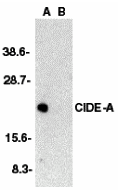 |
| Western blot analysis of CIDE-A in human brain tissue lysate in the absence (A) or presence (B) of peptide (3080P) with CIDE-A antibody at 1:2000 dilution. |
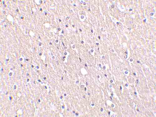 |
| Immunohistochemistry of CIDE-A in human brain tissue with CIDE-A antibody at 5 µg/mL. |
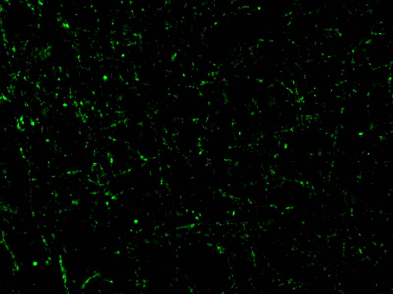 |
| Immunofluorescence of CIDE-A in Human Brain cells with CIDE-A antibody at 20 µg/mL. |
FAQ & Publications
Frequently Asked Questions
What applications is the rabbit anti-CIDE-A (CT) polyclonal antibody (SKU 3080) validated for?
This antibody is validated for use in ELISA, Western blot (WB), immunohistochemistry on paraffin-embedded tissue (IHC-P), and immunofluorescence (IF). Recommended dilutions are 0.5 µg/mL for immunoblotting and 5 µg/mL for immunohistochemistry, though optimal concentrations should be determined by the end user.
How should the rabbit anti-CIDE-A (CT) polyclonal antibody be stored to maintain stability?
The antibody should be stored at -20°C and is stable for at least one year under these conditions. It is recommended to avoid multiple freeze-thaw cycles to preserve antibody integrity.
Publications
| pmid | title | authors | citation |
|---|---|---|---|
| We haven't added any publications to our database yet. | |||
Published literature highly relevant to the biological target of this product and referencing this antibody or clone are retrieved from the PubMed database provided by the United States National Library of Medicine at the National Institutes of Health.
Protocols
| relevant to this product |
|---|
| Western blot IHC ICC |
Documents
| Batch Number | QC File | SDS |
|---|---|---|
| To view batch-specific Safety Datasheets and Quality Certificates associated with your account, please Log In. | ||
Only logged in customers who have purchased this product may leave a review.


Reviews
There are no reviews yet.