| Weight | 1 lbs |
|---|---|
| Dimensions | 9 × 5 × 2 in |
| host | rabbit |
| isotype | IgG |
| clonality | polyclonal |
| concentration | 1 mg/mL |
| applications | ICC/IF, WB |
| reactivity | Caspase-9 (I20) |
| available sizes | 100 µg |


rabbit anti-Caspase-9 (I120) polyclonal antibody 4094
$445.00
Antibody summary
- Rabbit polyclonal to Caspase-9 (I120)
- Suitable for: ELISA,WB,ICC,IF,IP
- Isotype: IgG
- 100 µg
rabbit anti-Caspase-9 (I120) polyclonal antibody 4094
| antibody |
|---|
| Tested applications WB,IHC,IHC,ICC/IF,ELISA |
| Recommended dilutions Immunoblotting: use at 1ug/mL. Immunocytochemistry: use at 2ug/mL. These are recommended concentrations. Enduser should determine optimal concentrations for their applications. Positive control: Whole cell lysate from HeLa cells. |
| Immunogen Peptide corresponding to aa 299- 318 of human caspase-9 (accession no. P55211). |
| Size and concentration 100µg and lot specific |
| Form liquid |
| Storage Instructions This antibody is stable for at least one (1) year at -20°C. Avoid multiple freeze-thaw cycles. |
| Storage buffer PBS, pH 7.4. |
| Purity peptide affinity purification |
| Clonality polyclonal |
| Isotype IgG |
| Compatible secondaries goat anti-rabbit IgG, H&L chain specific, peroxidase conjugated, conjugated polyclonal antibody 9512 goat anti-rabbit IgG, H&L chain specific, biotin conjugated polyclonal antibody 2079 goat anti-rabbit IgG, H&L chain specific, FITC conjugated polyclonal antibody 7863 goat anti-rabbit IgG, H&L chain specific, Cross Absorbed polyclonal antibody 2371 goat anti-rabbit IgG, H&L chain specific, biotin conjugated polyclonal antibody, crossabsorbed 1715 goat anti-rabbit IgG, H&L chain specific, FITC conjugated polyclonal antibody, crossabsorbed 1720 |
| Isotype control Rabbit polyclonal - Isotype Control |
| target relevance |
|---|
| Homo sapiens CASP9 Caspase-9 |
| Protein names Caspase-9 |
| Alternative names Apoptotic protease Mch-6, Apoptotic protease-activating factor 3, ICE-like apoptotic protease 6 |
| Gene names CASP9 |
| Protein family Belongs to the peptidase C14A family |
| Function Involved in the activation cascade of caspases responsible for apoptosis execution. Binding of caspase-9 to Apaf-1 leads to activation of the protease which then cleaves and activates effector caspases caspase-3 (CASP3) or caspase-7 (CASP7). Promotes DNA damage-induced apoptosis in a ABL1/c-Abl-dependent manner. Proteolytically cleaves poly(ADP-ribose) polymerase (PARP). Cleaves BIRC6 following inhibition of BIRC6-caspase binding by DIABLO/SMAC (PubMed:36758105, PubMed:36758106) |
| Catalytic activity Strict requirement for an Asp residue at position P1 and with a marked preference for His at position P2. It has a preferred cleavage sequence of Leu-Gly-His-Asp-|-Xaa. |
| Structure Heterotetramer that consists of two anti-parallel arranged heterodimers, each one formed by a 35 kDa (p35) and a 10 kDa (p10) subunit. Caspase-9 and APAF1 bind to each other via their respective NH2-terminal CED-3 homologous domains in the presence of cytochrome C and ATP. Interacts (inactive form) with EFHD2. Interacts with HAX1. Interacts with BIRC2/c-IAP1, XIAP/BIRC4, BIRC5/survivin, BIRC6/bruce and BIRC7/livin. Interacts with ABL1 (via SH3 domain); the interaction is direct and increases in the response of cells to genotoxic stress and ABL1/c-Abl activation. Interacts with BCL2L10 (PubMed:19255499). Interacts with NleF from pathogenic E.coli |
| Post-translational modification Cleavages at Asp-315 by granzyme B and at Asp-330 by caspase-3 generate the two active subunits. Caspase-8 and -10 can also be involved in these processing events Phosphorylated at Thr-125 by MAPK1/ERK2. Phosphorylation at Thr-125 is sufficient to block caspase-9 processing and subsequent caspase-3 activation. Phosphorylation on Tyr-153 by ABL1/c-Abl; occurs in the response of cells to DNA damage (Microbial infection) ADP-riboxanation by C.violaceum CopC blocks CASP9 processing, preventing CASP9 activation and ability to mediate intrinsic apoptosis Ubiquitinated by BIRC6; this activity is inhibited by DIABLO/SMAC |
| Keywords 3D-structure, Alternative splicing, Apoptosis, Hydrolase, Phosphoprotein, Protease, Proteomics identification, Reference proteome, Thiol protease, Ubl conjugation, Zymogen |
| Sequence MDEADRRLLRRCRLRLVEELQVDQLWDALLSRELFRPHMIEDIQRAGSGSRRDQARQLII DLETRGSQALPLFISCLEDTGQDMLASFLRTNRQAAKLSKPTLENLTPVVLRPEIRKPEV LRPETPRPVDIGSGGFGDVGALESLRGNADLAYILSMEPCGHCLIINNVNFCRESGLRTR TGSNIDCEKLRRRFSSLHFMVEVKGDLTAKKMVLALLELAQQDHGALDCCVVVILSHGCQ ASHLQFPGAVYGTDGCPVSVEKIVNIFNGTSCPSLGGKPKLFFIQACGGEQKDHGFEVAS TSPEDESPGSNPEPDATPFQEGLRTFDQLDAISSLPTPSDIFVSYSTFPGFVSWRDPKSG SWYVETLDDIFEQWAHSEDLQSLLLRVANAVSVKGIYKQMPGCFNFLRKKLFFKTS |
| UniProt accession: P55211 |
Data
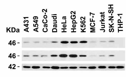 |
| Independent Antibody Validation (IAV) via Protein Expression Profile in Human Cell Lines Loading: 15 µg of lysates per lane. Antibodies: Caspase 9, 2417 (1 µg/mL), Caspase 9, 4094 (1 µg/mL) and beta-actin (1.5 µg/mL), 1h incubation at RT in 5% NFDM/TBST.Secondary: Goat anti-rabbit IgG HRP conjugate at 1:10000 dilution. |
 |
| Western Blot Validation in Human Cell Lines Loading: 15 µg of lysates per lane. Antibodies: : Caspase 9, 4094 (1 µg/mL)), 1h incubation at RT in 5% NFDM/TBST.Secondary: Goat anti-rabbit IgG HRP conjugate at 1:10000 dilution. |
 |
| Western Blot Validation in Mouse Cell Line Loading: 15 µg of 3T3/NIH cell lysate. Antibodies: : Caspase 9, 4094 (1 µg/mL)), 1h incubation at RT in 5% NFDM/TBST.Secondary: Goat anti-rabbit IgG HRP conjugate at 1:10000 dilution. |
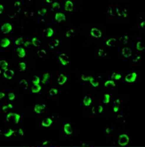 |
| Immunofluorescence Validation of Caspase 9 in K562 Cells Immunofluorescent analysis of 4% paraformaldehyde-fixed K562 cells labeling Caspase 9 with 4094 at 20 µg/mL, followed by goat anti-rabbit IgG secondary antibody at 1/500 dilution (green). |
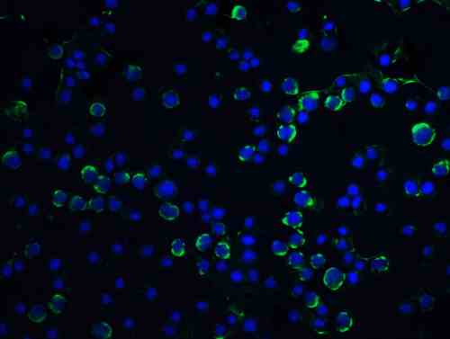 |
| Immunofluorescence Validation of Caspase 9 in HeLa Cells Immunofluorescent analysis of 4% paraformaldehyde-fixed HeLa cells labeling Caspase 9 with 4094 at 5 µg/mL, followed by goat anti-rabbit IgG secondary antibody at 1/500 dilution (green) and DAPI staining (blue). |
 |
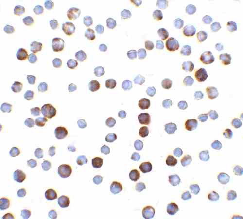
| Immunocytochemistry Validation of Caspase 9 in HeLa Cells Immunocytochemical analysis of HeLa cells using anti-Caspase 9 antibody (4094) at 5 µg/mL. Cells was fixed with formaldehyde and blocked with 10% serum for 1 h at RT; antigen retrieval was by heat mediation with a citrate buffer (pH6). Samples were incubated with primary antibody overnight at 4C. A goat anti-rabbit IgG H&L (HRP) at 1/250 was used as secondary. Counter stained with Hematoxylin. |
FAQ & Publications
Frequently Asked Questions
What applications has the rabbit anti-Caspase-9 (I120) polyclonal antibody 4094 been validated for?
This antibody is suitable and has been tested for Western Blot (WB), Immunohistochemistry (IHC), Immunocytochemistry/Immunofluorescence (ICC/IF), ELISA, and Immunoprecipitation (IP). Recommended dilutions include 1 µg/mL for immunoblotting and 2 µg/mL for immunocytochemistry.
How should the rabbit anti-Caspase-9 (I120) antibody be stored to maintain stability?
The antibody should be stored at -20°C and is stable for at least one year under these conditions. It is important to avoid multiple freeze-thaw cycles to preserve antibody integrity.
What is the host species and clonality of the Caspase-9 (I120) antibody 4094?
The antibody is a rabbit polyclonal IgG, generated against a peptide corresponding to amino acids 299-318 of human Caspase-9.
Which secondary antibodies are compatible with the rabbit anti-Caspase-9 (I120) polyclonal antibody 4094?
Compatible secondary antibodies include goat anti-rabbit IgG antibodies that are H&L chain specific and conjugated with peroxidase, biotin, or FITC. Variants include cross-absorbed forms to reduce species cross-reactivity.
Publications
| pmid | title | authors | citation |
|---|---|---|---|
| We haven't added any publications to our database yet. | |||
Published literature highly relevant to the biological target of this product and referencing this antibody or clone are retrieved from the PubMed database provided by the United States National Library of Medicine at the National Institutes of Health.
Protocols
| relevant to this product |
|---|
| Western blot IHC ICC |
Documents
| Batch Number | QC File | SDS |
|---|---|---|
| To view batch-specific Safety Datasheets and Quality Certificates associated with your account, please Log In. | ||
Only logged in customers who have purchased this product may leave a review.


Reviews
There are no reviews yet.