| Weight | 1 lbs |
|---|---|
| Dimensions | 9 × 5 × 2 in |
| host | rabbit |
| isotype | IgG |
| clonality | polyclonal |
| concentration | 1 mg/mL |
| applications | ICC/IF, WB |
| reactivity | Bim (IN) |
| available sizes | 100 µg |


rabbit anti-Bim (IN) polyclonal antibody 1410
$445.00
Antibody summary
- Rabbit polyclonal to Bim (IN)
- Suitable for: ELISA,WB,IHC-P,IF
- Isotype: IgG
- 100 µg
rabbit anti-Bim (IN) polyclonal antibody 1410
| antibody |
|---|
| Tested applications WB,IHC,IHC,ICC/IF,ELISA |
| Recommended dilutions Immunoblotting: use at 1ug/mL. Immunocytochemistry: use a 10-20ug/mL These are recommended concentrations. Enduser should determine optimal concentration for their application. Positive control: Whole cell lysate of human K562 cells. |
| Immunogen Peptide corresponding to aa 22-40 of human Bim. The sequence is identical to that of mouse and is different by one amino acid from that of rat. |
| Size and concentration 100µg and lot specific |
| Form liquid |
| Storage Instructions This antibody is stable for at least one (1) year at -20°C. Avoid multiple freeze-thaw cycles. |
| Storage buffer PBS, pH 7.4. |
| Purity peptide affinity purification |
| Clonality polyclonal |
| Isotype IgG |
| Compatible secondaries goat anti-rabbit IgG, H&L chain specific, peroxidase conjugated, conjugated polyclonal antibody 9512 goat anti-rabbit IgG, H&L chain specific, biotin conjugated polyclonal antibody 2079 goat anti-rabbit IgG, H&L chain specific, FITC conjugated polyclonal antibody 7863 goat anti-rabbit IgG, H&L chain specific, Cross Absorbed polyclonal antibody 2371 goat anti-rabbit IgG, H&L chain specific, biotin conjugated polyclonal antibody, crossabsorbed 1715 goat anti-rabbit IgG, H&L chain specific, FITC conjugated polyclonal antibody, crossabsorbed 1720 |
| Isotype control Rabbit polyclonal - Isotype Control |
| target relevance |
|---|
| Homo sapiens BCL2L11 Bcl-2-like protein 11 |
| Protein names Bcl-2-like protein 11 |
| Alternative names Bcl2-interacting mediator of cell death |
| Gene names BCL2L11 |
| Protein family Belongs to the Bcl-2 family |
| Function Induces apoptosis and anoikis. Isoform BimL is more potent than isoform BimEL. Isoform Bim-alpha1, isoform Bim-alpha2 and isoform Bim-alpha3 induce apoptosis, although less potent than isoform BimEL, isoform BimL and isoform BimS. Isoform Bim-gamma induces apoptosis. Isoform Bim-alpha3 induces apoptosis possibly through a caspase-mediated pathway. Isoform BimAC and isoform BimABC lack the ability to induce apoptosis |
| Subcellular location Mitochondrion |
| Structure Interacts with BAX; the interaction may lead to BAX activation through conformational change |
| Post-translational modification Phosphorylation at Ser-69 by MAPK1/MAPK3 leads to interaction with TRIM2 and polyubiquitination, followed by proteasomal degradation (PubMed:15486195, PubMed:21478148). Deubiquitination catalyzed by USP27X stabilizes the protein (By similarity) Ubiquitination by TRIM2 following phosphorylation by MAPK1/MAPK3 leads to proteasomal degradation. Conversely, deubiquitination catalyzed by USP27X stabilizes the protein |
| Keywords 3D-structure, Alternative splicing, Apoptosis, Membrane, Mitochondrion, Phosphoprotein, Proteomics identification, Reference proteome, Ubl conjugation |
| Sequence MAKQPSDVSSECDREGRQLQPAERPPQLRPGAPTSLQTEPQGNPEGNHGGEGDSCPHGSP QGPLAPPASPGPFATRSPLFIFMRRSSLLSRSSSGYFSFDTDRSPAPMSCDKSTQTPSPP CQAFNHYLSAMASMRQAEPADMRPEIWIAQELRRIGDEFNAYYARRVFLNNYQAAEDHPR MVILRLLRYIVRLVWRMH |
| UniProt accession: O43521 |
Data
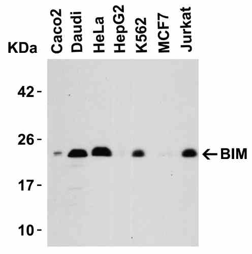 |
| Western Blot Validation in Human Cell Lines Loading: 15 µg of lysates per lane. Antibodies: BIM 1410, (0.5 µg/mL), 1h incubation at RT in 5% NFDM/TBST.Secondary: Goat anti-rabbit IgG HRP conjugate at 1:10000 dilution. |
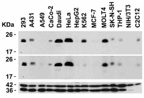 |
| Independent Antibody Validation (IAV) via Protein Expression Profile in Cell Lines Loading: 15 µg of lysates per lane. Antibodies: BIM 1410, (0.5 µg/mL), BIM 3405, (5 µg/mL), beta-actin (1 µg/mL) and GAPDH (0.02 µg/mL), 1h incubation at RT in 5% NFDM/TBST.Secondary: Goat anti-rabbit IgG HRP conjugate at 1:10000 dilution. |
 |
| Western Blot Validation in Human Tissue Loading: 15 µg of lysates per lane. Antibodies: BIM 1410, (0.5 µg/mL), 1h incubation at RT in 5% NFDM/TBST.Secondary: Goat anti-rabbit IgG HRP conjugate at 1:10000 dilution.Lane 1: Human thymusLane 2: Human colon |
 |
| Western Blot Validation in HeLa Cells Loading: 15 µg of lysate per lane. Antibodies: BIM 1410, (0.5 µg/mL), 1h incubation at RT in 5% NFDM/TBST.Secondary: Goat anti-rabbit IgG HRP conjugate at 1:10000 dilution.BIM 1410 can detect all three human isoforms, including EL, L and S isoforms. |
 |
| Western Blot Validation in Rat Myeloma Cell line Loading: 15 µg of rat myeloma YB2/0 cell lysate per lane. Antibodies: BIM 1410, (0.5 µg/mL), 1h incubation at RT in 5% NFDM/TBST.Secondary: Goat anti-rabbit IgG HRP conjugate at 1:10000 dilution. |
 |
| Immunofluorescence Validation of BIM in K562 Cells Immunofluorescent analysis of 4% paraformaldehyde-fixed K562 cells labeling Bim with 1410 at 20 µg/mL, followed by goat anti-rabbit IgG secondary antibody at 1/500 dilution (red). |
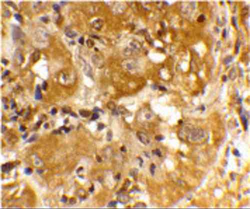 |
| Immunohistochemistry Validation of BIM in Human Skin Cancer Cells Immunohistochemical analysis of paraffin-embedded human spleen tissue using anti-BIM antibody (1410) at 20 µg/mL. Tissue was fixed with formaldehyde and blocked with 10% serum for 1 h at RT; antigen retrieval was by heat mediation with a citrate buffer (pH6). Samples were incubated with primary antibody overnight at 4C. A goat anti-rabbit IgG H&L (HRP) at 1/250 was used as secondary. Counter stained with Hematoxylin. |
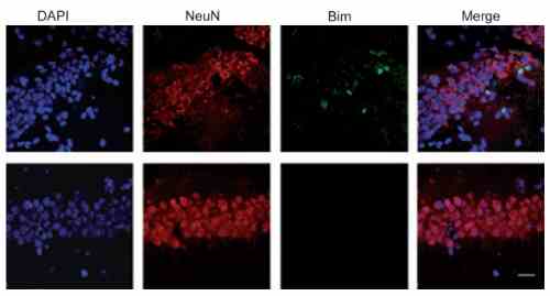 |
| Induced Expression Validation of BIM in Mouse Hippocampus (Tsuchiya et al., 2011) The induction of Bim protein was detected by immunohistochemical analysis of mice after i.h. injection of epoxomicin with anti-BIM antibodies. Sections from epoxomicin-treated animals exhibited cells staining positive for Bim expression within the NeuN-positive population of neurons in the CA1 of the ipsilateral side. In contrast, Bim-positive cells were absent within the NeuNpositiveCA1 neurons on the contralateral side. |
 |
| KD Validation of BIM in 293 Cells (Han et al., 2010) Immunofluorescence analysis with anti-BIM antibodies was performed for BIM in 293 cells transfected with control siRNA or BIM siRNA. BIM expression was disrupted after BIM siRNA knockdown. |
FAQ & Publications
Frequently Asked Questions
What are the recommended applications and dilutions for the rabbit anti-Bim (IN) polyclonal antibody 1410?
This antibody is suitable for ELISA, Western Blot (WB), Immunohistochemistry (IHC-P), and Immunofluorescence (IF). Recommended dilutions are 1 µg/mL for immunoblotting and 10-20 µg/mL for immunocytochemistry. Users should optimize concentrations for their specific applications.
How should the rabbit anti-Bim (IN) polyclonal antibody 1410 be stored to maintain stability?
The antibody is supplied in liquid form at 1 mg/mL concentration in PBS buffer at pH 7.4. It is stable for at least one year when stored at -20°C. Avoid multiple freeze-thaw cycles to maintain antibody integrity.
What is the immunogen used to generate the rabbit anti-Bim (IN) polyclonal antibody 1410, and does it cross-react with other species?
The immunogen is a peptide corresponding to amino acids 22-40 of human Bim, which is identical to mouse Bim and differs by one amino acid from rat Bim, suggesting cross-reactivity with these species.
What is the host species, clonality, and isotype of the anti-Bim antibody 1410?
The antibody is a rabbit polyclonal antibody of the IgG isotype.
Publications
| pmid | title | authors | citation |
|---|---|---|---|
| We haven't added any publications to our database yet. | |||
Published literature highly relevant to the biological target of this product and referencing this antibody or clone are retrieved from the PubMed database provided by the United States National Library of Medicine at the National Institutes of Health.
Protocols
| relevant to this product |
|---|
| Western blot IHC ICC |
Documents
| Batch Number | QC File | SDS |
|---|---|---|
| To view batch-specific Safety Datasheets and Quality Certificates associated with your account, please Log In. | ||
Only logged in customers who have purchased this product may leave a review.


Reviews
There are no reviews yet.