| Weight | 1 lbs |
|---|---|
| Dimensions | 9 × 5 × 2 in |
| host | rabbit |
| isotype | IgG |
| clonality | polyclonal |
| concentration | 1 mg/mL |
| applications | ICC/IF, WB |
| reactivity | BAFF (CT) |
| available sizes | 100 µg |


rabbit anti-BAFF (CT) polyclonal antibody 4041
$445.00
Antibody summary
- Rabbit polyclonal to BAFF (CT)
- Suitable for: ELISA,WB,ICC,IF,IHC
- Isotype: IgG
- 100 µg
rabbit anti-BAFF (CT) polyclonal antibody 4041
| antibody |
|---|
| Tested applications WB,IHC,IHC,ICC/IF,ELISA |
| Recommended dilutions Immunoblotting: use at 1ug/mL. Immunocytochemistry: use at 1ug/mL. These are recommended concentrations. Enduser should determine optimal concentration for their application. Positive control: Whole cell lysate from HL60 cells. |
| Immunogen Peptide corresponding to aa 254-269 at the C-terminus of human BAFF. |
| Size and concentration 100µg and lot specific |
| Form liquid |
| Storage Instructions This antibody is stable for at least one (1) year at -20°C. Avoid multiple freeze-thaw cycles. |
| Storage buffer PBS, pH 7.4. |
| Purity peptide affinity purification |
| Clonality polyclonal |
| Isotype IgG |
| Compatible secondaries goat anti-rabbit IgG, H&L chain specific, peroxidase conjugated, conjugated polyclonal antibody 9512 goat anti-rabbit IgG, H&L chain specific, biotin conjugated polyclonal antibody 2079 goat anti-rabbit IgG, H&L chain specific, FITC conjugated polyclonal antibody 7863 goat anti-rabbit IgG, H&L chain specific, Cross Absorbed polyclonal antibody 2371 goat anti-rabbit IgG, H&L chain specific, biotin conjugated polyclonal antibody, crossabsorbed 1715 goat anti-rabbit IgG, H&L chain specific, FITC conjugated polyclonal antibody, crossabsorbed 1720 |
| Isotype control Rabbit polyclonal - Isotype Control |
| target relevance |
|---|
| Homo sapiens TNFSF13B Tumor necrosis factor ligand superfamily member 13B |
| Protein names Tumor necrosis factor ligand superfamily member 13B |
| Alternative names B lymphocyte stimulator, B-cell-activating factor, BAFF, Dendritic cell-derived TNF-like molecule, TNF- and APOL-related leukocyte expressed ligand 1 |
| Gene names TNFSF13B |
| Protein family Belongs to the tumor necrosis factor family |
| Function Cytokine that binds to TNFRSF13B/TACI and TNFRSF17/BCMA. TNFSF13/APRIL binds to the same 2 receptors. Together, they form a 2 ligands -2 receptors pathway involved in the stimulation of B- and T-cell function and the regulation of humoral immunity. A third B-cell specific BAFF-receptor (BAFFR/BR3) promotes the survival of mature B-cells and the B-cell response |
| Subcellular location Secreted |
| Structure Homotrimer. Isoform 2 heteromultimerizes with isoform 1, probably limiting the amount of functional isoform 1 on the cell surface. Isoform 3 is unlikely form trimers or bind to BAFF receptors |
| Post-translational modification The soluble form derives from the membrane form by proteolytic processing Isoform 2 is not efficiently shed from the membrane unlike isoform 1 N-glycosylated |
| Keywords 3D-structure, Alternative splicing, Cell membrane, Cleavage on pair of basic residues, Cytokine, Direct protein sequencing, Disulfide bond, Glycoprotein, Immunity, Membrane, Proteomics identification, Reference proteome, Secreted, Signal-anchor, Transmembrane, Transmembrane helix |
| Sequence MDDSTEREQSRLTSCLKKREEMKLKECVSILPRKESPSVRSSKDGKLLAATLLLALLSCC LTVVSFYQVAALQGDLASLRAELQGHHAEKLPAGAGAPKAGLEEAPAVTAGLKIFEPPAP GEGNSSQNSRNKRAVQGPEETVTQDCLQLIADSETPTIQKGSYTFVPWLLSFKRGSALEE KENKILVKETGYFFIYGQVLYTDKTYAMGHLIQRKKVHVFGDELSLVTLFRCIQNMPETL PNNSCYSAGIAKLEEGDELQLAIPRENAQISLDGDVTFFGALKLL |
| UniProt accession: Q9Y275 |
Data
 |
| Western Blot Validation in Human HL60 Cell Lysate (H) and Mouse Spleen Lysate (M) Loading: 15 µg of lysates per lane. Antibodies: BAFF 4041 (1 µg/mL), 1h incubation at RT in 5% NFDM/TBST.Secondary: Goat anti-rabbit IgG HRP conjugate at 1:10000 dilution. |
 |
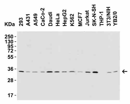
| Western Blot Validation in Human, Mouse and Rat Cell Lines Loading: 15 µg of lysates per lane. Antibodies: BAFF 4041 (1 µg/mL), 1h incubation at RT in 5% NFDM/TBST.Secondary: Goat anti-rabbit IgG HRP conjugate at 1:10000 dilution. |
 |
| Western Blot Validation with Recombinant Protein Loading: 30 ng of human BAFF recombinant protein per lane. Antibodies: BAFF 4041 (Lane 1: 0.25 µg/mL; Lane 2: 0.5 µg/mL and Lane 3: 1 µg/mL), 1h incubation at RT in 5% NFDM/TBST.Secondary: Goat anti-rabbit IgG HRP conjugate at 1:10000 dilution.Observed at around 18kD. |
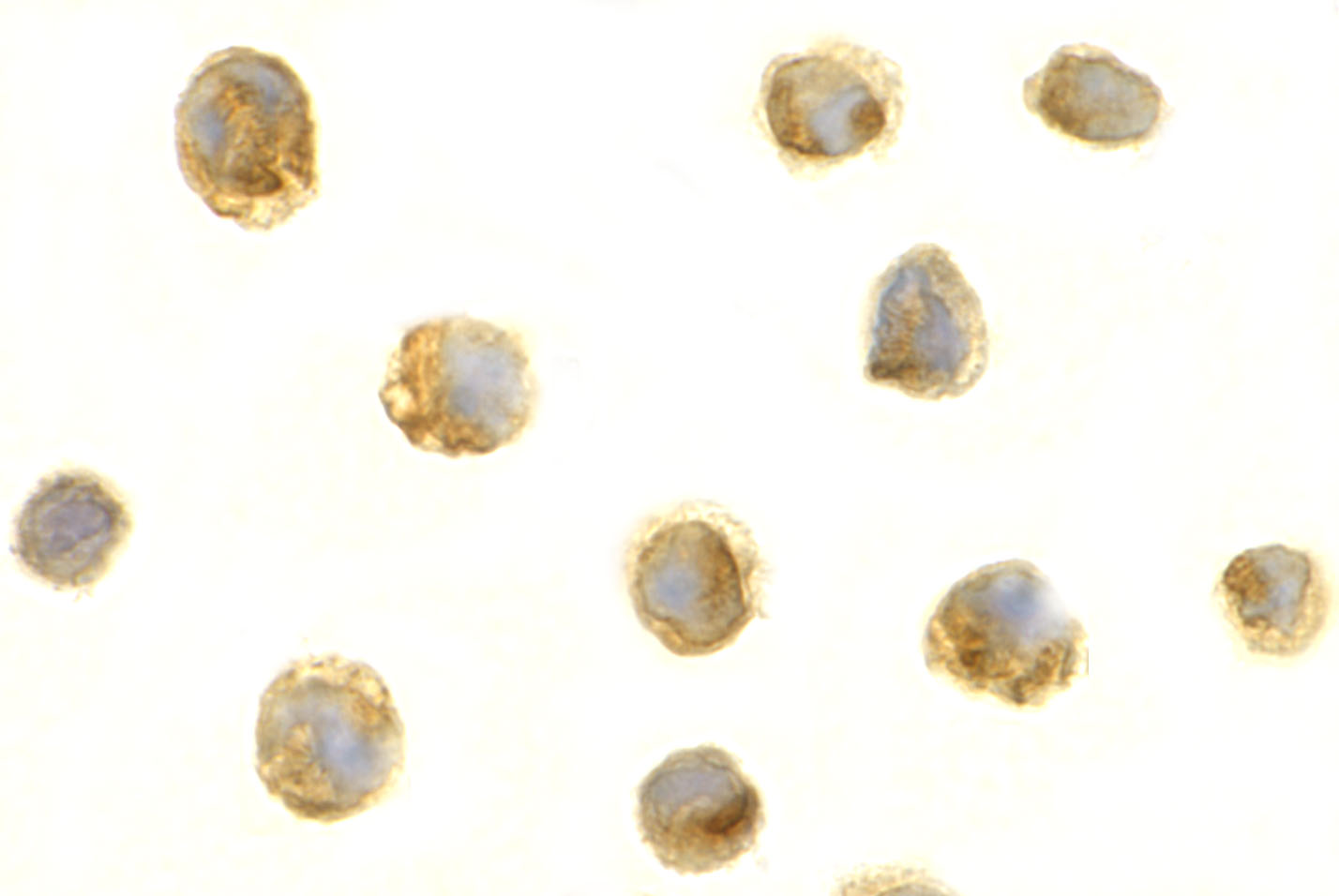 |
| Immunocytochemistry Validation of BAFF in HL60 Cells Immunocytochemical analysis of HL60 cells using anti-BAFF antibody (4041) at 1 µg/mL. Cells was fixed with formaldehyde and blocked with 10% serum for 1 h at RT; antigen retrieval was by heat mediation with a citrate buffer (pH6). Samples were incubated with primary antibody overnight at 4C. A goat anti-rabbit IgG H&L (HRP) at 1/250 was used as secondary. Counter stained with Hematoxylin. |
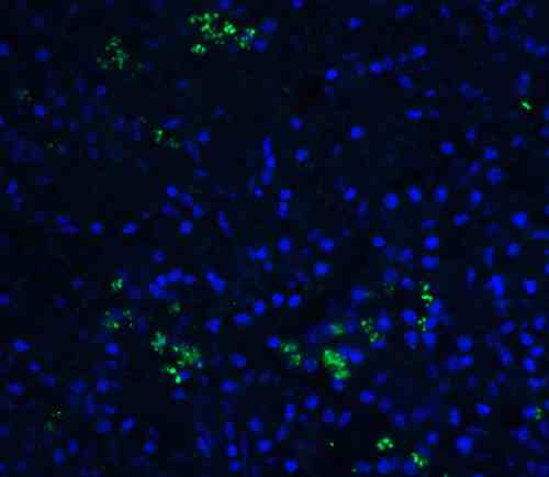 |
| Immunofluorescence Validation of BAFF in Human Spleen Tissue Immunofluorescent analysis of 4% paraformaldehyde-fixed human spleen tissue labeling BAFF with 4041 at 20 µg/mL, followed by goat anti-rabbit IgG secondary antibody at 1/500 dilution (green) and DAPI staining (blue). |
 |
| Regulated Expression Validation of BAFF in Myeloma Patients (Tai et al., 2006) Immunoblot analysis was performed to monitor protein expression of BAFF with anti-BAFF antibodies in multiple myeloma cells with or without BMSCs. BAFF expression in cocultures at 8hr or 24hr was up-regulated by ~3.5-fold relative to BMSCs alone. |
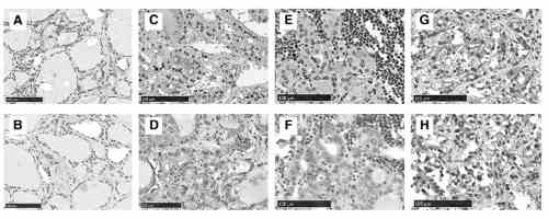 |
| Immunohistochemistry Validation of BAFF in Thyroid of Patients with Graves Diseases (Campi et al., 2015) BAFF expression detected by anti-BAFF antibodies (4041) was remarkably increased in thyrocytes from multinodular goiter (-) compared with either Hashimoto's thyroiditis (-) or Graves' disease (-) while no staining was found in normal thyroid tissue (-). |
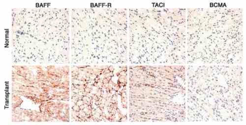 |
| Immunohistochemistry Validation of BAFF in Murine Cardiac Transplants at Rejection (Ye et al., 2004) BAFF expression detected by anti-BAFF antibodies (4041) was upregulated in intragraft leukocytes due to rejection at 7 days after heart transplant. |
FAQ & Publications
Frequently Asked Questions
What species reactivity does the rabbit anti-BAFF (CT) polyclonal antibody 4041 exhibit?
This antibody is reactive to human BAFF (CT) and has been validated using human, mouse, and rat cell lines.
Which applications are recommended for use with the rabbit anti-BAFF (CT) polyclonal antibody 4041?
The antibody is suitable for ELISA, Western Blot (WB), Immunocytochemistry/Immunofluorescence (ICC/IF), and Immunohistochemistry (IHC). Recommended dilutions are typically 1 µg/mL for immunoblotting and immunocytochemistry.
How should the rabbit anti-BAFF (CT) polyclonal antibody 4041 be stored to maintain stability?
The antibody should be stored at -20°C and is stable for at least one year under these conditions. It is important to avoid multiple freeze-thaw cycles.
What is the immunogen used to generate the rabbit anti-BAFF (CT) polyclonal antibody 4041?
The immunogen is a peptide corresponding to amino acids 254-269 at the C-terminus of human BAFF.
Which secondary antibodies are compatible with the rabbit anti-BAFF (CT) polyclonal antibody 4041 for detection?
Compatible secondary antibodies include goat anti-rabbit IgG H&L chain specific polyclonal antibodies conjugated to peroxidase, biotin, or FITC, including cross-absorbed versions for enhanced specificity.
Publications
| pmid | title | authors | citation |
|---|---|---|---|
| We haven't added any publications to our database yet. | |||
Published literature highly relevant to the biological target of this product and referencing this antibody or clone are retrieved from the PubMed database provided by the United States National Library of Medicine at the National Institutes of Health.
Protocols
| relevant to this product |
|---|
| Western blot IHC ICC |
Documents
| Batch Number | QC File | SDS |
|---|---|---|
| To view batch-specific Safety Datasheets and Quality Certificates associated with your account, please Log In. | ||
Only logged in customers who have purchased this product may leave a review.


Reviews
There are no reviews yet.