| Weight | 1 lbs |
|---|---|
| Dimensions | 9 × 5 × 2 in |
| host | rabbit |
| isotype | IgG |
| clonality | polyclonal |
| concentration | 1 mg/mL |
| applications | ICC/IF, WB |
| reactivity | AIF (CT) |
| available sizes | 100 µg |


rabbit anti-AIF (CT) polyclonal antibody 5029
$445.00
Antibody summary
- Rabbit polyclonal to AIF (CT)
- Suitable for: ELISA,WB,ICC
- Isotype: IgG
- 100 µg
rabbit anti-AIF (CT) polyclonal antibody 5029
| antibody |
|---|
| Tested applications WB,ICC/IF,ELISA |
| Recommended dilutions Immunoblotting: use at 0.5-2ug/mL. Immunocytochemistry: use at 5ug/mL. These are recommended concentrations. Enduser should determine optimal concentrations for their applications. Positive control: K562 cells or whole cell lysate. |
| Immunogen Peptide corresponding to aa 593- 606 at the C-terminus of human AIF. This sequence is identical to those of mouse and rat AIF. |
| Size and concentration 100µg and lot specific |
| Form liquid |
| Storage Instructions This antibody is stable for at least one (1) year at -20°C. Avoid multiple freeze-thaw cycles. |
| Storage buffer PBS, pH 7.4. |
| Purity peptide affinity purification |
| Clonality polyclonal |
| Isotype IgG |
| Compatible secondaries goat anti-rabbit IgG, H&L chain specific, peroxidase conjugated, conjugated polyclonal antibody 9512 goat anti-rabbit IgG, H&L chain specific, biotin conjugated polyclonal antibody 2079 goat anti-rabbit IgG, H&L chain specific, FITC conjugated polyclonal antibody 7863 goat anti-rabbit IgG, H&L chain specific, Cross Absorbed polyclonal antibody 2371 goat anti-rabbit IgG, H&L chain specific, biotin conjugated polyclonal antibody, crossabsorbed 1715 goat anti-rabbit IgG, H&L chain specific, FITC conjugated polyclonal antibody, crossabsorbed 1720 |
| Isotype control Rabbit polyclonal - Isotype Control |
| target relevance |
|---|
| Homo sapiens CNKSR1 Connector enhancer of kinase suppressor of ras 1 |
| Protein names Connector enhancer of kinase suppressor of ras 1 |
| Alternative names CNK homolog protein 1, Connector enhancer of KSR-like |
| Gene names CNKSR1 |
| Protein family Belongs to the CNKSR family |
| Function May function as an adapter protein or regulator of Ras signaling pathways |
| Subcellular location Cytoplasm, Membrane |
| Structure Interacts with RHO and RALGDS |
| Post-translational modification Phosphorylated on tyrosine |
| Keywords 3D-structure, Alternative splicing, Coiled coil, Cytoplasm, Membrane, Phosphoprotein, Proteomics identification, Reference proteome |
| Sequence MEPVETWTPGKVATWLRGLDDSLQDYPFEDWQLPGKNLLQLCPQSLEALAVRSLGHQELI LGGVEQLQALSSRLQTENLQSLTEGLLGATHDFQSIVQGCLGDCAKTPIDVLCAAVELLH EADALLFWLSRYLFSHLNDFSACQEIRDLLEELSQVLHEDGPAAEKEGTVLRICSHVAGI CHNILVCCPKELLEQKAVLEQVQLDSPLGLEIHTTSNCQHFVSQVDTQVPTDSRLQIQPG DEVVQINEQVVVREERDMVGWPRKNMVRELLREPAGLSLVLKKIPIPETPPQTPPQVLDS PHQRSPSLSLAPLSPRAPSEDVFAFDLSSNPSPGPSPAWTDSASLGPEPLPIPPEPPAIL PAGVAGTPGLPESPDKSPVGRKKSKGLATRLSRRRVSCRELGRPDCDGWLLLRKAPGGFM GPRWRRRWFVLKGHTLYWYRQPQDEKAEGLINVSNYSLESGHDQKKKYVFQLTHDVYKPF IFAADTLTDLSMWVRHLITCISKYQSPGRAPPPREEDCYSETEAEDPDDEAGSHSASPSP AQAGSPLHGDTSPAATPTQRSPRTSFGSLTDSSEEALEGMVRGLRQGGVSLLGQPQPLTQ EQWRSSFMRRNRDPQLNERVHRVRALQSTLKAKLQELQVLEEVLGDPELTGEKFRQWKEQ NRELYSEGLGAWGVAQAEGSSHILTSDSTEQSPHSLPSDPEEHSHLCPLTSESSLRPPDL |
| UniProt accession: Q969H4 |
Data
 |
| Western Blot Validation in Human Cell Lines Loading: 15 µg of lysates per lane. Antibodies: AIF 5029, (2 µg /mL), 1h incubation at RT in 5% NFDM/TBST.Secondary: Goat anti-rabbit IgG HRP conjugate at 1:10000 dilution. |
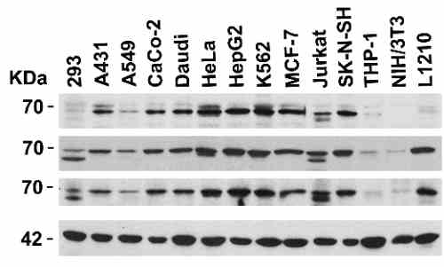 |
| Independent Antibody Validation (IAV) via Protein Expression Profile in Cell Lines Loading: 15 µg of lysates per lane. Antibodies: AIF 2267, (1 µg/mL), AIF 2239, (1 µg/mL), AIF 5029, (2 µg/mL), and beta-actin (1 µg/mL), 1h incubation at RT in 5% NFDM/TBST.Secondary: Goat anti-rabbit IgG HRP conjugate at 1:10000 dilution. |
 |
| Western Blot Validation in Mouse and Rat Cell Lines Loading: 15 µg of lysates per lane. Antibodies: AIF 5029, (2 µg/mL), 1h incubation at RT in 5% NFDM/TBST.Secondary: Goat anti-rabbit IgG HRP conjugate at 1:10000 dilution. |
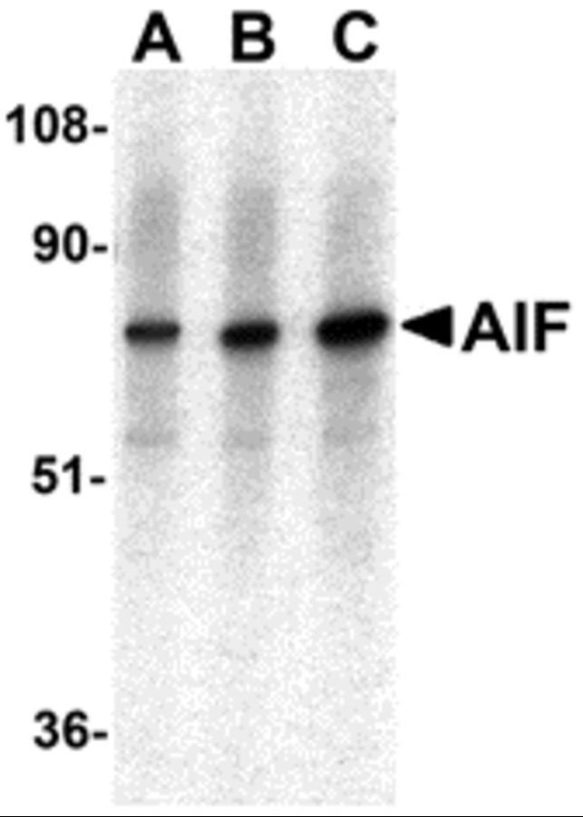 |
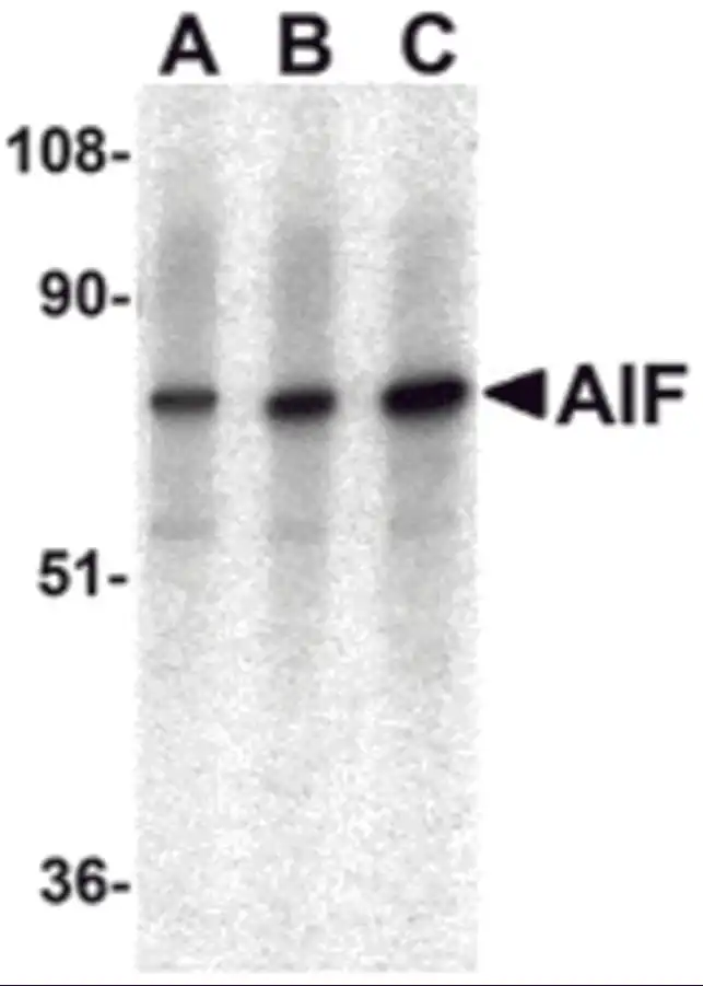
| Western Blot Validation in K562 Cell Line Loading: 15 µg of lysates per lane. Antibodies: AIF 5029, (A: 0.5 µg/mL, B: 1 µg/mL, C: 2 µg/mL), 1h incubation at RT in 5% NFDM/TBST.Secondary: Goat anti-rabbit IgG HRP conjugate at 1:10000 dilution. |
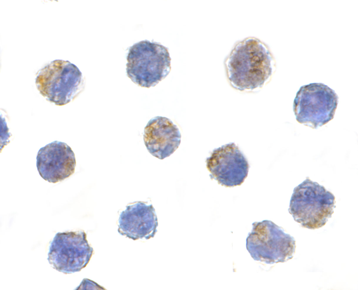 |
| Immunocytochemistry Validation of AIF in K562 Cells Immunocytochemical analysis of K562 cells using anti-AIF antibody (5029) at 5 µg/mL. Cells was fixed with formaldehyde and blocked with 10% serum for 1 h at RT; antigen retrieval was by heat mediation with a citrate buffer (pH6). Samples were incubated with primary antibody overnight at 4C. A goat anti-rabbit IgG H&L (HRP) at 1/250 was used as secondary. Counter stained with Hematoxylin. |
 |
| KD and Induced Validation of AIF in H1299 Cells(Stambolsky et al., 2006) Western blot analysis of AIF knockdown with anti-AIF antibodies in H1299 cells. AIF expression was disrupted in AIF knockdown cells (siRNA1 and siRNA4). An increased expression of AIF was induced by ZnCl2 treatment, which was not observed in AIF knockdown cells. |
 |
| KD Validation of AIF in AIF Silenced Stable Cells(Apostolova et al., 2006) AIF silencing is sustained in stable cell lines. Western blot analysis ofstable lines AIF-1-10, AIF-2-4 and pU6-2 using anti-AIF antibodies. AIF protein was disrupted after AIF silencing with AIF siRNA (AIF-1-10 and AIF-2-4) as compared to control (Hep3B and pU6-2). |
 |
| Immunofluorescence Validation of AIF in Rat Hippocampal Neurons (Hofer et al., 2011) (G-L) After exposure to bacterial components, AIF colocalized in mature neurons (MAP2; I, L), immature neurons (DcX;H, K), and stem/progenitor cells (Nestin; G, J). AIF expression was detected by anti-AIF antibodies. |
 |
| Subcellular Localization Validation of AIF in mononuclear cells (Gupta et al., 2424) A shows mononuclear cells (MNCs) alone, B shows MNCs transfected with control plasmid, C shows MNCs transfected with Bcl-2 expression plasmid. Overlay is of Mitotracker (red) and AIF (green). Hoechst 33258 dye is used to examine chromatin fragmentation. The release of AIF form mitochondria is detected by anti-AIF antibodies. |
FAQ & Publications
Frequently Asked Questions
What species does the rabbit anti-AIF (CT) polyclonal antibody 5029 cross-react with?
This antibody targets the C-terminal region of human AIF, with the immunogen sequence identical in mouse and rat, making it reactive in these species as well.
Which applications are validated for use with the rabbit anti-AIF (CT) polyclonal antibody 5029?
The antibody has been validated for ELISA, Western blot (WB), and immunocytochemistry/immunofluorescence (ICC/IF) applications.
How should the rabbit anti-AIF (CT) polyclonal antibody 5029 be stored to maintain stability?
Store the antibody at -20°C for at least one year and avoid multiple freeze-thaw cycles to preserve its stability.
What are the recommended working concentrations for immunoblotting and immunocytochemistry using this antibody?
For immunoblotting, use the antibody at 0.5 to 2 µg/mL; for immunocytochemistry, a concentration of 5 µg/mL is recommended. Users should optimize these concentrations for their specific applications.
Publications
| pmid | title | authors | citation |
|---|---|---|---|
| We haven't added any publications to our database yet. | |||
Published literature highly relevant to the biological target of this product and referencing this antibody or clone are retrieved from the PubMed database provided by the United States National Library of Medicine at the National Institutes of Health.
Protocols
| relevant to this product |
|---|
| Western blot IHC ICC |
Documents
| Batch Number | QC File | SDS |
|---|---|---|
| To view batch-specific Safety Datasheets and Quality Certificates associated with your account, please Log In. | ||
Only logged in customers who have purchased this product may leave a review.


Reviews
There are no reviews yet.