| Weight | 1 lbs |
|---|---|
| Dimensions | 9 × 5 × 2 in |
| host | mouse |
| isotype | IgG1 |
| clonality | monoclonal |
| concentration | 1 mg/mL |
| applications | ELISA, ICC/IF, WB |
| reactivity | tagged fusion proteins |
| available sizes | 1 mg, 100 µg, 25 µg |

mouse anti-RFP monoclonal antibody (RF5R) 1018
Price range: $100.00 through $2,600.00
Antibody summary
- Mouse monoclonal to RFP
- Suitable for: WB,ICC/IF,ELISA
- Reacts with: tagged fusion proteins
- Isotype: IgG1
- 100 µg
mouse anti-RFP monoclonal antibody (RF5R) 1018
| antibody |
|---|
| Database link: Q9U6Y8 |
| Tested applications WB,ICC/IF,ELISA |
| Recommended dilutions WB: 1:1000-3000 ICC/IF: 1:500-2000 For best results with other assays (e.g.: Dot, ELISA, IP, etc), please determine optimal working dilution by titration test. |
| Immunogen RFP from the?Discosoma sp.?(sea anemone) N-terminal peptide-KLH conjugates |
| Size and concentration 25, 100, 1000µg and 1 mg/mL |
| Form liquid |
| Storage Instructions -20°C for 2 years or more. °Centrifuge after first thaw to maximize product recovery. Aliquot to avoid repeated freeze-thaw cycles. Store aliquots at 4°C for several days to weeks. |
| Storage buffer PBS, pH 7.2, 0.05% NaN3 |
| Purity affinity purified |
| Clonality monoclonal |
| Isotype IgG1 |
| Compatible secondaries goat anti-mouse IgG, H&L chain specific, peroxidase conjugated polyclonal antibody 5486 goat anti-mouse IgG, H&L chain specific, biotin conjugated, Conjugate polyclonal antibody 2685 goat anti-mouse IgG, H&L chain specific, FITC conjugated polyclonal antibody 7854 goat anti-mouse IgG, H&L chain specific, peroxidase conjugated polyclonal antibody, crossabsorbed 1706 goat anti-mouse IgG, H&L chain specific, biotin conjugated polyclonal antibody, crossabsorbed 1716 goat anti-mouse IgG, H&L chain specific, FITC conjugated polyclonal antibody, crossabsorbed 1721 |
| Isotype control Mouse monocolonal IgG1 - Isotype Control |
| target relevance |
|---|
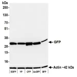
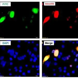
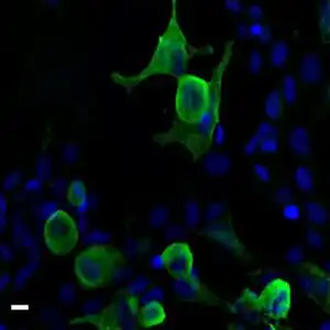
| Protein expression of RFP and RFP tagged proteins can be checked and quantified using this antibody in Western blotting. When imaging in situ, RFP fluorescence can be amplified by this antibody when used in conjunction with a suitable fluorescent label or secondary antibody. Click for more on: epitope tags and RFP |
| Protein names Red fluorescent protein drFP583 (DsRed) |
| Protein family GFP family |
| Mass 25931Da |
| Function FUNCTION: Thought to play a role in photoprotection of the coral's resident symbiont microalgae's photosystems from photoinhibition caused by high light levels found near the surface of coral reefs. In deeper water, the fluorescence may be to convert blue light into longer wavelengths more suitable for use in photosynthesis by the microalgal symbionts. |
| Post-translational modification PTM: Contains a chromophore consisting of modified amino acid residues. The chromophore is formed by autocatalytic backbone condensation between Xaa-N and Gly-(N+2), oxidation of Tyr-(N+1) to didehydrotyrosine, and formation of a double bond to the alpha-amino nitrogen of residue Xaa-N. Maturation of the chromophore requires nothing other than molecular oxygen. |
| Biotechnology Red fluorescent proteins (RFPs) have become invaluable tools in scientific research, particularly in the fields of cell biology, molecular imaging, and fluorescence microscopy. RFPs are a group of genetically engineered proteins that emit red light when excited by specific wavelengths of light. Due to their spectral properties, RFPs are highly compatible with commonly used green fluorescent proteins (GFPs) and yellow fluorescent proteins (YFPs), allowing for the simultaneous visualization of multiple cellular structures or processes. Researchers utilize RFPs as versatile molecular tags to track and study specific proteins, organelles, or cellular processes in living cells and organisms. By fusing RFPs to proteins of interest, scientists can monitor their subcellular localization, dynamics, and interactions in real-time, providing crucial insights into cellular mechanisms and protein function. RFPs have also proven valuable in tracking cell migration, tissue development, and gene expression in vivo. The versatility, brightness, and photostability of red fluorescent proteins have revolutionized fluorescence-based imaging techniques, enabling a deeper understanding of complex biological processes and advancing our knowledge across various research disciplines. |
| Target Relevance information above includes information from UniProt accession: Q9U6Y8 |
| The UniProt Consortium |
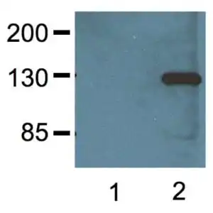
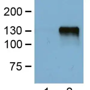
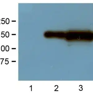
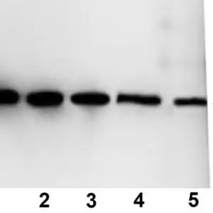
Data
 |
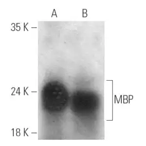
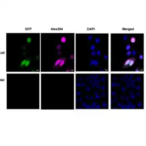
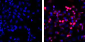
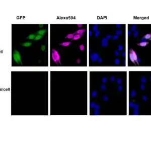
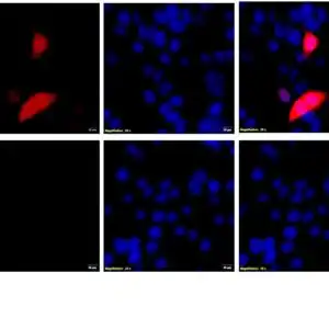
| 1:1000 (1µg/mL) Ab dilution probed against HEK293 cells transfected with RFP-tagged protein vector; untransfected control (1), transfected with TurboRFP (2), and transfected with dsRed (3). |
FAQ & Publications
Frequently Asked Questions
What applications is the mouse anti-RFP monoclonal antibody (RF5R) 1018 validated for, and what are the recommended dilutions?
This antibody is validated for Western blotting (WB), immunocytochemistry/immunofluorescence (ICC/IF), and ELISA. Recommended dilutions are 1:1000-3000 for WB and 1:500-2000 for ICC/IF. For other assays, such as dot blot or immunoprecipitation, optimal dilutions should be determined by titration.
How should the mouse anti-RFP monoclonal antibody (RF5R) 1018 be stored to maintain stability and activity?
The antibody should be stored at -20°C for long-term storage (up to 2 years or more). After the first thaw, centrifuge to maximize product recovery, aliquot to avoid repeated freeze-thaw cycles, and store aliquots at 4°C for several days to weeks. The storage buffer is PBS, pH 7.2, containing 0.05% sodium azide.
Publications
| pmid | title | authors | citation |
|---|---|---|---|
| 26071555 | Visualization of Fra-1/AP-1 activation during LPS-induced inflammatory lung injury using fluorescence optical imaging. | Subbiah Rajasekaran, Chandramohan R Tamatam, Haranatha R Potteti, Venu Raman, Jae-Woo Lee, Michael A Matthay, Dolly Mehta, Narsa M Reddy, Sekhar P Reddy | Am J Physiol Lung Cell Mol Physiol 309:L414-24 |
Published literature highly relevant to the biological target of this product and referencing this antibody or clone are retrieved from the PubMed database provided by the United States National Library of Medicine at the National Institutes of Health.
Protocols
| relevant to this product |
|---|
| Western blot ICC |
Documents
| Batch Number | QC File | SDS |
|---|---|---|
| To view batch-specific Safety Datasheets and Quality Certificates associated with your account, please Log In. | ||
Only logged in customers who have purchased this product may leave a review.


Reviews
There are no reviews yet.