| Weight | 1 lbs |
|---|---|
| Dimensions | 9 × 5 × 2 in |
| host | mouse |
| isotype | IgG1 |
| clonality | monoclonal |
| concentration | 1 mg/mL |
| applications | ICC/IF, WB |
| reactivity | Rad50 |
| available sizes | 100 µg |



mouse anti-Rad50 monoclonal antibody (2C6) 6741
$503.00
Antibody summary
- Mouse monoclonal to Rad50
- Suitable for: WB,ICC/IF,IHC-P,IP,IHC,in vitro
- Isotype: IgG1
- 100 µg
mouse anti-Rad50 monoclonal antibody (2C6) 6741
| antibody |
|---|
| Tested applications WB,IHC,IHC,ICC/IF |
| Recommended dilutions Immunoblotting, Immunoprecipitation: use at 0.1-2 ug/mL. Positive control: Raji cells. |
| Immunogen GST fusion protein corresponding to the complete coding region (aa 1-425) of Rad50 expressed in E. coli. |
| Size and concentration 100µg and lot specific |
| Form liquid |
| Storage Instructions This antibody is stable for at least one (1) year at -70°C. Avoid multiple freeze- thaw cycles. |
| Storage buffer PBS, pH 7.4 |
| Purity protein affinity purification |
| Clonality monoclonal |
| Isotype IgG1 |
| Compatible secondaries goat anti-mouse IgG, H&L chain specific, peroxidase conjugated polyclonal antibody 5486 goat anti-mouse IgG, H&L chain specific, biotin conjugated, Conjugate polyclonal antibody 2685 goat anti-mouse IgG, H&L chain specific, FITC conjugated polyclonal antibody 7854 goat anti-mouse IgG, H&L chain specific, peroxidase conjugated polyclonal antibody, crossabsorbed 1706 goat anti-mouse IgG, H&L chain specific, biotin conjugated polyclonal antibody, crossabsorbed 1716 goat anti-mouse IgG, H&L chain specific, FITC conjugated polyclonal antibody, crossabsorbed 1721 |
| Isotype control Mouse monocolonal IgG1 - Isotype Control |
| target relevance |
|---|
| Homo sapiens RAD50 DNA repair protein RAD50 |
| Protein names DNA repair protein RAD50 |
| Gene names RAD50 |
| Protein family Belongs to the SMC family. RAD50 subfamily |
| Function Component of the MRN complex, which plays a central role in double-strand break (DSB) repair, DNA recombination, maintenance of telomere integrity and meiosis (PubMed:15064416, PubMed:21757780, PubMed:27889449, PubMed:28134932, PubMed:28867292, PubMed:9590181, PubMed:9651580, PubMed:9705271). The MRN complex is involved in the repair of DNA double-strand breaks (DSBs) via homologous recombination (HR), an error-free mechanism which primarily occurs during S and G2 phases (PubMed:15064416, PubMed:21757780, PubMed:27889449, PubMed:28867292, PubMed:9590181, PubMed:9651580, PubMed:9705271). The complex (1) mediates the end resection of damaged DNA, which generates proper single-stranded DNA, a key initial steps in HR, and is (2) required for the recruitment of other repair factors and efficient activation of ATM and ATR upon DNA damage (PubMed:15064416, PubMed:27889449, PubMed:28867292, PubMed:9590181, PubMed:9651580, PubMed:9705271). The MRN complex possesses single-strand endonuclease activity and double-strand-specific 3'-5' exonuclease activity, which are provided by MRE11, to initiate end resection, which is required for single-strand invasion and recombination (PubMed:11741547, PubMed:9590181, PubMed:9651580, PubMed:9705271). Within the complex, RAD50 is both required to bind DNA ends and hold them in close proximity and regulate the activity of MRE11 (PubMed:11741547, PubMed:12805565, PubMed:28134932). RAD50 provides an ATP-dependent control of MRE11 by positioning DNA ends into the MRE11 active site: ATP-binding induces a large structural change from an open form with accessible MRE11 nuclease sites into a closed form (By similarity). The MRN complex is also required for DNA damage signaling via activation of the ATM and ATR kinases: the nuclease activity of MRE11 is not required to activate ATM and ATR (PubMed:15064416, PubMed:15790808, PubMed:16622404). The MRN complex is also required for the processing of R-loops (PubMed:31537797). In telomeres the MRN complex may modulate t-loop formation (PubMed:10888888) |
| Catalytic activity ATP + H2O = ADP + phosphate + H(+) |
| Subcellular location Nucleus, Chromosome, telomere, Chromosome |
| Structure (Microbial infection) Interacts with herpes simplex virus 1 protein UL12 (PubMed:20943970) |
| Post-translational modification Phosphorylation at Ser-635 by ATM in response to DNA damage is required for double-strand break (DSB) repair |
| Involvement in disease Nijmegen breakage syndrome-like disorder A disorder similar to Nijmegen breakage syndrome and characterized by chromosomal instability, radiation sensitivity, microcephaly, growth retardation, short stature and bird-like face. Immunodeficiency is absent. |
| Keywords 3D-structure, Acetylation, Alternative splicing, ATP-binding, Cell cycle, Chromosome, Coiled coil, DNA damage, DNA repair, Host-virus interaction, Hydrolase, Magnesium, Meiosis, Metal-binding, Nucleotide-binding, Nucleus, Phosphoprotein, Proteomics identification, Reference proteome, Telomere, Zinc |
| Sequence MSRIEKMSILGVRSFGIEDKDKQIITFFSPLTILVGPNGAGKTTIIECLKYICTGDFPPG TKGNTFVHDPKVAQETDVRAQIRLQFRDVNGELIAVQRSMVCTQKSKKTEFKTLEGVITR TKHGEKVSLSSKCAEIDREMISSLGVSKAVLNNVIFCHQEDSNWPLSEGKALKQKFDEIF SATRYIKALETLRQVRQTQGQKVKEYQMELKYLKQYKEKACEIRDQITSKEAQLTSSKEI VKSYENELDPLKNRLKEIEHNLSKIMKLDNEIKALDSRKKQMEKDNSELEEKMEKVFQGT DEQLNDLYHNHQRTVREKERKLVDCHRELEKLNKESRLLNQEKSELLVEQGRLQLQADRH QEHIRARDSLIQSLATQLELDGFERGPFSERQIKNFHKLVRERQEGEAKTANQLMNDFAE KETLKQKQIDEIRDKKTGLGRIIELKSEILSKKQNELKNVKYELQQLEGSSDRILELDQE LIKAERELSKAEKNSNVETLKMEVISLQNEKADLDRTLRKLDQEMEQLNHHTTTRTQMEM LTKDKADKDEQIRKIKSRHSDELTSLLGYFPNKKQLEDWLHSKSKEINQTRDRLAKLNKE LASSEQNKNHINNELKRKEEQLSSYEDKLFDVCGSQDFESDLDRLKEEIEKSSKQRAMLA GATAVYSQFITQLTDENQSCCPVCQRVFQTEAELQEVISDLQSKLRLAPDKLKSTESELK KKEKRRDEMLGLVPMRQSIIDLKEKEIPELRNKLQNVNRDIQRLKNDIEEQETLLGTIMP EEESAKVCLTDVTIMERFQMELKDVERKIAQQAAKLQGIDLDRTVQQVNQEKQEKQHKLD TVSSKIELNRKLIQDQQEQIQHLKSTTNELKSEKLQISTNLQRRQQLEEQTVELSTEVQS LYREIKDAKEQVSPLETTLEKFQQEKEELINKKNTSNKIAQDKLNDIKEKVKNIHGYMKD IENYIQDGKDDYKKQKETELNKVIAQLSECEKHKEKINEDMRLMRQDIDTQKIQERWLQD NLTLRKRNEELKEVEEERKQHLKEMGQMQVLQMKSEHQKLEENIDNIKRNHNLALGRQKG YEEEIIHFKKELREPQFRDAEEKYREMMIVMRTTELVNKDLDIYYKTLDQAIMKFHSMKM EEINKIIRDLWRSTYRGQDIEYIEIRSDADENVSASDKRRNYNYRVVMLKGDTALDMRGR CSAGQKVLASLIIRLALAETFCLNCGIIALDEPTTNLDRENIESLAHALVEIIKSRSQQR NFQLLVITHDEDFVELLGRSEYVEKFYRIKKNIDQCSEIVKCSVSSLGFNVH |
| UniProt accession: Q92878 |
Data
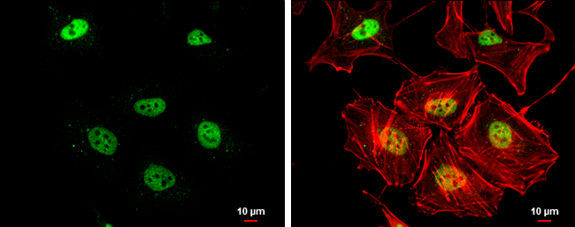 |
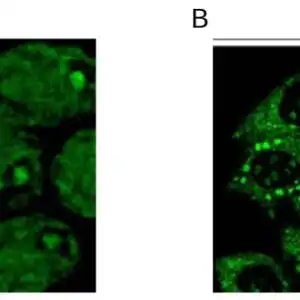
| Rad50 antibody [13B3] detects Rad50 protein at nucleus by immunofluorescent analysis. Sample: HeLa cells were fixed in 4% paraformaldehyde at RT for 15 min. Green: Rad50 protein stained by Rad50 antibody [13B3] (6741) diluted at 1:200. Red: phalloidin, a cytoskeleton marker, diluted at 1:200. Scale bar = 10 um. |
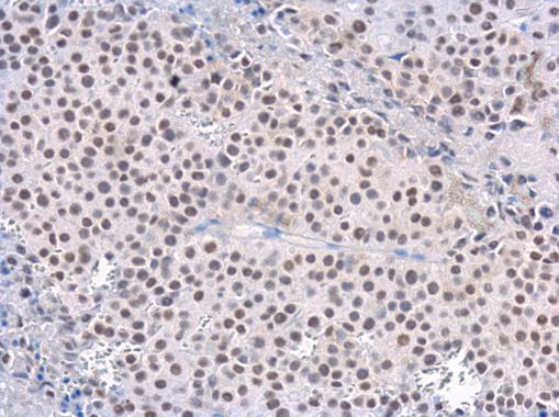 |
| Rad50 antibody [13B3] detects Rad50 protein at nucleus in CAL 27 xenograft by immunohistochemical analysis. Sample: Paraffin-embedded CAL 27 xenograft. Rad50 antibody [13B3] (6741) diluted at 1:200. Antigen Retrieval: Citrate buffer, pH 6.0, 15 min |
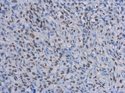 |
| Rad50 antibody [13B3] detects Rad50 protein at nucleus in PC-3 xenograft by immunohistochemical analysis. Sample: Paraffin-embedded PC-3 xenograft. Rad50 antibody [13B3] (6741) diluted at 1:200. Antigen Retrieval: Citrate buffer, pH 6.0, 15 min |
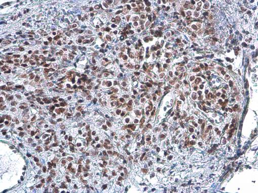 |
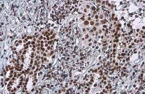
| Rad50 antibody [13B3] detects Rad50 protein at nucleus by immunohistochemical analysis.Sample: Paraffin-embedded human lung cancer.Rad50 stained by Rad50 antibody [13B3] (6741) diluted at 1:100.Antigen Retrieval: Citrate buffer, pH 6.0, 15 min |
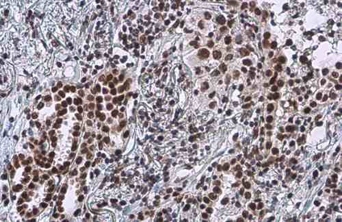 |
| Rad50 antibody [13B3] detects Rad50 protein at nucleus by immunohistochemical analysis.Sample: Paraffin-embedded human lung cancer.Rad50 stained by Rad50 antibody [13B3] (6741) diluted at 1:100.Antigen Retrieval: Citrate buffer, pH 6.0, 15 min |
 |
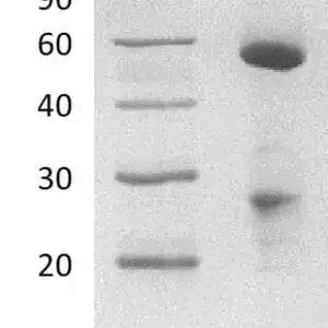
| HeLa whole cell and nuclear extracts (30 µg) were separated by 5% SDS-PAGE, and the membrane was blotted with Rad50 antibody [13B3] (6741) diluted at 1:1000. The HRP-conjugated anti-mouse IgG antibody was used to detect the primary antibody. |
FAQ & Publications
Frequently Asked Questions
What applications has the mouse anti-Rad50 monoclonal antibody (2C6) been validated for?
This antibody is suitable and has been tested for Western blotting (WB), immunocytochemistry/immunofluorescence (ICC/IF), immunohistochemistry on paraffin-embedded sections (IHC-P), immunoprecipitation (IP), and general immunohistochemistry (IHC). Recommended dilutions for immunoprecipitation range from 0.1 to 2 µg/mL.
How should the mouse anti-Rad50 monoclonal antibody (2C6) be stored to maintain stability?
The antibody is supplied in liquid form at a concentration of 1 mg/mL in PBS buffer at pH 7.4. It is stable for at least one year when stored at -70°C. It is recommended to avoid multiple freeze-thaw cycles to preserve antibody integrity.
What is the immunogen used to produce the mouse anti-Rad50 monoclonal antibody (2C6)?
The immunogen is a GST fusion protein corresponding to the complete coding region (amino acids 1-425) of Rad50 expressed in Escherichia coli. This ensures specificity for the Rad50 protein target.
Publications
| pmid | title | authors | citation |
|---|---|---|---|
| We haven't added any publications to our database yet. | |||
Published literature highly relevant to the biological target of this product and referencing this antibody or clone are retrieved from the PubMed database provided by the United States National Library of Medicine at the National Institutes of Health.
Protocols
| relevant to this product |
|---|
| Western blot IHC ICC |
Documents
| Batch Number | QC File | SDS |
|---|---|---|
| To view batch-specific Safety Datasheets and Quality Certificates associated with your account, please Log In. | ||
Only logged in customers who have purchased this product may leave a review.


Reviews
There are no reviews yet.