| Weight | 1 lbs |
|---|---|
| Dimensions | 9 × 5 × 2 in |
| host | mouse |
| isotype | IgG1 |
| clonality | monoclonal |
| concentration | concentrate, predilute |
| applications | IHC |
| reactivity | human |
| available size | 0.1 mL, 0.5 mL, 1 mL concentrated, 7 mL prediluted |

mouse anti-Podoplanin monoclonal antibody (D2-40) 6341
Price range: $160.00 through $528.00
Antibody summary
- Mouse monoclonal to Podoplanin
- Suitable for: Immunohistochemistry (formalin-fixed, paraffin-embedded tissues)
- Reacts with: Human
- Isotype:IgG1
- Control: Germ cell tumor
- Visualization: Cytoplasmic
- 0.1, 0.5, 1.0 mL concentrated, 7 mL prediluted
mouse anti-Podoplanin monoclonal antibody D2-40 6341
| target relevance |
|---|
| Homo sapiens PDPN Podoplanin |
| Protein names Podoplanin |
| Alternative names Aggrus, Glycoprotein 36, PA2.26 antigen, T1-alpha |
| Gene names PDPN |
| Protein family Belongs to the podoplanin family |
| Function Mediates effects on cell migration and adhesion through its different partners. During development plays a role in blood and lymphatic vessels separation by binding CLEC1B, triggering CLEC1B activation in platelets and leading to platelet activation and/or aggregation (PubMed:14522983, PubMed:15231832, PubMed:17222411, PubMed:17616532, PubMed:18215137). Interaction with CD9, on the contrary, attenuates platelet aggregation induced by PDPN (PubMed:18541721). Through MSN or EZR interaction promotes epithelial-mesenchymal transition (EMT) leading to ERZ phosphorylation and triggering RHOA activation leading to cell migration increase and invasiveness (PubMed:17046996, PubMed:21376833). Interaction with CD44 promotes directional cell migration in epithelial and tumor cells (PubMed:20962267). In lymph nodes (LNs), controls fibroblastic reticular cells (FRCs) adhesion to the extracellular matrix (ECM) and contraction of the actomyosin by maintaining ERM proteins (EZR; MSN and RDX) and MYL9 activation through association with unknown transmembrane proteins. Engagement of CLEC1B by PDPN promotes FRCs relaxation by blocking lateral membrane interactions leading to reduction of ERM proteins (EZR; MSN and RDX) and MYL9 activation (By similarity). Through binding with LGALS8 may participate in connection of the lymphatic endothelium to the surrounding extracellular matrix (PubMed:19268462). In keratinocytes, induces changes in cell morphology showing an elongated shape, numerous membrane protrusions, major reorganization of the actin cytoskeleton, increased motility and decreased cell adhesion (PubMed:15515019). Controls invadopodia stability and maturation leading to efficient degradation of the extracellular matrix (ECM) in tumor cells through modulation of RHOC activity in order to activate ROCK1/ROCK2 and LIMK1/LIMK2 and inactivation of CFL1 (PubMed:25486435). Required for normal lung cell proliferation and alveolus formation at birth (By similarity). Does not function as a water channel or as a regulator of aquaporin-type water channels (PubMed:9651190). Does not have any effect on folic acid or amino acid transport (By similarity) |
| Subcellular location Cytoplasm, cytosol |
| Structure Homodimer (PubMed:21376833). Interacts with CLEC1B; the interaction is independent of CLEC1B glycosylation and activates CLEC1B; the interaction is dependent of sialic acid on O-glycans (PubMed:17616532, PubMed:18215137, PubMed:25458834). Interacts with CD9; this interaction is homophilic and attenuates platelet aggregation and pulmonary metastasis induced by PDPN (PubMed:18541721). Interacts with LGALS8; the interaction is glycosylation-dependent; may participate in connection of the lymphatic endothelium to the surrounding extracellular matrix (PubMed:19268462). Interacts with HSPA9 (PubMed:23541579). Interacts (via extracellular domain) with CD44; this interaction is required for PDPN-mediated directional migration and regulation of lamellipodia extension/stabilization during cell spreading and migration (PubMed:20962267). Interacts (via cytoplasmic domain) with MSN and EZR; activates RHOA and promotes epithelial-mesenchymal transition (PubMed:17046996). Interacts with CCL21; relocalized PDPN to the basolateral membrane (PubMed:14978162) |
| Post-translational modification Extensively O-glycosylated. Contains sialic acid residues. O-glycosylation is necessary for platelet aggregation activity. Disialylated at Thr-52; sialic acid is critical for platelet-aggregating activity and for CLEC1B interaction (PubMed:17222411, PubMed:25458834) The N-terminus is blocked Cleaved by a metalloprotease within its extracellular (EC) domain, generating a membrane-bound C-terminal fragment (PCTF33) and an extracellular fragment. The resulting membrane-bound C-terminal fragment (PCTF33) is further processed between Val-150 and Val-151 by PSEN1/gamma-secretase generating the intracellular domain of podoplanin (PICD) |
| Keywords 3D-structure, Alternative splicing, Cell junction, Cell membrane, Cell projection, Cell shape, Cytoplasm, Developmental protein, Direct protein sequencing, Glycoprotein, Membrane, Proteomics identification, Reference proteome, Sialic acid, Signal, Transmembrane, Transmembrane helix |
| Sequence MWKVSALLFVLGSASLWVLAEGASTGQPEDDTETTGLEGGVAMPGAEDDVVTPGTSEDRY KSGLTTLVATSVNSVTGIRIEDLPTSESTVHAQEQSPSATASNVATSHSTEKVDGDTQTT VEKDGLSTVTLVGIIVGVLLAIGFIGAIIVVVMRKMSGRYSP |
| UniProt accession: Q86YL7 |
Data
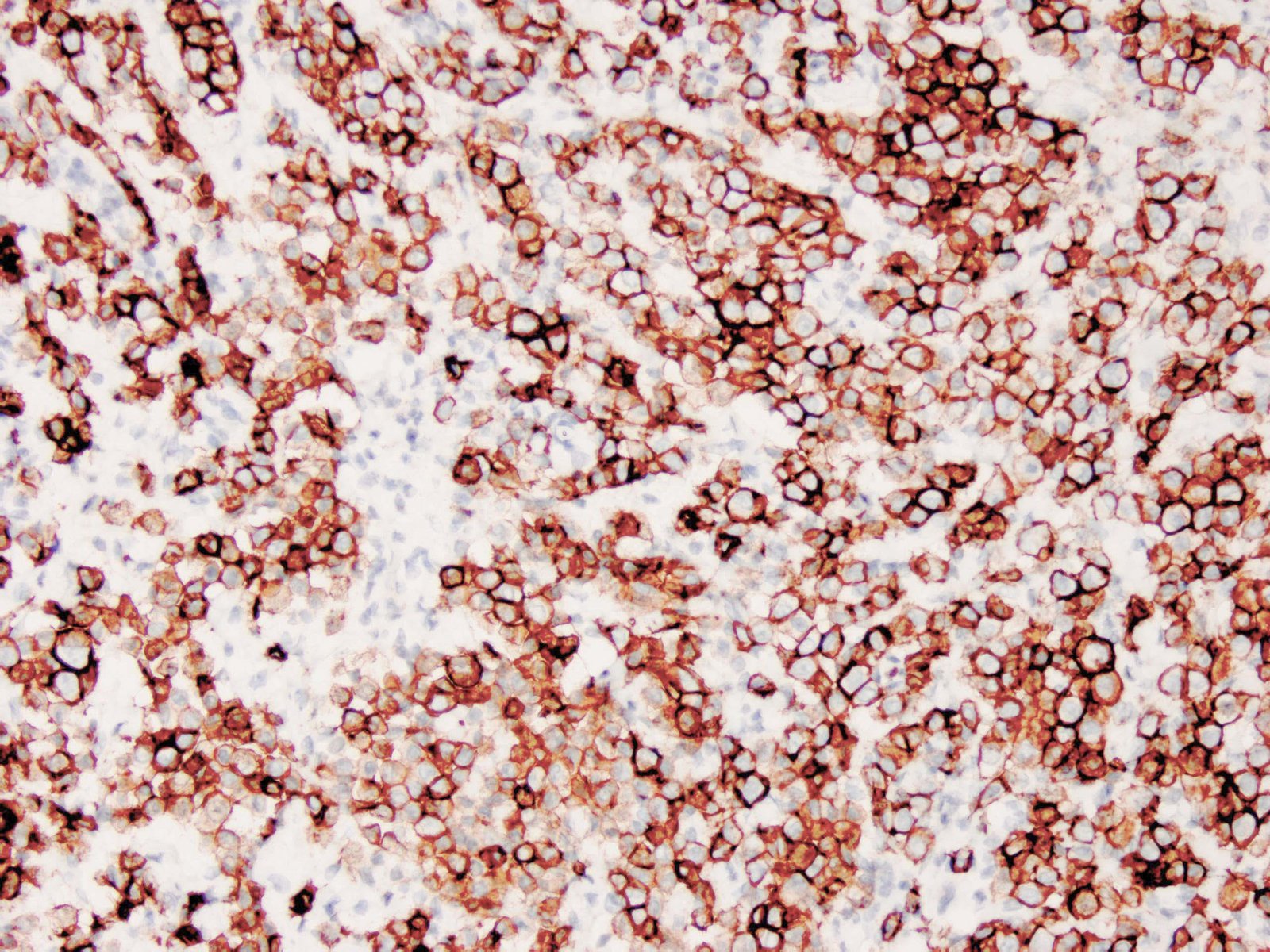 |
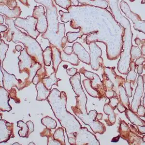
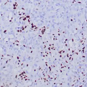
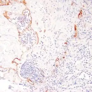
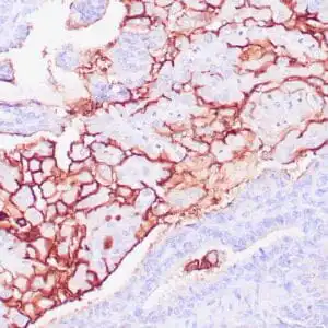
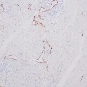
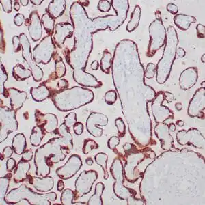
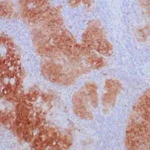
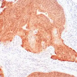
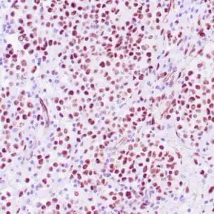
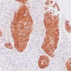
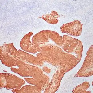
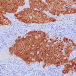
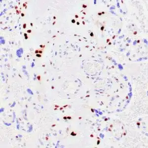
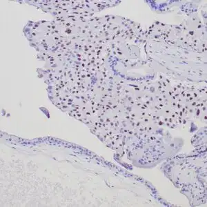
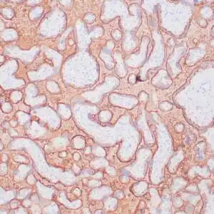
| Human testicular seminoma stained with anti-D2-40 antibody using peroxidase-conjugate and DAB chromogen. Note cytoplasmic staining of tumor cells and endothelial cells of lymphatic channels. |
FAQ & Publications
Frequently Asked Questions
What is the recommended application and dilution range for the mouse anti-Podoplanin monoclonal antibody (D2-40)?
This antibody is suitable for immunohistochemistry (IHC) on formalin-fixed, paraffin-embedded human tissues. The recommended dilution range for the concentrated antibody is 1:20 to 1:100.
How should the mouse anti-Podoplanin monoclonal antibody (D2-40) be stored to maintain its stability?
For short-term storage, keep the antibody at 2-8°C. For long-term storage, it should be kept at -20°C. Avoid repeated freeze/thaw cycles to preserve antibody quality.
What is the host species and isotype of the anti-Podoplanin monoclonal antibody (D2-40)?
The antibody is a mouse monoclonal antibody of the IgG1 isotype.
Which secondary antibodies are compatible with the mouse anti-Podoplanin monoclonal antibody (D2-40) for detection?
Compatible secondary antibodies include goat anti-mouse IgG, heavy and light chain specific, available as peroxidase conjugated polyclonal antibody (catalog 5486), biotin conjugated polyclonal antibody (catalog 2685), FITC conjugated polyclonal antibody (catalog 7854), and their cross-absorbed variants.
Publications
| pmid | title | authors | citation |
|---|---|---|---|
| We haven't added any publications to our database yet. | |||
Published literature highly relevant to the biological target of this product and referencing this antibody or clone are retrieved from the PubMed database provided by the United States National Library of Medicine at the National Institutes of Health.
Protocols
| relevant to this product |
|---|
| IHC |
Documents
| Batch Number | QC File | SDS |
|---|---|---|
| To view batch-specific Safety Datasheets and Quality Certificates associated with your account, please Log In. | ||
Only logged in customers who have purchased this product may leave a review.
Reviews
There are no reviews yet.