| Weight | 1 lbs |
|---|---|
| Dimensions | 9 × 5 × 2 in |
| host | mouse |
| isotype | IgG1 |
| clonality | monoclonal |
| concentration | 1 mg/mL |
| applications | ELISA, ICC/IF, WB |
| reactivity | human, mouse, rat |
| available sizes | 1 mg, 100 µg, 25 µg |


mouse anti-GAPDH monoclonal antibody (GA1R) 1937
Price range: $100.00 through $2,600.00
Antibody summary
- Mouse monoclonal to GAPDH
- Suitable for: WB,ICC/IF,ELISA
- Reacts with: human, mouse, rat
- Isotype: IgG1
- 100 µg, 25 µg, 1 mg
mouse anti-GAPDH monoclonal antibody (GA1R) 1937
| antibody |
|---|
| Database link: human P04406 mouse P16858 rat P04797 |
| Tested applications WB,ICC/IF,ELISA |
| Recommended dilutions WB: 1:1000-100000 |
| Immunogen Recombinant GAPDH protein (human). |
| Size and concentration 25, 100, 1000µg and 1 mg/mL |
| Form liquid |
| Storage Instructions -20°C for 2 years or more. Centrifuge after first thaw to maximize product recovery. Aliquot to avoid repeated freeze-thaw cycles. Store aliquots at 4°C for several days to weeks. |
| Storage buffer PBS, pH 7.2, 0.05% NaN3 |
| Purity affinity purified |
| Clonality monoclonal |
| Isotype IgG1 |
| Compatible secondaries goat anti-mouse IgG, H&L chain specific, peroxidase conjugated polyclonal antibody 5486 goat anti-mouse IgG, H&L chain specific, biotin conjugated, Conjugate polyclonal antibody 2685 goat anti-mouse IgG, H&L chain specific, FITC conjugated polyclonal antibody 7854 goat anti-mouse IgG, H&L chain specific, peroxidase conjugated polyclonal antibody, crossabsorbed 1706 goat anti-mouse IgG, H&L chain specific, biotin conjugated polyclonal antibody, crossabsorbed 1716 goat anti-mouse IgG, H&L chain specific, FITC conjugated polyclonal antibody, crossabsorbed 1721 |
| Isotype control Mouse monocolonal IgG1 - Isotype Control |
| target relevance |
|---|
| Homo sapiens GAPDH Glyceraldehyde-3-phosphate dehydrogenase |
| Protein names Glyceraldehyde-3-phosphate dehydrogenase |
| Alternative names Peptidyl-cysteine S-nitrosylase GAPDH |
| Gene names GAPDH |
| Protein family Belongs to the glyceraldehyde-3-phosphate dehydrogenase family |
| Function Catalyzes the conversion of D-glyceraldehyde 3-phosphate (G3P) into 3-phospho-D-glyceroyl phosphate in glycolysis and the reverse reaction in gluconeogenesis (PubMed:11724794, PubMed:3170585). Also shows nitrosylase activity, thereby playing a role in nuclear functions (PubMed:11724794, PubMed:3170585). Modulates the organization and assembly of the cytoskeleton (By similarity). Facilitates the CHP1-dependent microtubule and membrane associations through its ability to stimulate the binding of CHP1 to microtubules (By similarity). Component of the GAIT (gamma interferon-activated inhibitor of translation) complex which mediates interferon-gamma-induced transcript-selective translation inhibition in inflammation processes (PubMed:23071094). Upon interferon-gamma treatment assembles into the GAIT complex which binds to stem loop-containing GAIT elements in the 3'-UTR of diverse inflammatory mRNAs (such as ceruplasmin) and suppresses their translation (PubMed:23071094). Also plays a role in innate immunity by promoting TNF-induced NF-kappa-B activation and type I interferon production, via interaction with TRAF2 and TRAF3, respectively (PubMed:23332158, PubMed:27387501). Participates in nuclear events including transcription, RNA transport, DNA replication and apoptosis (By similarity). Nuclear functions are probably due to the nitrosylase activity that mediates cysteine S-nitrosylation of nuclear target proteins such as SIRT1, HDAC2 and PRKDC (By similarity) |
| Catalytic activity D-glyceraldehyde 3-phosphate + phosphate + NAD(+) = (2R)-3-phospho-glyceroyl phosphate + NADH + H(+) S-nitroso-L-cysteinyl-[GAPDH] + L-cysteinyl-[protein] = L-cysteinyl-[GAPDH] + S-nitroso-L-cysteinyl-[protein] |
| Subcellular location Cytoplasm, cytosol, Nucleus, Cytoplasm, perinuclear region, Membrane, Cytoplasm, cytoskeleton |
| Structure Homotetramer (PubMed:16239728, PubMed:16510976). Interacts with TPPP; the interaction is direct (By similarity). Interacts (when S-nitrosylated) with SIAH1; leading to nuclear translocation (By similarity). Interacts with RILPL1/GOSPEL, leading to prevent the interaction between GAPDH and SIAH1 and prevent nuclear translocation (By similarity). Interacts with CHP1; the interaction increases the binding of CHP1 with microtubules (By similarity). Associates with microtubules (By similarity). Interacts with EIF1AD, USP25, PRKCI and WARS1 (PubMed:11724794, PubMed:15628863, PubMed:16501887, PubMed:20644585). Interacts with phosphorylated RPL13A; inhibited by oxidatively-modified low-densitity lipoprotein (LDL(ox)) (PubMed:22771119). Component of the GAIT complex (PubMed:15479637). Interacts with FKBP6; leading to inhibit GAPDH catalytic activity (PubMed:19001379). Interacts with TRAF2, promoting TRAF2 ubiquitination (PubMed:23332158). Interacts with TRAF3, promoting TRAF3 ubiquitination (PubMed:27387501) |
| Post-translational modification S-nitrosylation of Cys-152 leads to interaction with SIAH1, followed by translocation to the nucleus (By similarity). S-nitrosylation of Cys-247 is induced by interferon-gamma and LDL(ox) implicating the iNOS-S100A8/9 transnitrosylase complex and seems to prevent interaction with phosphorylated RPL13A and to interfere with GAIT complex activity (PubMed:22771119, PubMed:25417112) ISGylated Sulfhydration at Cys-152 increases catalytic activity Oxidative stress can promote the formation of high molecular weight disulfide-linked GAPDH aggregates, through a process called nucleocytoplasmic coagulation. Such aggregates can be observed in vivo in the affected tissues of patients with Alzheimer disease or alcoholic liver cirrhosis, or in cell cultures during necrosis. Oxidation at Met-46 may play a pivotal role in the formation of these insoluble structures. This modification has been detected in vitro following treatment with free radical donor (+/-)-(E)-4-ethyl-2-[(E)-hydroxyimino]-5-nitro-3-hexenamide. It has been proposed to destabilize nearby residues, increasing the likelihood of secondary oxidative damages, including oxidation of Tyr-45 and Met-105. This cascade of oxidations may augment GAPDH misfolding, leading to intermolecular disulfide cross-linking and aggregation Succination of Cys-152 and Cys-247 by the Krebs cycle intermediate fumarate, which leads to S-(2-succinyl)cysteine residues, inhibits glyceraldehyde-3-phosphate dehydrogenase activity. Fumarate concentration as well as succination of cysteine residues in GAPDH is significantly increased in muscle of diabetic mammals. It was proposed that the S-(2-succinyl)cysteine chemical modification may be a useful biomarker of mitochondrial and oxidative stress in diabetes and that succination of GAPDH and other thiol proteins by fumarate may contribute to the metabolic changes underlying the development of diabetes complications (Microbial infection) Glycosylated by C.rodentium protein NleB, enteropathogenic E.coli protein NleB1 and S.typhimurium protein Ssek1: arginine GlcNAcylation prevents the interaction with TRAF2 and TRAF3 (PubMed:23332158, PubMed:27387501, PubMed:28522607). This leads to reduced ubiquitination of TRAF2 and TRAF3, and subsequent inhibition of NF-kappa-B signaling and type I interferon production, respectively (PubMed:23332158, PubMed:27387501) |
| Keywords 3D-structure, Acetylation, ADP-ribosylation, Alternative splicing, Apoptosis, Cytoplasm, Cytoskeleton, Direct protein sequencing, Glycolysis, Glycoprotein, Immunity, Innate immunity, Isopeptide bond, Membrane, Methylation, NAD, Nucleus, Oxidation, Oxidoreductase, Phosphoprotein, Proteomics identification, Reference proteome, S-nitrosylation, Transferase, Translation regulation, Ubl conjugation |
| Sequence MGKVKVGVNGFGRIGRLVTRAAFNSGKVDIVAINDPFIDLNYMVYMFQYDSTHGKFHGTV KAENGKLVINGNPITIFQERDPSKIKWGDAGAEYVVESTGVFTTMEKAGAHLQGGAKRVI ISAPSADAPMFVMGVNHEKYDNSLKIISNASCTTNCLAPLAKVIHDNFGIVEGLMTTVHA ITATQKTVDGPSGKLWRDGRGALQNIIPASTGAAKAVGKVIPELNGKLTGMAFRVPTANV SVVDLTCRLEKPAKYDDIKKVVKQASEGPLKGILGYTEHQVVSSDFNSDTHSSTFDAGAG IALNDHFVKLISWYDNEFGYSNRVVDLMAHMASKE |
| UniProt accession: P04406 |
Data
 |
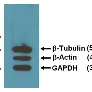
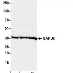
| Three loading control mAbs reacting against 10µg/lane of mouse brain tissue lysates. 50kDa band is Anti-β-Tubulin (#5007) at 1:2,000 dilution (0.5 µg/mL); 42kDa band is Anti-β-Actin (#5132) at 1:1,000 dilution (1 µg/mL); 37kDa band is Anti-GAPDH (#1937) at 1:5,000 dilution (0.2 µg/mL). |
 |
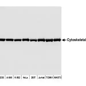
| LEFT: 1:2,000 (0.5µg/mL) Ab dilution used in WB of 5µg/lane tissue lysates from human (1), mouse (2), rat (3), rabbit (4), chicken (5), and hamster (6). RIGHT: WB from BL-21 bacteria (1), Sf9 insect (2), and Saccharomyces cerevisiae (3). |
FAQ & Publications
Frequently Asked Questions
What species does the mouse anti-GAPDH monoclonal antibody (GA1R) 1937 react with?
This antibody reacts with human, mouse, and rat GAPDH proteins.
Which applications has the mouse anti-GAPDH monoclonal antibody (GA1R) 1937 been validated for?
The antibody has been tested and is suitable for Western blotting (WB), immunocytochemistry/immunofluorescence (ICC/IF), and ELISA applications.
What are the recommended storage conditions for this GAPDH antibody?
Store the antibody at -20°C for long-term preservation of at least 2 years. After the first thaw, centrifuge to maximize recovery and aliquot to avoid repeated freeze-thaw cycles. Aliquots can be stored at 4°C for several days to weeks.
What is the concentration and format of the mouse anti-GAPDH monoclonal antibody (GA1R) 1937?
The antibody is supplied as a liquid at a concentration of 1 mg/mL, available in sizes of 25 µg, 100 µg, and 1 mg.
Publications
| pmid | title | authors | citation |
|---|---|---|---|
| 36833172 | Involvement of Mitochondrial Dysfunction in FOXG1 Syndrome. | Victoria A Bjerregaard, Amanda M Levy, Mille S Batz, Ravina Salehi, Mathis Hildonen, Trine B Hammer, Rikke S Møller, Claus Desler, Zeynep Tümer | Genes (Basel) 14:N/A |
| 36831065 | Low-Molecular-Weight β-1,3-1,6-Glucan Derived from Aureobasidium pullulans Exhibits Anticancer Activity by Inducing Apoptosis in Colorectal Cancer Cells. | Ji Hyeon Kim, Jeonghyeon Seo, Huiwon No, Takao Kuge, Takahiro Mori, Hisashi Kimoto, Jin-Kyung Kim | Biomedicines 11:N/A |
| 36830676 | LRRK2 Kinase Inhibition Attenuates Astrocytic Activation in Response to Amyloid β(1-42) Fibrils. | Alice Filippini, Valentina Salvi, Vincenzo Dattilo, Chiara Magri, Stefania Castrezzati, Robert Veerhuis, Daniela Bosisio, Massimo Gennarelli, Isabella Russo | Biomolecules 13:N/A |
| 36818283 | Role of human HSPE1 for OPA1 processing independent of HSPD1. | Nelson Yeung, Daisuke Murata, Miho Iijima, Hiromi Sesaki | iScience 26:106067 |
| 36685538 | Duck TRIM29 negatively regulates type I IFN production by targeting MAVS. | Weiqiang Li, Yating Song, Yuqing Du, Zhanhong Huang, Meng Zhang, Zuxian Chen, Zhuoliang He, Yangbao Ding, Junsheng Zhang, Luxiang Zhao, Hailiang Sun, Peirong Jiao | Front Immunol 13:1016214 |
Published literature highly relevant to the biological target of this product and referencing this antibody or clone are retrieved from the PubMed database provided by the United States National Library of Medicine at the National Institutes of Health.
Protocols
| relevant to this product |
|---|
| Western blot ICC |
7 reviews for mouse anti-GAPDH monoclonal antibody (GA1R) 1937
Only logged in customers who have purchased this product may leave a review.


verified user –
verified user –
verified user –
verified user –
verified user –
verified user –
verified user –