| Weight | 1 lbs |
|---|---|
| Dimensions | 9 × 5 × 2 in |
| host | mouse |
| isotype | IgG2a |
| clonality | monoclonal |
| concentration | concentrate, predilute |
| applications | IHC |
| reactivity | human |
| available size | 0.1 mL, 0.5 mL, 1 mL concentrated, 7 mL prediluted |

mouse anti-Cytokeratin monoclonal antibody (CAM 5.2) 6134
Price range: $160.00 through $528.00
Antibody summary
- Mouse monoclonal to Cytokeratin
- Suitable for: Immunohistochemistry (formalin-fixed, paraffin-embedded tissues)
- Reacts with: Human
- Isotype:IgG2a
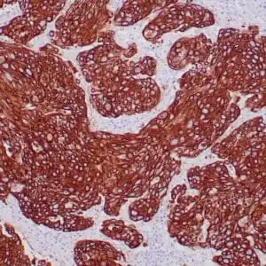
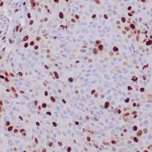
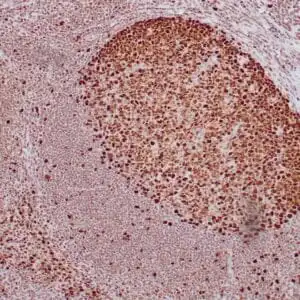
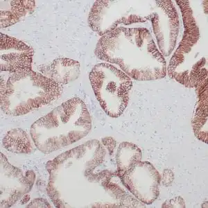
- Control: Ovarian carcinoma
- Visualization: Cytoplasmic
- 0.1, 0.5, 1.0 mL concentrated, 7 mL prediluted
mouse anti-Cytokeratin monoclonal antibody CAM 5.2 6134
| target relevance |
|---|
| Homo sapiens KRT1 Keratin, type II cytoskeletal 1 |
| Protein names Keratin, type II cytoskeletal 1 |
| Alternative names 67 kDa cytokeratin, Cytokeratin-1, Hair alpha protein, Keratin-1, Type-II keratin Kb1 |
| Gene names KRT1 |
| Protein family Belongs to the intermediate filament family |
| Function May regulate the activity of kinases such as PKC and SRC via binding to integrin beta-1 (ITB1) and the receptor of activated protein C kinase 1 (RACK1). In complex with C1QBP is a high affinity receptor for kininogen-1/HMWK |
| Subcellular location Cell membrane, Cytoplasm |
| Structure Heterotetramer of two type I and two type II keratins (PubMed:24940650, PubMed:27595935). Heterodimer with KRT10 (PubMed:24940650, PubMed:27595935). Two heterodimers of KRT1 and KRT10 form a heterotetramer (PubMed:27595935). Forms a heterodimer with KRT14; the interaction is more abundant in the absence of KRT5 (By similarity). Interacts with PLEC isoform 1C, when in a heterodimer with KRT10 (PubMed:24940650). Interacts with ITGB1 in the presence of RACK1 and SRC, and with RACK1 (PubMed:17956333). Interacts with C1QBP; the association represents a cell surface kininogen receptor (PubMed:21544310). Interacts with EPPK1; interaction is dependent of higher-order structure of intermediate filament (PubMed:16923132) |
| Post-translational modification Undergoes deimination of some arginine residues (citrullination) |
| Involvement in disease Epidermolytic hyperkeratosis 1 A skin disorder characterized by widespread blistering and an ichthyotic erythroderma at birth that persist into adulthood. Histologically there is a diffuse epidermolytic degeneration in the lower spinous layer of the epidermis. Within a few weeks from birth, erythroderma and blister formation diminish and hyperkeratoses develop. EHK1 inheritance is autosomal dominant or autosomal recessive. Ichthyosis hystrix, Curth-Macklin type A genodermatosis with severe verrucous hyperkeratosis. Affected individuals manifest congenital verrucous black scale on the scalp, neck, and limbs with truncal erythema, palmoplantar keratoderma and keratoses on the lips, ears, nipples and buttocks. Keratoderma, palmoplantar, non-epidermolytic A dermatological disorder characterized by well-demarcated hyperkeratosis is present over the palms and soles. A red band is frequently present at the periphery of the keratosis. It is usually non-transgredient, with a sharp demarcation of the lesions at the wrists. Ichthyosis, annular epidermolytic, 2 A form of annular epidermolytic ichthyosis, an autosomal dominant skin disorder characterized by polycyclic, migratory erythematous and scaly plaques. AEI2 patients manifest erythema and blistering of skin at birth that improves without scarring, as well as palmoplantar keratoderma. Keratoderma, palmoplantar, striate 3 A dermatological disorder characterized by thickening of the stratum corneum and epidermal layers on palms and soles. There is no involvement of non-palmoplantar skin, and both hair and nails are normal. Palmoplantar keratoderma, epidermolytic, 2 A form of epidermolytic palmoplantar keratoderma, a dermatological disorder characterized by diffuse thickening of the epidermis on the entire surface of palms and soles sharply bordered with erythematous margins. Some patients may present knuckle pads, thick pads of skin appearing over the proximal phalangeal joints. EPPK2 is an autosomal dominant form in which hyperkeratosis is restricted to palms and soles and is apparent from birth or childhood. |
| Keywords 3D-structure, Cell membrane, Citrullination, Coiled coil, Cytoplasm, Direct protein sequencing, Disease variant, Ichthyosis, Intermediate filament, Keratin, Membrane, Methylation, Palmoplantar keratoderma, Phosphoprotein, Proteomics identification, Reference proteome |
| Sequence MSRQFSSRSGYRSGGGFSSGSAGIINYQRRTTSSSTRRSGGGGGRFSSCGGGGGSFGAGG GFGSRSLVNLGGSKSISISVARGGGRGSGFGGGYGGGGFGGGGFGGGGFGGGGIGGGGFG GFGSGGGGFGGGGFGGGGYGGGYGPVCPPGGIQEVTINQSLLQPLNVEIDPEIQKVKSRE REQIKSLNNQFASFIDKVRFLEQQNQVLQTKWELLQQVDTSTRTHNLEPYFESFINNLRR RVDQLKSDQSRLDSELKNMQDMVEDYRNKYEDEINKRTNAENEFVTIKKDVDGAYMTKVD LQAKLDNLQQEIDFLTALYQAELSQMQTQISETNVILSMDNNRSLDLDSIIAEVKAQYED IAQKSKAEAESLYQSKYEELQITAGRHGDSVRNSKIEISELNRVIQRLRSEIDNVKKQIS NLQQSISDAEQRGENALKDAKNKLNDLEDALQQAKEDLARLLRDYQELMNTKLALDLEIA TYRTLLEGEESRMSGECAPNVSVSVSTSHTTISGGGSRGGGGGGYGSGGSSYGSGGGSYG SGGGGGGGRGSYGSGGSSYGSGGGSYGSGGGGGGHGSYGSGSSSGGYRGGSGGGGGGSSG GRGSGGGSSGGSIGGRGSSSGGVKSSGGSSSVKFVSTTYSGVTR |
| UniProt accession: P04264 |
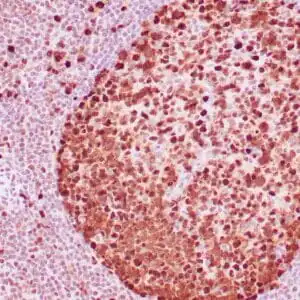
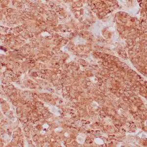
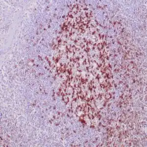
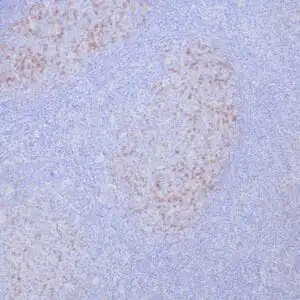
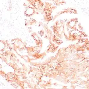
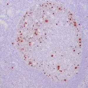
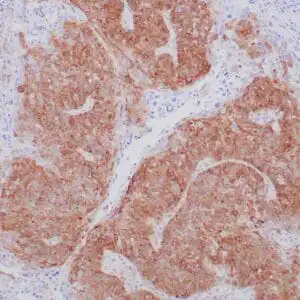
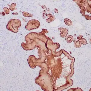
Data
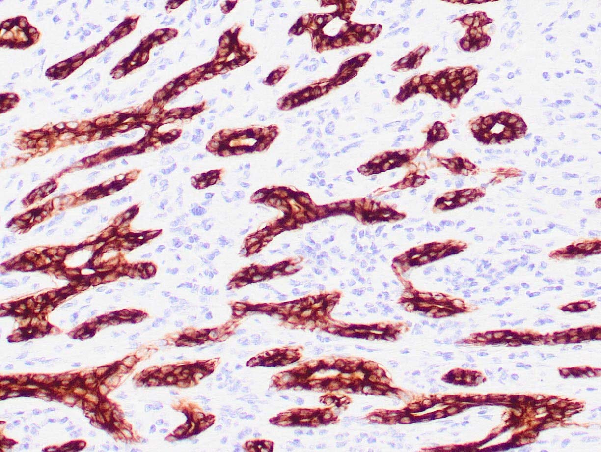 |
| Human breast stained with antibody CAM 5.2 using peroxidase-conjugate and DAB chromogen. Note the cytoplasmic staining of ductal cells. |
FAQ & Publications
Frequently Asked Questions
What species does the mouse anti-Cytokeratin monoclonal antibody (CAM 5.2) 6134 react with?
This antibody is reactive with human tissues and is suitable for immunohistochemistry on formalin-fixed, paraffin-embedded human tissue samples.
How should the CAM 5.2 antibody be stored to maintain its stability?
For short-term storage, keep the antibody at 2-8°C. For long-term storage, it should be stored at -20°C. Avoid repeated freeze/thaw cycles to preserve antibody integrity.
Publications
| pmid | title | authors | citation |
|---|---|---|---|
| We haven't added any publications to our database yet. | |||
Published literature highly relevant to the biological target of this product and referencing this antibody or clone are retrieved from the PubMed database provided by the United States National Library of Medicine at the National Institutes of Health.
Protocols
| relevant to this product |
|---|
| IHC |
Documents
| Batch Number | QC File | SDS |
|---|---|---|
| To view batch-specific Safety Datasheets and Quality Certificates associated with your account, please Log In. | ||
Only logged in customers who have purchased this product may leave a review.
Reviews
There are no reviews yet.