| Weight | 1 lbs |
|---|---|
| Dimensions | 9 × 5 × 2 in |
| host | mouse |
| isotype | IgG1 |
| clonality | monoclonal |
| concentration | concentrate, predilute |
| applications | IHC |
| reactivity | human |
| available size | 0.1 mL, 0.5 mL, 1 mL concentrated, 7 mL prediluted |

mouse anti-Cytokeratin 8 monoclonal antibody (ZM123) 6151
Price range: $160.00 through $528.00
Antibody summary
- Mouse monoclonal to Cytokeratin 8
- Suitable for: Immunohistochemistry (formalin-fixed, paraffin-embedded tissues)
- Reacts with: Human
- Isotype:IgG1
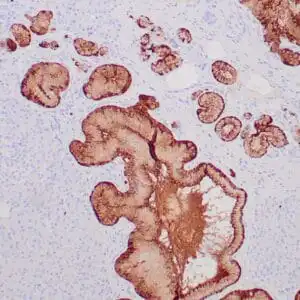
- Control: Breast carcinoma
- Visualization: Cytoplasmic
- 0.1, 0.5, 1.0 mL concentrated, 7 mL prediluted
mouse anti-Cytokeratin 8 monoclonal antibody ZM123 6151
| target relevance |
|---|
| Homo sapiens KRT8 Keratin, type II cytoskeletal 8 |
| Protein names Keratin, type II cytoskeletal 8 |
| Alternative names Cytokeratin-8, Keratin-8, Type-II keratin Kb8 |
| Gene names KRT8 |
| Protein family Belongs to the intermediate filament family |
| Function Required for the formation of KRT8/KRT18 filaments that are involved in ARHGEF40-mediated actin stress fiber formation and tensional force-induced stress fiber formation and reinforcement (PubMed:26823019). Together with KRT19, helps to link the contractile apparatus to dystrophin at the costameres of striated muscle |
| Subcellular location Cytoplasm, Nucleus, nucleoplasm, Nucleus matrix, Cytoplasm, cytoskeleton |
| Structure (Microbial infection) Interacts with hepatitis C virus/HCV core protein |
| Post-translational modification Phosphorylation on serine residues is enhanced during EGF stimulation and mitosis. Ser-74 phosphorylation plays an important role in keratin filament reorganization O-glycosylated. O-GlcNAcylation at multiple sites increases solubility, and decreases stability by inducing proteasomal degradation O-glycosylated (O-GlcNAcylated), in a cell cycle-dependent manner |
| Involvement in disease Cirrhosis A liver disease characterized by severe panlobular liver-cell swelling with Mallory body formation, prominent pericellular fibrosis, and marked deposits of copper. Clinical features include abdomen swelling, jaundice and pulmonary hypertension. |
| Keywords 3D-structure, Acetylation, Alternative splicing, Coiled coil, Cytoplasm, Cytoskeleton, Direct protein sequencing, Disease variant, Glycoprotein, Host-virus interaction, Intermediate filament, Isopeptide bond, Keratin, Methylation, Nucleus, Phosphoprotein, Proteomics identification, Reference proteome, Ubl conjugation |
| Sequence MSIRVTQKSYKVSTSGPRAFSSRSYTSGPGSRISSSSFSRVGSSNFRGGLGGGYGGASGM GGITAVTVNQSLLSPLVLEVDPNIQAVRTQEKEQIKTLNNKFASFIDKVRFLEQQNKMLE TKWSLLQQQKTARSNMDNMFESYINNLRRQLETLGQEKLKLEAELGNMQGLVEDFKNKYE DEINKRTEMENEFVLIKKDVDEAYMNKVELESRLEGLTDEINFLRQLYEEEIRELQSQIS DTSVVLSMDNSRSLDMDSIIAEVKAQYEDIANRSRAEAESMYQIKYEELQSLAGKHGDDL RRTKTEISEMNRNISRLQAEIEGLKGQRASLEAAIADAEQRGELAIKDANAKLSELEAAL QRAKQDMARQLREYQELMNVKLALDIEIATYRKLLEGEESRLESGMQNMSIHTKTTSGYA GGLSSAYGGLTSPGLSYSLGSSFGSGAGSSSFSRTSSSRAVVVKKIETRDGKLVSESSDV LPK |
| UniProt accession: P05787 |
Data
 |
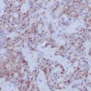
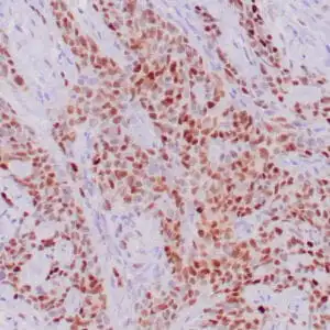
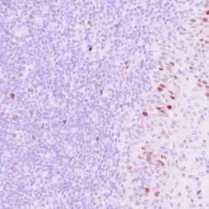
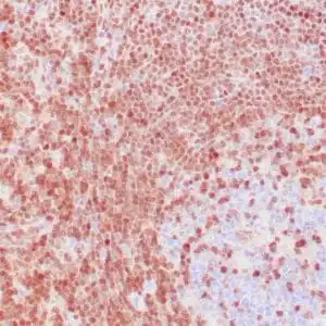
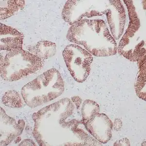
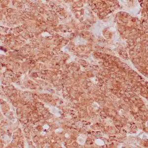
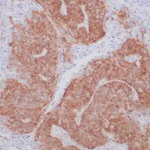
| Human basal cell carcinoma stained with anti-keratin 8 antibody using peroxidase-conjugate and DAB chromogen. Note the cytoplasmic staining of tumor cells. |
FAQ & Publications
Frequently Asked Questions
What is the recommended dilution range for the mouse anti-Cytokeratin 8 monoclonal antibody (ZM123) when used in immunohistochemistry?
For immunohistochemistry applications, the concentrated mouse anti-Cytokeratin 8 monoclonal antibody (ZM123) is recommended to be diluted between 1:100 and 1:200.
Which species does the mouse anti-Cytokeratin 8 antibody specifically react with, and what is a suitable positive control tissue?
This antibody specifically reacts with human Cytokeratin 8. Breast carcinoma tissue is recommended as a positive control for validating staining.
What are the storage conditions recommended for maintaining the stability of the mouse anti-Cytokeratin 8 monoclonal antibody?
For short-term storage, keep the antibody at 2-8°C. For long-term preservation, store at -20°C and avoid repeated freeze/thaw cycles to maintain antibody integrity.
Publications
| pmid | title | authors | citation |
|---|---|---|---|
| We haven't added any publications to our database yet. | |||
Published literature highly relevant to the biological target of this product and referencing this antibody or clone are retrieved from the PubMed database provided by the United States National Library of Medicine at the National Institutes of Health.
Protocols
| relevant to this product |
|---|
| IHC |
Documents
| Batch Number | QC File | SDS |
|---|---|---|
| To view batch-specific Safety Datasheets and Quality Certificates associated with your account, please Log In. | ||
Only logged in customers who have purchased this product may leave a review.
Reviews
There are no reviews yet.