| Weight | 1 lbs |
|---|---|
| Dimensions | 9 × 5 × 2 in |
| host | mouse |
| isotype | IgG2a |
| clonality | monoclonal |
| concentration | concentrate, predilute |
| applications | IHC |
| reactivity | human |
| available size | 0.1 mL, 0.5 mL, 1 mL concentrated, 7 mL prediluted |

mouse anti-Collagen IV monoclonal antibody (ZM177) 6128
Price range: $160.00 through $528.00
Antibody summary
- Mouse monoclonal to Collagen IV
- Suitable for: Immunohistochemistry (formalin-fixed, paraffin-embedded tissues)
- Reacts with: Human
- Isotype:IgG2a
- Control: Skin
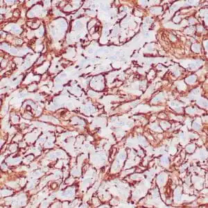
- Visualization: Cytoplasmic and membranous
- 0.1, 0.5, 1.0 mL concentrated, 7 mL prediluted
mouse anti-Collagen IV monoclonal antibody ZM177 6128
| target relevance |
|---|
| Homo sapiens COL4A1 Collagen alpha-1(IV) chain |
| Protein names Collagen alpha-1(IV) chain |
| Gene names COL4A1 |
| Protein family Belongs to the type IV collagen family |
| Function Type IV collagen is the major structural component of glomerular basement membranes (GBM), forming a 'chicken-wire' meshwork together with laminins, proteoglycans and entactin/nidogen |
| Subcellular location Secreted, extracellular space, extracellular matrix, basement membrane |
| Structure There are six type IV collagen isoforms, alpha 1(IV)-alpha 6(IV), each of which can form a triple helix structure with 2 other chains to generate type IV collagen network. Interacts with EFEMP2 (By similarity) |
| Post-translational modification Lysines at the third position of the tripeptide repeating unit (G-X-Y) are hydroxylated. The modified lysines can be O-glycosylated Contains 4-hydroxyproline (Probable). Prolines at the third position of the tripeptide repeating unit (G-X-Y) are hydroxylated in some or all of the chains (By similarity) Contains 3-hydroxyproline. This modification occurs on the first proline residue in the sequence motif Gly-Pro-Hyp, where Hyp is 4-hydroxyproline Type IV collagens contain numerous cysteine residues which are involved in inter- and intramolecular disulfide bonding (PubMed:2844531). 12 of these, located in the NC1 domain, are conserved in all known type IV collagens The trimeric structure of the NC1 domains is stabilized by covalent bonds (sulfilimine cross-links) between Lys and Met residues (PubMed:12011424). These cross-links are important for the mechanical stability of the basement membrane (By similarity). Sulfilimine cross-link is catalyzed by PXDN (By similarity) Proteolytic processing produces the C-terminal NC1 peptide, arresten |
| Involvement in disease Hereditary angiopathy with nephropathy aneurysms and muscle cramps The clinical renal manifestations include hematuria and bilateral large cysts. Histologic analysis revealed complex basement membrane defects in kidney and skin. The systemic angiopathy appears to affect both small vessels and large arteries. Brain small vessel disease 1 with or without ocular anomalies An autosomal dominant cerebrovascular disorder with variable manifestations reflecting the location and severity of the vascular defect. BSVD1 features include cerebral hemorrage, unilateral fluid-filled cysts or cavities within the cerebral hemispheres, leukoencephalopathy, hemiplegia, seizures, intellectual disability, and facial paresis. Affected individuals may manifest variable visual defects and ocular anomalies. Intracerebral hemorrhage A pathological condition characterized by bleeding into one or both cerebral hemispheres including the basal ganglia and the cerebral cortex. It is often associated with hypertension and craniocerebral trauma. Intracerebral bleeding is a common cause of stroke. Tortuosity of retinal arteries A disease characterized by marked tortuosity of second- and third-order retinal arteries with normal first-order arteries and venous system. Most patients manifest variable degrees of symptomatic transient vision loss due to retinal hemorrhage following minor stress or trauma. Schizencephaly Extremely rare human congenital disorder characterized by a full-thickness cleft within the cerebral hemispheres. These clefts are lined with gray matter and most commonly involve the parasylvian regions. Large portions of the cerebral hemispheres may be absent and replaced by cerebro-spinal fluid. Microangiopathy and leukoencephalopathy, pontine, autosomal dominant A form of cerebral small vessel disease characterized by the recurrence of ischemic strokes starting in the thirties or forties, and associated with progressive imbalance and cognitive impairment. MRI examination shows ischemic lacunas in the pons and cerebral hemispheres, and diffuse leukoencephalopathy affecting various brain regions. |
| Keywords 3D-structure, Alternative splicing, Angiogenesis, Basement membrane, Collagen, Direct protein sequencing, Disease variant, Disulfide bond, Extracellular matrix, Glycoprotein, Hydroxylation, Proteomics identification, Reference proteome, Repeat, Secreted, Signal |
| Sequence MGPRLSVWLLLLPAALLLHEEHSRAAAKGGCAGSGCGKCDCHGVKGQKGERGLPGLQGVI GFPGMQGPEGPQGPPGQKGDTGEPGLPGTKGTRGPPGASGYPGNPGLPGIPGQDGPPGPP GIPGCNGTKGERGPLGPPGLPGFAGNPGPPGLPGMKGDPGEILGHVPGMLLKGERGFPGI PGTPGPPGLPGLQGPVGPPGFTGPPGPPGPPGPPGEKGQMGLSFQGPKGDKGDQGVSGPP GVPGQAQVQEKGDFATKGEKGQKGEPGFQGMPGVGEKGEPGKPGPRGKPGKDGDKGEKGS PGFPGEPGYPGLIGRQGPQGEKGEAGPPGPPGIVIGTGPLGEKGERGYPGTPGPRGEPGP KGFPGLPGQPGPPGLPVPGQAGAPGFPGERGEKGDRGFPGTSLPGPSGRDGLPGPPGSPG PPGQPGYTNGIVECQPGPPGDQGPPGIPGQPGFIGEIGEKGQKGESCLICDIDGYRGPPG PQGPPGEIGFPGQPGAKGDRGLPGRDGVAGVPGPQGTPGLIGQPGAKGEPGEFYFDLRLK GDKGDPGFPGQPGMPGRAGSPGRDGHPGLPGPKGSPGSVGLKGERGPPGGVGFPGSRGDT GPPGPPGYGPAGPIGDKGQAGFPGGPGSPGLPGPKGEPGKIVPLPGPPGAEGLPGSPGFP GPQGDRGFPGTPGRPGLPGEKGAVGQPGIGFPGPPGPKGVDGLPGDMGPPGTPGRPGFNG LPGNPGVQGQKGEPGVGLPGLKGLPGLPGIPGTPGEKGSIGVPGVPGEHGAIGPPGLQGI RGEPGPPGLPGSVGSPGVPGIGPPGARGPPGGQGPPGLSGPPGIKGEKGFPGFPGLDMPG PKGDKGAQGLPGITGQSGLPGLPGQQGAPGIPGFPGSKGEMGVMGTPGQPGSPGPVGAPG LPGEKGDHGFPGSSGPRGDPGLKGDKGDVGLPGKPGSMDKVDMGSMKGQKGDQGEKGQIG PIGEKGSRGDPGTPGVPGKDGQAGQPGQPGPKGDPGISGTPGAPGLPGPKGSVGGMGLPG TPGEKGVPGIPGPQGSPGLPGDKGAKGEKGQAGPPGIGIPGLRGEKGDQGIAGFPGSPGE KGEKGSIGIPGMPGSPGLKGSPGSVGYPGSPGLPGEKGDKGLPGLDGIPGVKGEAGLPGT PGPTGPAGQKGEPGSDGIPGSAGEKGEPGLPGRGFPGFPGAKGDKGSKGEVGFPGLAGSP GIPGSKGEQGFMGPPGPQGQPGLPGSPGHATEGPKGDRGPQGQPGLPGLPGPMGPPGLPG IDGVKGDKGNPGWPGAPGVPGPKGDPGFQGMPGIGGSPGITGSKGDMGPPGVPGFQGPKG LPGLQGIKGDQGDQGVPGAKGLPGPPGPPGPYDIIKGEPGLPGPEGPPGLKGLQGLPGPK GQQGVTGLVGIPGPPGIPGFDGAPGQKGEMGPAGPTGPRGFPGPPGPDGLPGSMGPPGTP SVDHGFLVTRHSQTIDDPQCPSGTKILYHGYSLLYVQGNERAHGQDLGTAGSCLRKFSTM PFLFCNINNVCNFASRNDYSYWLSTPEPMPMSMAPITGENIRPFISRCAVCEAPAMVMAV HSQTIQIPPCPSGWSSLWIGYSFVMHTSAGAEGSGQALASPGSCLEEFRSAPFIECHGRG TCNYYANAYSFWLATIERSEMFKKPTPSTLKAGELRTHVSRCQVCMRRT |
| UniProt accession: P02462 |
Data
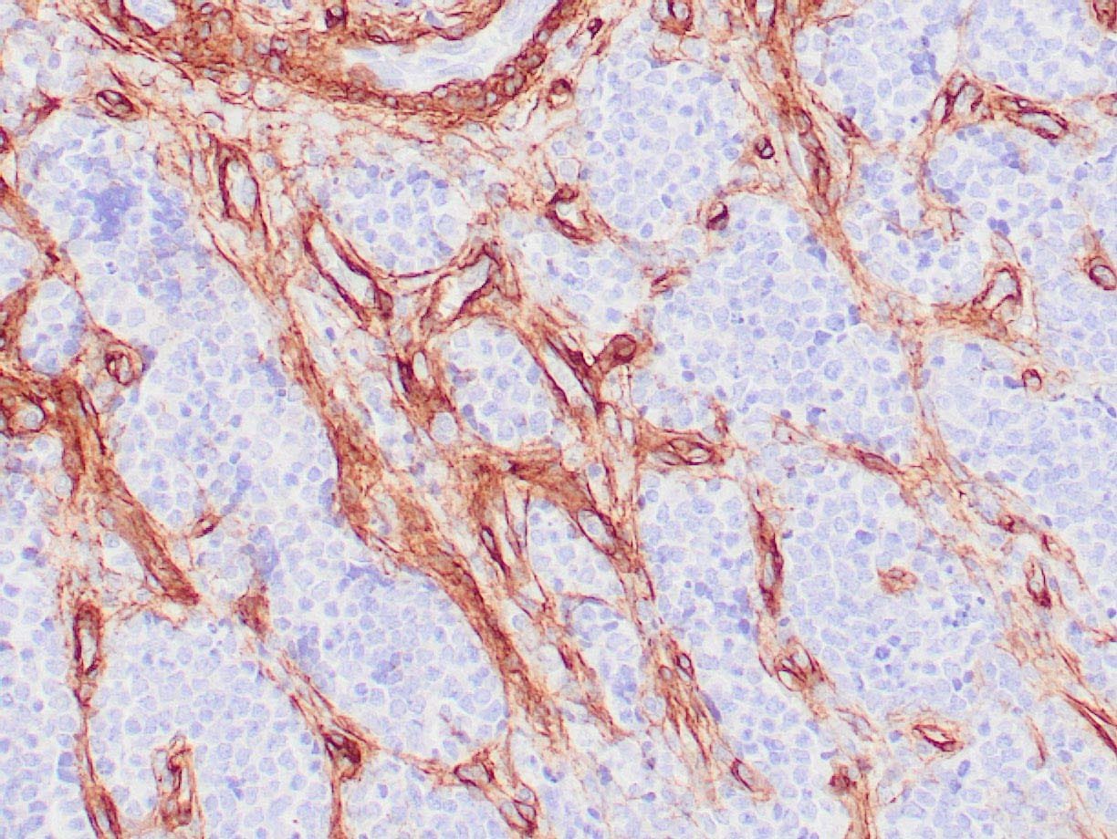 |
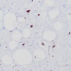
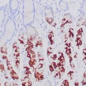
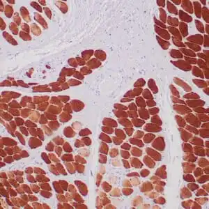
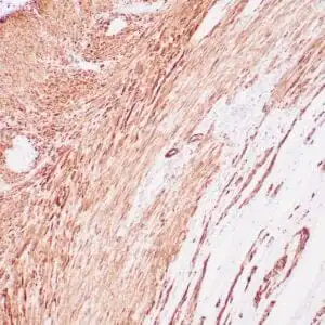
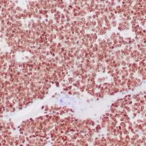
| Human glomus tumor stained with anti-Collagen IV using peroxidase-conjugate and DAB chromogen. Note basement membrane staining of around individual cells. |
FAQ & Publications
Frequently Asked Questions
What species reactivity does the mouse anti-Collagen IV monoclonal antibody (ZM177) exhibit?
This antibody specifically reacts with human Collagen IV.
What are the recommended storage conditions for the mouse anti-Collagen IV monoclonal antibody (ZM177)?
For short-term storage, keep the antibody at 2-8°C. For longer-term preservation, store it at -20°C and avoid repeated freeze/thaw cycles to maintain stability.
Publications
| pmid | title | authors | citation |
|---|---|---|---|
| We haven't added any publications to our database yet. | |||
Published literature highly relevant to the biological target of this product and referencing this antibody or clone are retrieved from the PubMed database provided by the United States National Library of Medicine at the National Institutes of Health.
Protocols
| relevant to this product |
|---|
| IHC |
Documents
| Batch Number | QC File | SDS |
|---|---|---|
| To view batch-specific Safety Datasheets and Quality Certificates associated with your account, please Log In. | ||
Only logged in customers who have purchased this product may leave a review.

Reviews
There are no reviews yet.