Showing all 5 results
6XHis tag antibodies
The 6x His tag is a widely used epitope tag in molecular biology that consists of six consecutive histidine residues. It is commonly incorporated into recombinant proteins to facilitate their purification, detection, and characterization. The His tag enables researchers to purify tagged proteins using nickel-affinity chromatography, as the histidine residues have a strong affinity for nickel ions. This method is efficient and allows for the isolation of proteins from complex mixtures with high specificity.
In Western blotting (WB), the 6x His tag is employed to detect and confirm the expression of recombinant proteins. Antibodies specific to the His tag can be used to visualize the tagged protein in various samples, providing insights into protein expression levels and post-translational modifications. The antibodies for the 6x His tag available on your website are also suitable for immunofluorescence (IF) and immunohistochemistry (IHC), allowing researchers to examine the localization of recombinant proteins within cells and tissues.
The use of the 6x His tag is especially valuable in protein engineering, structural biology, and functional studies, where tracking and purifying recombinant proteins is essential. By enabling easy detection and purification, the His tag facilitates a wide range of applications, including enzyme assays, protein-protein interaction studies, and the development of biopharmaceuticals. Its versatility and effectiveness make it an indispensable tool in protein research.
| target | type | reactivity | applications | ||||
|---|---|---|---|---|---|---|---|
| 1955 | 6X His tag | mouse monoclonal (7B8) | other | WB ELISA | |||
| 2966 | 6X His tag | mouse monoclonal (HIS.H8) | other | WB ICC ELISA | |||
| 7094 | 6X His tag | rabbit polyclonal | other | ELISA | |||
| 8278 | 6X His tag | chicken polyclonal | other | ELISA | |||
| 9027 | 6X His tag | goat polyclonal | other | WB ICC ELISA | |||

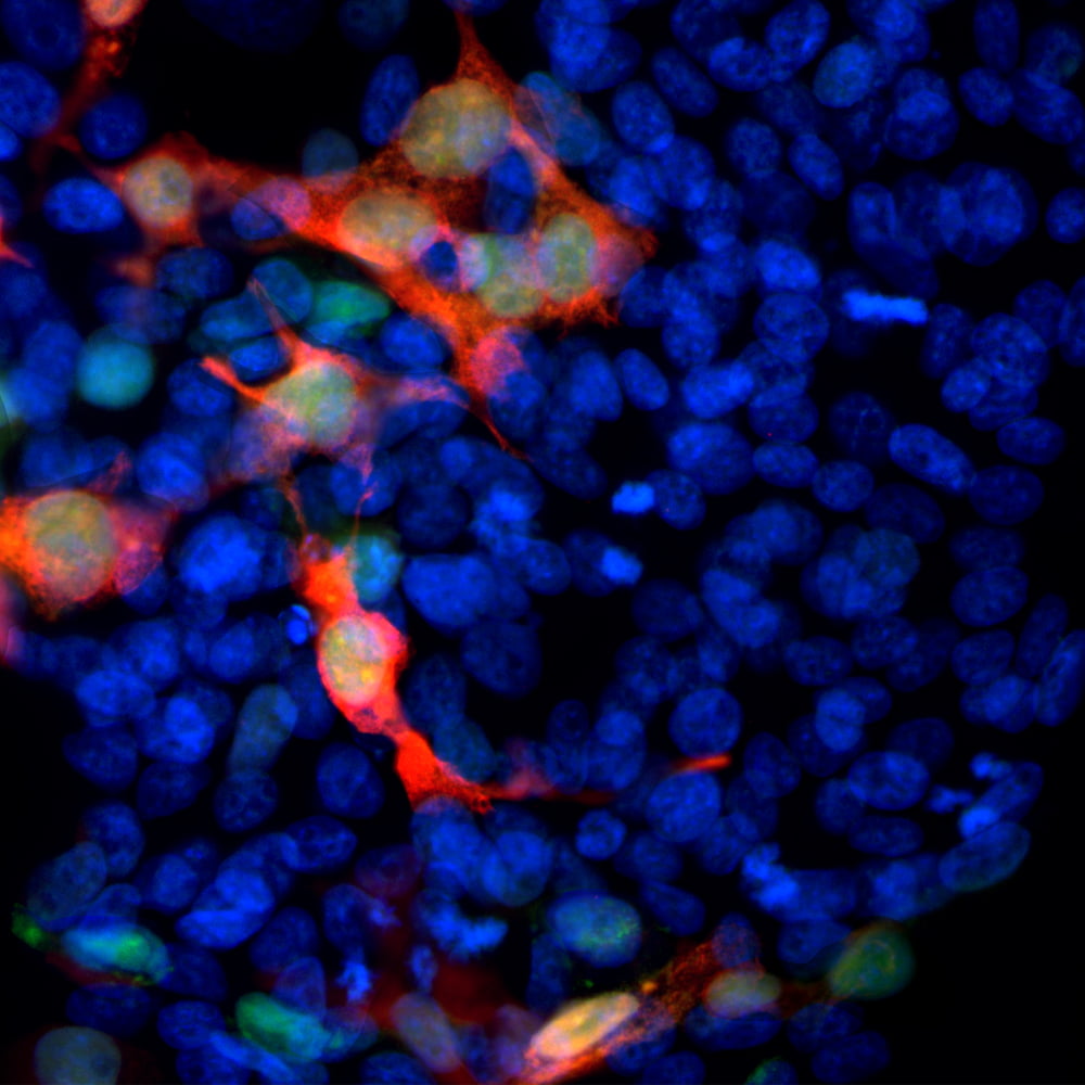
ICC/IF with antibody [3128] : Immunofluorescent analysis of transfected HEK293 cells with a GFP-construct in green stained with mouse mAb to GFP, 3128, dilution 1:1,000 in red. The blue is Hoechst staining of nuclear DNA. 3128 antibody reveals GFP protein expressed only in transfected cells, as a result cells are appeared in orange-golden color.

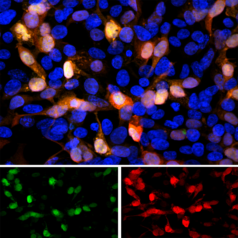
ICC/IF with antibody [1290] : Immunofluorescent analysis of transfected HEK293 cells with a GFP construct, in green, and stained with rabbit pAb to GFP, 1290, dilution 1:2,000 in red. The blue is Hoechst staining of nuclear DNA. The 1290 antibody reveals GFP protein expressed only in transfected cells, and as a result these cells appear orange-yellow in color.

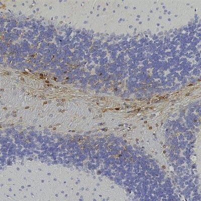
IHC with antibody [1379] : Chromogenic immunostaining of formalin fixed paraffin embedded GFP transduced mouse brain with goat pAb to GFP, 1379, dilution 1:5,000, detected with DAB (brown) using the Vector Elite 8718C-HRP detection and reagents with citra buffer retrieval. Hematoxylin (blue) was used as the counterstain. Antibody 1379 specifically detected GFP positive cells in the cerebellum as expected for this model.


