| Weight | 1 lbs |
|---|---|
| Dimensions | 9 × 5 × 2 in |
| host | rabbit |
| isotype | IgG |
| clonality | polyclonal |
| concentration | 1 mg/mL |
| applications | ICC/IF, WB |
| reactivity | EndoG |
| available sizes | 100 µg |


rabbit anti-EndoG polyclonal antibody 1570
$445.00
Antibody summary
- Rabbit polyclonal to EndoG
- Suitable for: ELISA,WB,IHC-P,IF
- Isotype: IgG
- 100 µg
rabbit anti-EndoG polyclonal antibody 1570
| antibody |
|---|
| Tested applications WB,IHC,IHC,ICC/IF,ELISA |
| Recommended dilutions Immunoblotting: use at 1-2ug/mL. A band of 35kDa is detected. Immunohistochemistry: use at 10-15ug/mL. Positive control: HepG2 cell lysate. These are recommended concentrations. Enduser should determine optimal concentrations for their applications |
| Immunogen Peptide corresponding to aa 55- 70 of human EndoG (accession no. NP_004426). |
| Size and concentration 100µg and lot specific |
| Form liquid |
| Storage Instructions This antibody is stable for at least one (1) year at -20°C. Avoid multiple freeze-thaw cycles. |
| Storage buffer PBS, pH 7.4. |
| Purity peptide affinity purification |
| Clonality polyclonal |
| Isotype IgG |
| Compatible secondaries goat anti-rabbit IgG, H&L chain specific, peroxidase conjugated, conjugated polyclonal antibody 9512 goat anti-rabbit IgG, H&L chain specific, biotin conjugated polyclonal antibody 2079 goat anti-rabbit IgG, H&L chain specific, FITC conjugated polyclonal antibody 7863 goat anti-rabbit IgG, H&L chain specific, Cross Absorbed polyclonal antibody 2371 goat anti-rabbit IgG, H&L chain specific, biotin conjugated polyclonal antibody, crossabsorbed 1715 goat anti-rabbit IgG, H&L chain specific, FITC conjugated polyclonal antibody, crossabsorbed 1720 |
| Isotype control Rabbit polyclonal - Isotype Control |
| target relevance |
|---|
| Protein names Endonuclease G, mitochondrial (Endo G) (EC 3.1.30.-) |
| Gene names ENDOG,ENDOG |
| Protein family DNA/RNA non-specific endonuclease family |
| Mass 32620Da |
| Function FUNCTION: Endonuclease that preferentially catalyzes the cleavage of double-stranded 5-hydroxymethylcytosine (5hmC)-modified DNA (PubMed:25355512). The 5hmC-modified nucleotide does not increase the binding affinity, but instead increases the efficiency of cutting and specifies the site of cleavage for the modified DNAs (By similarity). Shows significantly higher affinity for four-stranded Holliday junction over duplex and single-stranded DNAs (By similarity). Promotes conservative recombination when the DNA is 5hmC-modified (PubMed:25355512). Promotes autophagy through the suppression of mTOR by its phosphorylation-mediated interaction with YWHAG and its endonuclease activity-mediated DNA damage response (PubMed:33473107). GSK3-beta mediated phosphorylation of ENDOG enhances its interaction with YWHAG, leading to the release of TSC2 and PIK3C3 from YWHAG resulting in mTOR pathway suppression and autophagy initiation (PubMed:33473107). Promotes cleavage of mtDNA in response to oxidative and nitrosative stress, in turn inducing compensatory mtDNA replication (PubMed:29719607). {ECO:0000250|UniProtKB:O08600, ECO:0000269|PubMed:25355512, ECO:0000269|PubMed:29719607, ECO:0000269|PubMed:33473107}. |
| Subellular location SUBCELLULAR LOCATION: Mitochondrion {ECO:0000269|PubMed:33473107}. |
| Structure SUBUNIT: Homodimer; disulfide-linked (By similarity). Homodimerization is essential for enzyme activity (By similarity). Interacts with YWHAG (PubMed:33473107). {ECO:0000250|UniProtKB:O08600, ECO:0000269|PubMed:33473107}. |
| Post-translational modification PTM: GSK3-beta-mediated dual phosphorylations at Thr-128 and Ser-288 is necessary for its interaction with YWHAG and the induction of autophagy. {ECO:0000269|PubMed:33473107}. |
| Target Relevance information above includes information from UniProt accession: Q14249 |
| The UniProt Consortium |
Data
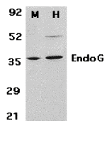 |
| Western Blot Validation in Mouse 3T3 (M) and Human HepG2 (H) Cell Lysates Loading: 15 µg of lysates per lane. Antibodies: EndoG 1570 (2 µg/mL), 1h incubation at RT in 5% NFDM/TBST.Secondary: Goat anti-rabbit IgG HRP conjugate at 1:10000 dilution. |
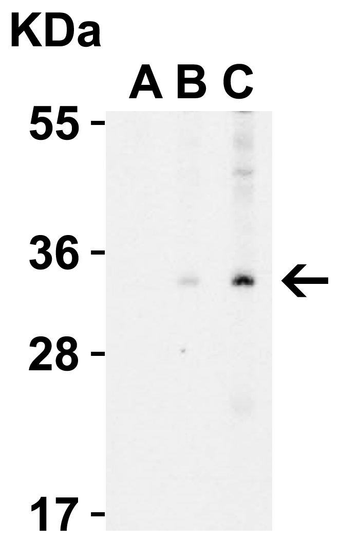 |
| Western Blot Validation in Human A431 Cell Lysate with the presence (A) or absence (B and C) of blocking peptide Loading: 15 µg of lysates per lane. Antibodies: EndoG 1570 (A: 0.5 µg/mL, B: 0.5 µg/mL, C: 1 µg/mL), 1h incubation at RT in 5% NFDM/TBST.Secondary: Goat anti-rabbit IgG HRP conjugate at 1:10000 dilution. |
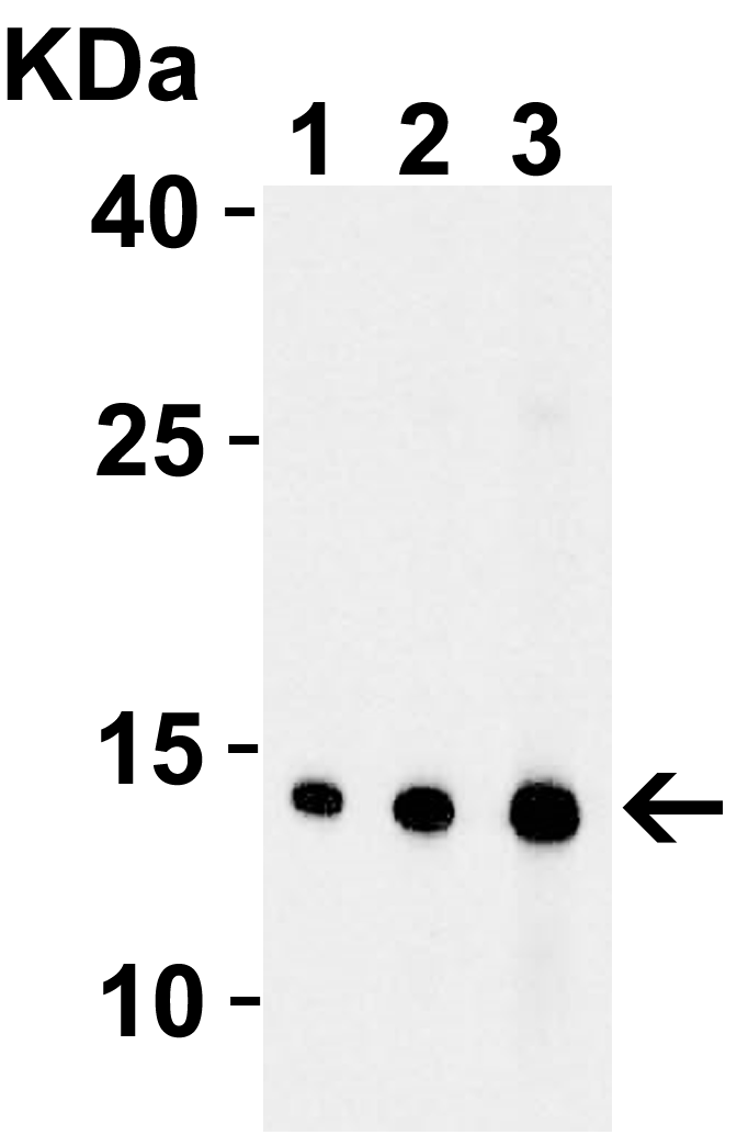 |
| Western Blot Validation with Recombinant Protein Loading: 30 ng of human EndoG recombinant protein per lane. Antibodies: EndoG 1570, 1h incubation at RT in 5% NFDM/TBST.Secondary: Goat anti-rabbit IgG HRP conjugate at 1:10000 dilution.Lane 1: 0.5 µg/mLLane 2: 1 µg/mLLane 3: 2 µg/mL |
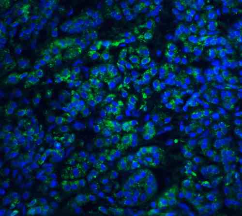 |
| Immunofluorescence Validation of EndoG in Human Pancreas Tissue Immunofluorescent analysis of 4% paraformaldehyde-fixed human pancreas tissue labeling EndoG with 1570 at 20 µg/mL, followed by goat anti-rabbit IgG secondary antibody at 1/500 dilution (green) and DAPI staining (blue). |
 |
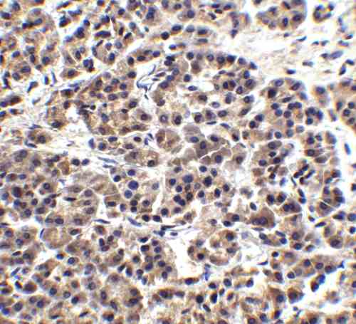
| Immunohistochemistry Validation of EndoG in Human Pancreas Tissue Immunohistochemical analysis of paraffin-embedded human pancreas tissue using anti-EndoG antibody (1570) at 15 µg/mL. Tissue was fixed with formaldehyde and blocked with 10% serum for 1 h at RT; antigen retrieval was by heat mediation with a citrate buffer (pH6). Samples were incubated with primary antibody overnight at 4C. A goat anti-rabbit IgG H&L (HRP) at 1/250 was used as secondary. Counter stained with Hematoxylin. |
 |
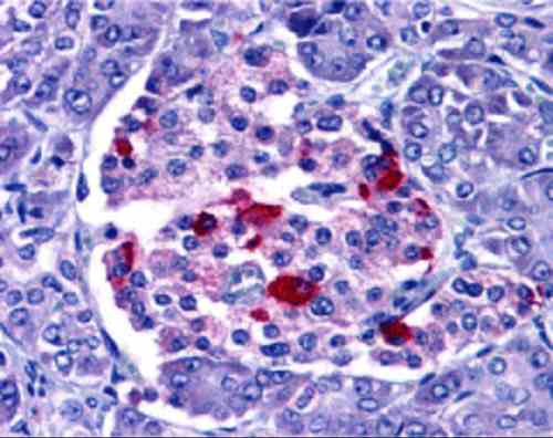
| Immunohistochemistry Validation of EndoG in Human Pancreas Tissue Immunohistochemical analysis of paraffin-embedded human pancreas tissue using anti-EndoG antibody (1570) at 2.5 µg/mL. Tissue was fixed with formaldehyde and blocked with 10% serum for 1 h at RT; antigen retrieval was by heat mediation with a citrate buffer (pH6). Samples were incubated with primary antibody overnight at 4C. A goat anti-rabbit IgG H&L (HRP) at 1/250 was used as secondary. Counter stained with Hematoxylin. |
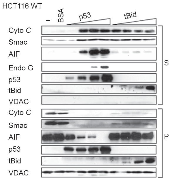 |
| Localization Validation of EndoG by Purified p53 in Human Colorectal Cancer (HCT116) WT Cells (Wolff et al., 2008) Immunoblot analysis of subcellular fraction enriched with supernatant (S) was used to determine EndoG protein levels with increasing amounts of p53 (10, 20, 40, 100nM) in HCT116 WT cells. The release of EndoG from mitochondria induced by p53 is detected by anti-EndoG antibodies. |
 |
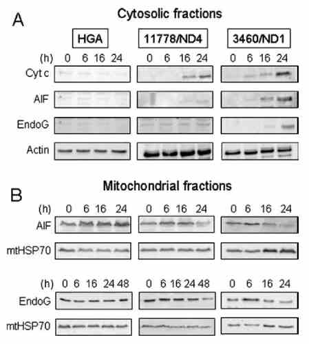
| Localization Validation of EndoG in LHON cybrids (Zanna et al., 2005) Immunoblots of subcellular fractions enriched for (A) cytosol and (B) mitochondria were used to determine EndoG protein levels with anti-EndoG antibodies in LHON cybrids. The release of EndoG from mitochondria into the cytosol of 3460/ND1 mutant is observed after 24hr. Control (HGA) was unaffected. |
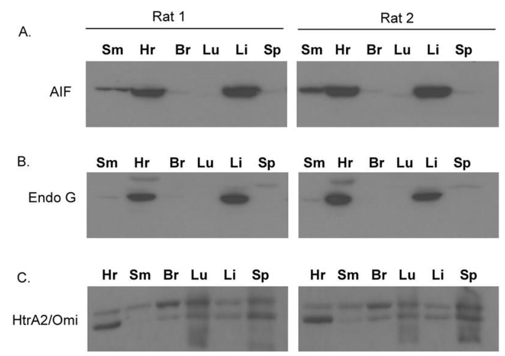 |
| Tissue Specificity of EndoG in Rat Organs ( Siu et al., 2416) WB analysis with anti-EndoG antibodies was performed for (B) EndoG in different organs of rats. EndoG was expressed very high in the heart and the liver of rats. |
FAQ & Publications
Frequently Asked Questions
What applications is the rabbit anti-EndoG polyclonal antibody 1570 validated for?
This antibody is suitable and tested for ELISA, Western Blot (WB), Immunohistochemistry (IHC), and Immunofluorescence (IF) applications.
How should the rabbit anti-EndoG antibody 1570 be stored to ensure stability?
The antibody should be stored at -20°C and is stable for at least one year. It is recommended to avoid multiple freeze-thaw cycles to maintain antibody integrity.
What is the immunogen used to generate the rabbit anti-EndoG polyclonal antibody 1570?
The immunogen is a peptide corresponding to amino acids 55-70 of human EndoG (accession number NP_004426).
Which secondary antibodies are compatible with the rabbit anti-EndoG polyclonal antibody 1570?
Compatible secondary antibodies include goat anti-rabbit IgG antibodies that are H&L chain specific, available conjugated with peroxidase, biotin, or FITC, including both regular and cross-absorbed polyclonal antibodies.
Publications
| pmid | title | authors | citation |
|---|---|---|---|
| We haven't added any publications to our database yet. | |||
Published literature highly relevant to the biological target of this product and referencing this antibody or clone are retrieved from the PubMed database provided by the United States National Library of Medicine at the National Institutes of Health.
Protocols
| relevant to this product |
|---|
| Western blot IHC ICC |
Documents
| Batch Number | QC File | SDS |
|---|---|---|
| To view batch-specific Safety Datasheets and Quality Certificates associated with your account, please Log In. | ||
Only logged in customers who have purchased this product may leave a review.


Reviews
There are no reviews yet.