| Weight | 1 lbs |
|---|---|
| Dimensions | 9 × 5 × 2 in |
| host | rabbit |
| isotype | IgG |
| clonality | polyclonal |
| concentration | 1 mg/mL |
| applications | ICC/IF, WB |
| reactivity | ZIP Kinase |
| available sizes | 100 µg |
rabbit anti-ZIP Kinase polyclonal antibody 2387
$445.00
Antibody summary
- Rabbit polyclonal to ZIP Kinase
- Suitable for: ELISA,WB,ICC,IF
- Isotype: IgG
- 100 µg
rabbit anti-ZIP Kinase polyclonal antibody 2387
| antibody |
|---|
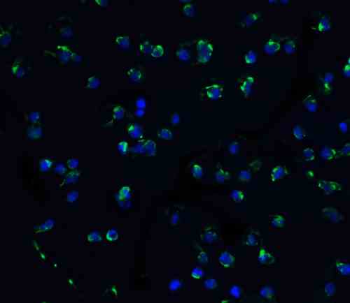
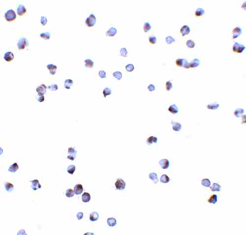
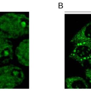
| Tested applications WB,ICC/IF,ELISA |
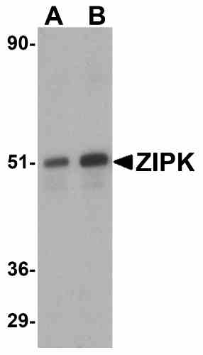
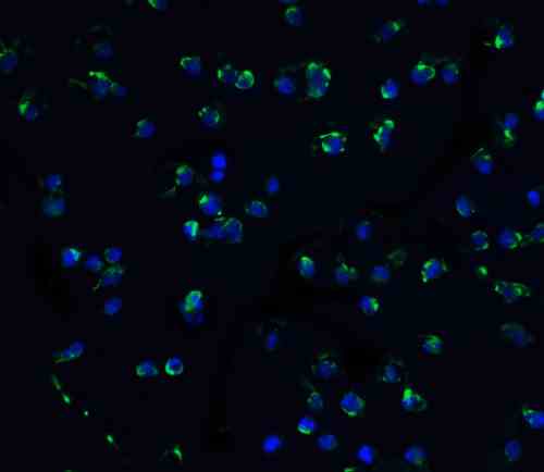
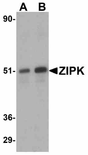
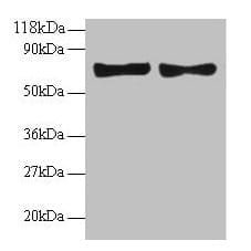
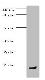
| Recommended dilutions Immunoblotting: use at 1ug/mL. Positive control: HeLa cell lysate. Immunocytochemistry: use at 10ug/mL. These are recommended concentrations. Enduser should determine optimal concentrations for their applications. |
| Immunogen Peptide corresponding to aa 279- 298 of human ZIP kinase (accession no. BAA81746). |
| Size and concentration 100µg and lot specific |
| Form liquid |
| Storage Instructions This antibody is stable for at least one (1) year at -20°C. Avoid multiple freeze-thaw cycles. |
| Storage buffer PBS, pH 7.4. |
| Purity peptide affinity purification |
| Clonality polyclonal |
| Isotype IgG |
| Compatible secondaries goat anti-rabbit IgG, H&L chain specific, peroxidase conjugated, conjugated polyclonal antibody 9512 goat anti-rabbit IgG, H&L chain specific, biotin conjugated polyclonal antibody 2079 goat anti-rabbit IgG, H&L chain specific, FITC conjugated polyclonal antibody 7863 goat anti-rabbit IgG, H&L chain specific, Cross Absorbed polyclonal antibody 2371 goat anti-rabbit IgG, H&L chain specific, biotin conjugated polyclonal antibody, crossabsorbed 1715 goat anti-rabbit IgG, H&L chain specific, FITC conjugated polyclonal antibody, crossabsorbed 1720 |
| Isotype control Rabbit polyclonal - Isotype Control |
| target relevance |
|---|
| Protein names Death-associated protein kinase 3 (DAP kinase 3) (EC 2.7.11.1) (DAP-like kinase) (Dlk) (MYPT1 kinase) (Zipper-interacting protein kinase) (ZIP-kinase) |
| Gene names DAPK3,DAPK3 ZIPK |
| Protein family Protein kinase superfamily, CAMK Ser/Thr protein kinase family, DAP kinase subfamily |
| Mass 52536Da |
| Function FUNCTION: Serine/threonine kinase which is involved in the regulation of apoptosis, autophagy, transcription, translation and actin cytoskeleton reorganization. Involved in the regulation of smooth muscle contraction. Regulates both type I (caspase-dependent) apoptotic and type II (caspase-independent) autophagic cell deaths signal, depending on the cellular setting. Involved in regulation of starvation-induced autophagy. Regulates myosin phosphorylation in both smooth muscle and non-muscle cells. In smooth muscle, regulates myosin either directly by phosphorylating MYL12B and MYL9 or through inhibition of smooth muscle myosin phosphatase (SMPP1M) via phosphorylation of PPP1R12A; the inhibition of SMPP1M functions to enhance muscle responsiveness to Ca(2+) and promote a contractile state. Phosphorylates MYL12B in non-muscle cells leading to reorganization of actin cytoskeleton. Isoform 2 can phosphorylate myosin, PPP1R12A and MYL12B. Overexpression leads to condensation of actin stress fibers into thick bundles. Involved in actin filament focal adhesion dynamics. The function in both reorganization of actin cytoskeleton and focal adhesion dissolution is modulated by RhoD. Positively regulates canonical Wnt/beta-catenin signaling through interaction with NLK and TCF7L2. Phosphorylates RPL13A on 'Ser-77' upon interferon-gamma activation which is causing RPL13A release from the ribosome, RPL13A association with the GAIT complex and its subsequent involvement in transcript-selective translation inhibition. Enhances transcription from AR-responsive promoters in a hormone- and kinase-dependent manner. Involved in regulation of cell cycle progression and cell proliferation. May be a tumor suppressor. {ECO:0000269|PubMed:10356987, ECO:0000269|PubMed:11384979, ECO:0000269|PubMed:11781833, ECO:0000269|PubMed:12917339, ECO:0000269|PubMed:15096528, ECO:0000269|PubMed:15367680, ECO:0000269|PubMed:16219639, ECO:0000269|PubMed:17126281, ECO:0000269|PubMed:17158456, ECO:0000269|PubMed:18084323, ECO:0000269|PubMed:18995835, ECO:0000269|PubMed:21169990, ECO:0000269|PubMed:21408167, ECO:0000269|PubMed:21454679, ECO:0000269|PubMed:21487036, ECO:0000269|PubMed:23454120, ECO:0000269|PubMed:38009294}. |
| Catalytic activity CATALYTIC ACTIVITY: Reaction=L-seryl-[protein] + ATP = O-phospho-L-seryl-[protein] + ADP + H(+); Xref=Rhea:RHEA:17989, Rhea:RHEA-COMP:9863, Rhea:RHEA-COMP:11604, ChEBI:CHEBI:15378, ChEBI:CHEBI:29999, ChEBI:CHEBI:30616, ChEBI:CHEBI:83421, ChEBI:CHEBI:456216; EC=2.7.11.1; Evidence={ECO:0000269|PubMed:10356987, ECO:0000269|PubMed:11384979, ECO:0000269|PubMed:11781833, ECO:0000269|PubMed:15096528, ECO:0000269|PubMed:17126281, ECO:0000269|PubMed:18995835}; CATALYTIC ACTIVITY: Reaction=L-threonyl-[protein] + ATP = O-phospho-L-threonyl-[protein] + ADP + H(+); Xref=Rhea:RHEA:46608, Rhea:RHEA-COMP:11060, Rhea:RHEA-COMP:11605, ChEBI:CHEBI:15378, ChEBI:CHEBI:30013, ChEBI:CHEBI:30616, ChEBI:CHEBI:61977, ChEBI:CHEBI:456216; EC=2.7.11.1; Evidence={ECO:0000269|PubMed:10356987, ECO:0000269|PubMed:11384979, ECO:0000269|PubMed:11781833, ECO:0000269|PubMed:15096528, ECO:0000269|PubMed:17126281, ECO:0000269|PubMed:18995835}; |
| Subellular location SUBCELLULAR LOCATION: Nucleus {ECO:0000269|PubMed:15367680, ECO:0000269|PubMed:15910542, ECO:0000269|PubMed:20854903}. Nucleus, PML body {ECO:0000250|UniProtKB:O54784}. Cytoplasm, cytoskeleton, microtubule organizing center, centrosome {ECO:0000250|UniProtKB:O54784}. Chromosome, centromere {ECO:0000269|PubMed:38009294}. Cytoplasm {ECO:0000269|PubMed:15367680, ECO:0000269|PubMed:15611134, ECO:0000269|PubMed:17953487, ECO:0000269|PubMed:20854903}. Cytoplasm, cytoskeleton, spindle {ECO:0000269|PubMed:38009294}. Midbody {ECO:0000269|PubMed:38009294}. Note=Predominantly localizes to the cytoplasm but can shuttle between the nucleus and cytoplasm; cytoplasmic localization is promoted by phosphorylation at Thr-299 and involves Rho/Rock signaling. {ECO:0000269|PubMed:17953487, ECO:0000269|PubMed:20854903}.; SUBCELLULAR LOCATION: [Isoform 1]: Nucleus {ECO:0000269|PubMed:17126281}. Cytoplasm {ECO:0000269|PubMed:17126281}.; SUBCELLULAR LOCATION: [Isoform 2]: Nucleus {ECO:0000269|PubMed:17126281}. Cytoplasm {ECO:0000269|PubMed:17126281}. |
| Tissues TISSUE SPECIFICITY: Widely expressed. Isoform 1 and isoform 2 are expressed in the bladder smooth muscle. {ECO:0000269|PubMed:15292222, ECO:0000269|PubMed:17126281}. |
| Structure SUBUNIT: Homooligomer in its kinase-active form (homotrimers and homodimers are reported); monomeric in its kinase-inactive form. Homodimerization is required for activation segment autophosphorylation (Probable). Isoform 1 and isoform 2 interact with myosin and PPP1R12A; interaction of isoform 1 with PPP1R12A is inhibited by RhoA dominant negative form. Interacts with NLK, DAXX, STAT3, RHOD (GTP-bound form) and TCP10L. Interacts with PAWR; the interaction is reported conflictingly: according to PubMed:17953487 does not interact with PAWR. Interacts with ULK1; may be a substrate of ULK1. Interacts with LUZP1; the interaction is likely to occur throughout the cell cycle and reduces the LUZP1-mediated suppression of MYL9 phosphorylation (PubMed:38009294). {ECO:0000269|PubMed:12917339, ECO:0000269|PubMed:15292222, ECO:0000269|PubMed:15910542, ECO:0000269|PubMed:16219639, ECO:0000269|PubMed:17126281, ECO:0000269|PubMed:18239682, ECO:0000269|PubMed:21169990, ECO:0000269|PubMed:21408167, ECO:0000269|PubMed:21454679, ECO:0000269|PubMed:23454120, ECO:0000269|PubMed:38009294, ECO:0000303|PubMed:18239682}. |
| Post-translational modification PTM: The phosphorylation status is critical for kinase activity, oligomerization and intracellular localization. Phosphorylation at Thr-180, Thr-225 and Thr-265 is essential for activity. The phosphorylated form is localized in the cytoplasm promoted by phosphorylation at Thr-299; nuclear translocation or retention is maximal when it is not phosphorylated. Phosphorylation increases the trimeric form, and its dephosphorylation favors a kinase-inactive monomeric form. Both isoform 1 and isoform 2 can undergo autophosphorylation. {ECO:0000269|PubMed:15367680, ECO:0000269|PubMed:15611134, ECO:0000269|PubMed:17158456, ECO:0000269|PubMed:20854903}. |
| Target Relevance information above includes information from UniProt accession: O43293 |
| The UniProt Consortium |
Data
Publications
| pmid | title | authors | citation |
|---|---|---|---|
| We haven't added any publications to our database yet. | |||
Protocols
| relevant to this product |
|---|
| Western blot IHC ICC |
Documents
| # | SDS | Certificate | |
|---|---|---|---|
| Please enter your product and batch number here to retrieve product datasheet, SDS, and QC information. | |||
Only logged in customers who have purchased this product may leave a review.

Reviews
There are no reviews yet.