| Weight | 1 lbs |
|---|---|
| Dimensions | 9 × 5 × 2 in |
| host | rabbit |
| isotype | IgG |
| clonality | polyclonal |
| concentration | 1 mg/mL |
| applications | ICC/IF, WB |
| reactivity | APP (Aβ-NT) |
| available sizes | 100 µg |


rabbit anti-APP (Abeta-NT) polyclonal antibody 5729
$445.00
Antibody summary
- Rabbit polyclonal to APP (Abeta-NT)
- Suitable for: ELISA,WB,IHC-P
- Isotype: IgG
- 100 µg
rabbit anti-APP (Abeta-NT) polyclonal antibody 5729
| antibody |
|---|
| Tested applications WB,IHC,IHC,ELISA |
| Recommended dilutions Immunoblotting: use at 1:500-1:1,000 dilution. Positive control: Mouse brain lysate. |
| Immunogen Peptide corresponding to aa 653- 662 of human amyloid protein precursor (APP) or aa 1-10 of the 4kD Ab peptide generated by b- and g-secretases. The sequences are identical to those of rabbit, pig, cow, guinea pig, and chicken. |
| Size and concentration 100µg and lot specific |
| Form liquid |
| Storage Instructions This antibody is stable for at least one (1) year at -20°C. Avoid multiple freeze-thaw cycles. |
| Storage buffer PBS, pH 7.4. |
| Purity immunogen affinity purification |
| Clonality polyclonal |
| Isotype IgG |
| Compatible secondaries goat anti-rabbit IgG, H&L chain specific, peroxidase conjugated, conjugated polyclonal antibody 9512 goat anti-rabbit IgG, H&L chain specific, biotin conjugated polyclonal antibody 2079 goat anti-rabbit IgG, H&L chain specific, FITC conjugated polyclonal antibody 7863 goat anti-rabbit IgG, H&L chain specific, Cross Absorbed polyclonal antibody 2371 goat anti-rabbit IgG, H&L chain specific, biotin conjugated polyclonal antibody, crossabsorbed 1715 goat anti-rabbit IgG, H&L chain specific, FITC conjugated polyclonal antibody, crossabsorbed 1720 |
| Isotype control Rabbit polyclonal - Isotype Control |
| target relevance |
|---|
| Homo sapiens APP Amyloid-beta precursor protein |
| Protein names Amyloid-beta precursor protein |
| Alternative names ABPP, APPI, Alzheimer disease amyloid A4 protein homolog, Alzheimer disease amyloid protein, Amyloid precursor protein, Amyloid-beta (A4) precursor protein, Amyloid-beta A4 protein, Cerebral vascular amyloid peptide, PreA4, Protease nexin-II |
| Gene names APP |
| Protein family Belongs to the APP family |
| Function Functions as a cell surface receptor and performs physiological functions on the surface of neurons relevant to neurite growth, neuronal adhesion and axonogenesis. Interaction between APP molecules on neighboring cells promotes synaptogenesis (PubMed:25122912). Involved in cell mobility and transcription regulation through protein-protein interactions. Can promote transcription activation through binding to APBB1-KAT5 and inhibits Notch signaling through interaction with Numb. Couples to apoptosis-inducing pathways such as those mediated by G(o) and JIP. Inhibits G(o) alpha ATPase activity (By similarity). Acts as a kinesin I membrane receptor, mediating the axonal transport of beta-secretase and presenilin 1 (By similarity). By acting as a kinesin I membrane receptor, plays a role in axonal anterograde transport of cargo towards synapses in axons (PubMed:17062754, PubMed:23011729). Involved in copper homeostasis/oxidative stress through copper ion reduction. In vitro, copper-metallated APP induces neuronal death directly or is potentiated through Cu(2+)-mediated low-density lipoprotein oxidation. Can regulate neurite outgrowth through binding to components of the extracellular matrix such as heparin and collagen I and IV. The splice isoforms that contain the BPTI domain possess protease inhibitor activity. Induces a AGER-dependent pathway that involves activation of p38 MAPK, resulting in internalization of amyloid-beta peptide and leading to mitochondrial dysfunction in cultured cortical neurons. Provides Cu(2+) ions for GPC1 which are required for release of nitric oxide (NO) and subsequent degradation of the heparan sulfate chains on GPC1 |
| Subcellular location Nucleus, Cytoplasm |
| Structure Interacts with SORL1 |
| Post-translational modification Proteolytically processed under normal cellular conditions. Cleavage either by alpha-secretase, beta-secretase or theta-secretase leads to generation and extracellular release of soluble APP peptides, S-APP-alpha and S-APP-beta, and the retention of corresponding membrane-anchored C-terminal fragments, C80, C83 and C99. Subsequent processing of C80 and C83 by gamma-secretase yields P3 peptides. This is the major secretory pathway and is non-amyloidogenic. Alternatively, presenilin/nicastrin-mediated gamma-secretase processing of C99 releases the amyloid-beta proteins, amyloid-beta protein 40 and amyloid-beta protein 42, major components of amyloid plaques, and the cytotoxic C-terminal fragments, gamma-CTF(50), gamma-CTF(57) and gamma-CTF(59). PSEN1 cleavage is more efficient with C83 than with C99 as substrate (in vitro) (PubMed:30630874). Amyloid-beta protein 40 and Amyloid-beta protein 42 are cleaved by ACE (PubMed:11604391, PubMed:16154999). Many other minor amyloid-beta peptides, amyloid-beta 1-X peptides, are found in cerebral spinal fluid (CSF) including the amyloid-beta X-15 peptides, produced from the cleavage by alpha-secretase and all terminating at Gln-686 Proteolytically cleaved by caspases during neuronal apoptosis. Cleavage at Asp-739 by either CASP6, CASP8 or CASP9 results in the production of the neurotoxic C31 peptide and the increased production of amyloid-beta peptides N-glycosylated (PubMed:2900137). N- and O-glycosylated (PubMed:2649245). O-glycosylation on Ser and Thr residues with core 1 or possibly core 8 glycans. Partial tyrosine glycosylation (Tyr-681) is found on some minor, short amyloid-beta peptides (amyloid-beta 1-15, 1-16, 1-17, 1-18, 1-19 and 1-20) but not found on amyloid-beta protein 38, amyloid-beta protein 40 nor on amyloid-beta protein 42. Modification on a tyrosine is unusual and is more prevelant in AD patients. Glycans had Neu5AcHex(Neu5Ac)HexNAc-O-Tyr, Neu5AcNeu5AcHex(Neu5Ac)HexNAc-O-Tyr and O-AcNeu5AcNeu5AcHex(Neu5Ac)HexNAc-O-Tyr structures, where O-Ac is O-acetylation of Neu5Ac. Neu5AcNeu5Ac is most likely Neu5Ac 2,8Neu5Ac linked. O-glycosylations in the vicinity of the cleavage sites may influence the proteolytic processing. Appicans are L-APP isoforms with O-linked chondroitin sulfate Phosphorylation in the C-terminal on tyrosine, threonine and serine residues is neuron-specific (PubMed:10341243). Phosphorylation can affect APP processing, neuronal differentiation and interaction with other proteins (PubMed:10341243). Phosphorylated on Thr-743 in neuronal cells by Cdc5 kinase and Mapk10, in dividing cells by Cdc2 kinase in a cell-cycle dependent manner with maximal levels at the G2/M phase and, in vitro, by GSK-3-beta (PubMed:11146006, PubMed:8131745). The Thr-743 phosphorylated form causes a conformational change which reduces binding of Fe65 family members (PubMed:11517218). In dopaminergic (DA) neurons, phosphorylation on Thr-743 by LRKK2 promotes the production and the nuclear translocation of the APP intracellular domain (AICD) which induces DA neuron apoptosis (PubMed:28720718). Phosphorylation on Tyr-757 is required for SHC binding (PubMed:11877420). Phosphorylated in the extracellular domain by casein kinases on both soluble and membrane-bound APP. This phosphorylation is inhibited by heparin (PubMed:8999878) Extracellular binding and reduction of copper, results in a corresponding oxidation of Cys-144 and Cys-158, and the formation of a disulfide bond. In vitro, the APP-Cu(+) complex in the presence of hydrogen peroxide results in an increased production of amyloid-beta-containing peptides Trophic-factor deprivation triggers the cleavage of surface APP by beta-secretase to release sAPP-beta which is further cleaved to release an N-terminal fragment of APP (N-APP) Amyloid-beta peptides are degraded by IDE Sulfated on tyrosine residues |
| Involvement in disease Alzheimer disease 1 A form of Alzheimer disease, a neurodegenerative disorder characterized by progressive dementia, loss of cognitive abilities, and deposition of fibrillar amyloid proteins as intraneuronal neurofibrillary tangles, extracellular amyloid plaques and vascular amyloid deposits. The major constituents of these plaques are neurotoxic amyloid-beta protein 40 and amyloid-beta protein 42, that are produced by the proteolysis of the transmembrane APP protein. The cytotoxic C-terminal fragments (CTFs) and the caspase-cleaved products, such as C31, are also implicated in neuronal death. It can be associated with cerebral amyloid angiopathy. Alzheimer disease can be associated with cerebral amyloid angiopathy. Cerebral amyloid angiopathy, APP-related A hereditary localized amyloidosis due to amyloid-beta A4 peptide(s) deposition in the cerebral vessels. The principal clinical characteristics are recurrent cerebral and cerebellar hemorrhages, recurrent strokes, cerebral ischemia, cerebral infarction, and progressive mental deterioration. Patients develop cerebral hemorrhage because of the severe cerebral amyloid angiopathy. Parenchymal amyloid deposits are rare and largely in the form of pre-amyloid lesions or diffuse plaque-like structures. They are Congo red negative and lack the dense amyloid cores commonly present in Alzheimer disease. Some affected individuals manifest progressive aphasic dementia, leukoencephalopathy, and occipital calcifications. |
| Keywords 3D-structure, Alternative splicing, Alzheimer disease, Amyloid, Amyloidosis, Apoptosis, Cell adhesion, Cell membrane, Cell projection, Coated pit, Copper, Cytoplasm, Cytoplasmic vesicle, Direct protein sequencing, Disease variant, Disulfide bond, Endocytosis, Endoplasmic reticulum, Endosome, Glycoprotein, Golgi apparatus, Heparin-binding, Iron, Isopeptide bond, Membrane, Metal-binding, Neurodegeneration, Notch signaling pathway, Nucleus, Oxidation, Phosphoprotein, Protease inhibitor, Proteoglycan, Proteomics identification, Reference proteome, Secreted, Serine protease inhibitor, Signal, Sulfation, Transmembrane, Transmembrane helix, Ubl conjugation, Zinc |
| Sequence MLPGLALLLLAAWTARALEVPTDGNAGLLAEPQIAMFCGRLNMHMNVQNGKWDSDPSGTK TCIDTKEGILQYCQEVYPELQITNVVEANQPVTIQNWCKRGRKQCKTHPHFVIPYRCLVG EFVSDALLVPDKCKFLHQERMDVCETHLHWHTVAKETCSEKSTNLHDYGMLLPCGIDKFR GVEFVCCPLAEESDNVDSADAEEDDSDVWWGGADTDYADGSEDKVVEVAEEEEVAEVEEE EADDDEDDEDGDEVEEEAEEPYEEATERTTSIATTTTTTTESVEEVVREVCSEQAETGPC RAMISRWYFDVTEGKCAPFFYGGCGGNRNNFDTEEYCMAVCGSAMSQSLLKTTQEPLARD PVKLPTTAASTPDAVDKYLETPGDENEHAHFQKAKERLEAKHRERMSQVMREWEEAERQA KNLPKADKKAVIQHFQEKVESLEQEAANERQQLVETHMARVEAMLNDRRRLALENYITAL QAVPPRPRHVFNMLKKYVRAEQKDRQHTLKHFEHVRMVDPKKAAQIRSQVMTHLRVIYER MNQSLSLLYNVPAVAEEIQDEVDELLQKEQNYSDDVLANMISEPRISYGNDALMPSLTET KTTVELLPVNGEFSLDDLQPWHSFGADSVPANTENEVEPVDARPAADRGLTTRPGSGLTN IKTEEISEVKMDAEFRHDSGYEVHHQKLVFFAEDVGSNKGAIIGLMVGGVVIATVIVITL VMLKKKQYTSIHHGVVEVDAAVTPEERHLSKMQQNGYENPTYKFFEQMQN |
| UniProt accession: P05067 |
Data
 |
| Western blot analysis of APP in (A) human, (B) mouse, and (C) rat brain tissue lysates with APP antibody at 1 µg/mL. |
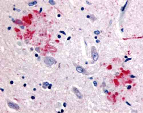 |
| Immunohistochemistry of APP in human brain (Alzheimer'sdisease) tissue with APP antibody at 10 µg/mL. |
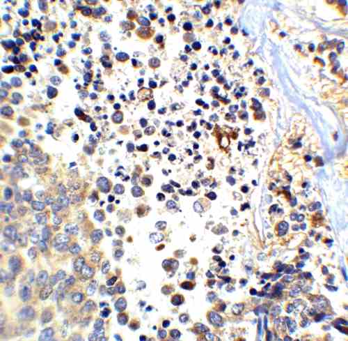 |
| Immunohistochemistry of APP in human brain tissue with APP antibody at 2.5 µg/mL. |
 |
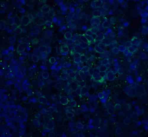
| Immunofluorescence of ASAH1 in rat heart tissue with ASAH1 antibody at 20 µg/mL.
Green: APP Antibody (5729) Blue: DAPI staining |
FAQ & Publications
Frequently Asked Questions
What applications has the rabbit anti-APP (Abeta-NT) polyclonal antibody 5729 been validated for?
This antibody has been tested and is suitable for use in ELISA, Western Blot (WB), and Immunohistochemistry on paraffin-embedded tissues (IHC-P). Recommended dilutions for immunoblotting are 1:500 to 1:1000, with mouse brain lysate as a positive control.
How should the rabbit anti-APP (Abeta-NT) polyclonal antibody 5729 be stored to maintain stability?
The antibody should be stored at -20°C and is stable for at least one year under these conditions. To preserve its activity, avoid multiple freeze-thaw cycles. The antibody is supplied in PBS buffer at pH 7.4.
What is the immunogen used to generate the rabbit anti-APP (Abeta-NT) polyclonal antibody 5729?
The immunogen is a peptide corresponding to amino acids 653-662 of the human amyloid protein precursor (APP) or amino acids 1-10 of the 4 kDa amyloid beta peptide generated by beta- and gamma-secretase cleavage. This sequence is identical in rabbit, pig, cow, guinea pig, and chicken.
Publications
| pmid | title | authors | citation |
|---|---|---|---|
| We haven't added any publications to our database yet. | |||
Published literature highly relevant to the biological target of this product and referencing this antibody or clone are retrieved from the PubMed database provided by the United States National Library of Medicine at the National Institutes of Health.
Protocols
| relevant to this product |
|---|
| Western blot IHC ICC |
Documents
| Batch Number | QC File | SDS |
|---|---|---|
| To view batch-specific Safety Datasheets and Quality Certificates associated with your account, please Log In. | ||
Only logged in customers who have purchased this product may leave a review.


Reviews
There are no reviews yet.