| Weight | 1 lbs |
|---|---|
| Dimensions | 9 × 5 × 2 in |
| host | rabbit |
| isotype | IgG |
| clonality | polyclonal |
| concentration | 1 mg/mL |
| applications | ICC/IF, WB |
| reactivity | CX3CR1 (NT) |
| available sizes | 100 µg |


rabbit anti-CX3CR1 (NT) polyclonal antibody 1398
$445.00
Antibody summary
- Rabbit polyclonal to CX3CR1 (NT)
- Suitable for: ELISA,WB,IHC-P,IF,FC
- Isotype: IgG
- 100 µg
rabbit anti-CX3CR1 (NT) polyclonal antibody 1398
| antibody |
|---|
| Tested applications WB,IHC,IHC,ICC/IF,ELISA |
| Recommended dilutions Immunoblotting: use at 1:500-1:2,000 dilution. Positive control: Tissue lysate from human spleen. |
| Immunogen Peptide corresponding to aa 2-21 of human CX3CR1. The sequence differs from mouse and rat CX3CR1 by four amino acids. |
| Size and concentration 100µg and lot specific |
| Form liquid |
| Storage Instructions This antibody is stable for at least one (1) year at -20°C. Avoid multiple freeze- thaw cycles. |
| Storage buffer PBS, pH 7.4. |
| Purity peptide affinity purification |
| Clonality polyclonal |
| Isotype IgG |
| Compatible secondaries goat anti-rabbit IgG, H&L chain specific, peroxidase conjugated, conjugated polyclonal antibody 9512 goat anti-rabbit IgG, H&L chain specific, biotin conjugated polyclonal antibody 2079 goat anti-rabbit IgG, H&L chain specific, FITC conjugated polyclonal antibody 7863 goat anti-rabbit IgG, H&L chain specific, Cross Absorbed polyclonal antibody 2371 goat anti-rabbit IgG, H&L chain specific, biotin conjugated polyclonal antibody, crossabsorbed 1715 goat anti-rabbit IgG, H&L chain specific, FITC conjugated polyclonal antibody, crossabsorbed 1720 |
| Isotype control Rabbit polyclonal - Isotype Control |
| target relevance |
|---|
| Protein names CX3C chemokine receptor 1 (C-X3-C CKR-1) (CX3CR1) (Beta chemokine receptor-like 1) (CMK-BRL-1) (CMK-BRL1) (Fractalkine receptor) (G-protein coupled receptor 13) (V28) |
| Gene names CX3CR1,CX3CR1 CMKBRL1 GPR13 |
| Protein family G-protein coupled receptor 1 family |
| Mass 40396Da |
| Function FUNCTION: Receptor for the C-X3-C chemokine fractalkine (CX3CL1) present on many early leukocyte cells; CX3CR1-CX3CL1 signaling exerts distinct functions in different tissue compartments, such as immune response, inflammation, cell adhesion and chemotaxis (PubMed:12055230, PubMed:23125415, PubMed:9390561, PubMed:9782118). CX3CR1-CX3CL1 signaling mediates cell migratory functions (By similarity). Responsible for the recruitment of natural killer (NK) cells to inflamed tissues (By similarity). Acts as a regulator of inflammation process leading to atherogenesis by mediating macrophage and monocyte recruitment to inflamed atherosclerotic plaques, promoting cell survival (By similarity). Involved in airway inflammation by promoting interleukin 2-producing T helper (Th2) cell survival in inflamed lung (By similarity). Involved in the migration of circulating monocytes to non-inflamed tissues, where they differentiate into macrophages and dendritic cells (By similarity). Acts as a negative regulator of angiogenesis, probably by promoting macrophage chemotaxis (PubMed:14581400, PubMed:18971423). Plays a key role in brain microglia by regulating inflammatory response in the central nervous system (CNS) and regulating synapse maturation (By similarity). Required to restrain the microglial inflammatory response in the CNS and the resulting parenchymal damage in response to pathological stimuli (By similarity). Involved in brain development by participating in synaptic pruning, a natural process during which brain microglia eliminates extra synapses during postnatal development (By similarity). Synaptic pruning by microglia is required to promote the maturation of circuit connectivity during brain development (By similarity). Acts as an important regulator of the gut microbiota by controlling immunity to intestinal bacteria and fungi (By similarity). Expressed in lamina propria dendritic cells in the small intestine, which form transepithelial dendrites capable of taking up bacteria in order to provide defense against pathogenic bacteria (By similarity). Required to initiate innate and adaptive immune responses against dissemination of commensal fungi (mycobiota) component of the gut: expressed in mononuclear phagocytes (MNPs) and acts by promoting induction of antifungal IgG antibodies response to confer protection against disseminated C.albicans or C.auris infection (PubMed:29326275). Also acts as a receptor for C-C motif chemokine CCL26, inducing cell chemotaxis (PubMed:20974991). {ECO:0000250|UniProtKB:Q9Z0D9, ECO:0000269|PubMed:12055230, ECO:0000269|PubMed:14581400, ECO:0000269|PubMed:18971423, ECO:0000269|PubMed:20974991, ECO:0000269|PubMed:23125415, ECO:0000269|PubMed:29326275, ECO:0000269|PubMed:9390561, ECO:0000269|PubMed:9782118}.; FUNCTION: [Isoform 1]: (Microbial infection) Acts as a coreceptor with CD4 for HIV-1 virus envelope protein. {ECO:0000269|PubMed:14607932, ECO:0000269|PubMed:9726990}.; FUNCTION: [Isoform 2]: (Microbial infection) Acts as a coreceptor with CD4 for HIV-1 virus envelope protein (PubMed:14607932). May have more potent HIV-1 coreceptothr activity than isoform 1 (PubMed:14607932). {ECO:0000269|PubMed:14607932}.; FUNCTION: [Isoform 3]: (Microbial infection) Acts as a coreceptor with CD4 for HIV-1 virus envelope protein (PubMed:14607932). May have more potent HIV-1 coreceptor activity than isoform 1 (PubMed:14607932). {ECO:0000269|PubMed:14607932}. |
| Subellular location SUBCELLULAR LOCATION: Cell membrane {ECO:0000269|PubMed:12055230, ECO:0000269|PubMed:28791023, ECO:0000269|PubMed:9390561}; Multi-pass membrane protein {ECO:0000255}. |
| Tissues TISSUE SPECIFICITY: Expressed in lymphoid and neural tissues (PubMed:7590284). Expressed in lymphocyte subsets, such as natural killer (NK) cells, gamma-delta T-cells and terminally differentiated CD8(+) T-cells (PubMed:12055230). Expressed in smooth muscle cells in atherosclerotic plaques (PubMed:14581400). {ECO:0000269|PubMed:12055230, ECO:0000269|PubMed:14581400, ECO:0000269|PubMed:7590284}. |
| Structure SUBUNIT: Found in a ternary complex with CX3CL1 and ITGAV:ITGB3 or ITGA4:ITGB1. {ECO:0000269|PubMed:23125415}.; SUBUNIT: (Microbial infection) Interacts with human respiratory syncytial virus (HRSV) protein G; this interaction modulates host immune response. {ECO:0000269|PubMed:11477410}.; SUBUNIT: (Microbial infection) Interacts with HIV-1 envelope polyprotein gp160. {ECO:0000269|PubMed:9726990}. |
| Post-translational modification PTM: This protein is not N-glycosylated which is unusual for G-protein-coupled receptors. {ECO:0000250|UniProtKB:P35411}. |
| Involvement in disease DISEASE: Macular degeneration, age-related, 12 (ARMD12) [MIM:613784]: A form of age-related macular degeneration, a multifactorial eye disease and the most common cause of irreversible vision loss in the developed world. In most patients, the disease is manifest as ophthalmoscopically visible yellowish accumulations of protein and lipid that lie beneath the retinal pigment epithelium and within an elastin-containing structure known as Bruch membrane. {ECO:0000269|PubMed:15208270}. Note=Disease susceptibility is associated with variants affecting the gene represented in this entry. |
| Target Relevance information above includes information from UniProt accession: P49238 |
| The UniProt Consortium |
Data
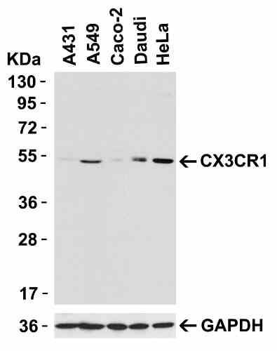 |
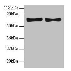
| Western Blot Validation in Human Cells Loading: 15 µg of lysates per lane. Antibodies: CX3CR1 1398 (0.5 µg/mL), 1h incubation at RT in 5% NFDM/TBST.Secondary: Goat anti-rabbit IgG HRP conjugate at 1:10000 dilution. |
 |
| KD Validation in 293 Cells Loading: 15 µg of lysates per lane. Antibodies: CX3CR1 1398 (0.5 µg/mL), 1h incubation at RT in 5% NFDM/TBST.Secondary: Goat anti-rabbit IgG HRP conjugate at 1:10000 dilution.Lane 1: 293 cells transfected with control siRNAs.Lane 2: 293 cells transfected with CX3CR1 siRNAs. |
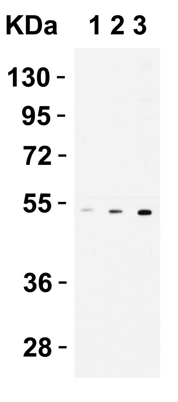 |
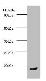
| Western Blot Validation in THP1 Cells Loading: 15 µg of lysates per lane. Antibodies: CX3CR1 1398 (1 µg/mL), 1h incubation at RT in 5% NFDM/TBST.Secondary: Goat anti-rabbit IgG HRP conjugate at 1:10000 dilution.Lane 1: 0.2 µg/mLLane 1: 0.5 µg/mLLane 1: 1 µg/mL |
 |
| Western Blot Validation in Human Spleen Lysates Loading: 15 µg of lysates per lane. Antibodies: CX3CR1 1398 (1 µg/mL) in the absence (lane 1) or presence of blocking peptide (lane 2), 1h incubation at RT in 5% NFDM/TBST.Secondary: Goat anti-rabbit IgG HRP conjugate at 1:10000 dilution. |
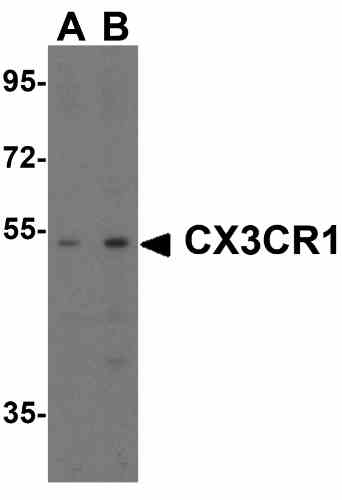 |
| Western Blot Validation in Rat Spleen Tissue Loading: 15 µg of lysates per lane. Antibodies: CX3CR1 1398, (A; 1 µg/mL, B; 2 µg/mL), 1h incubation at RT in 5% NFDM/TBST.Secondary: Goat anti-rabbit IgG HRP conjugate at 1:10000 dilution. |
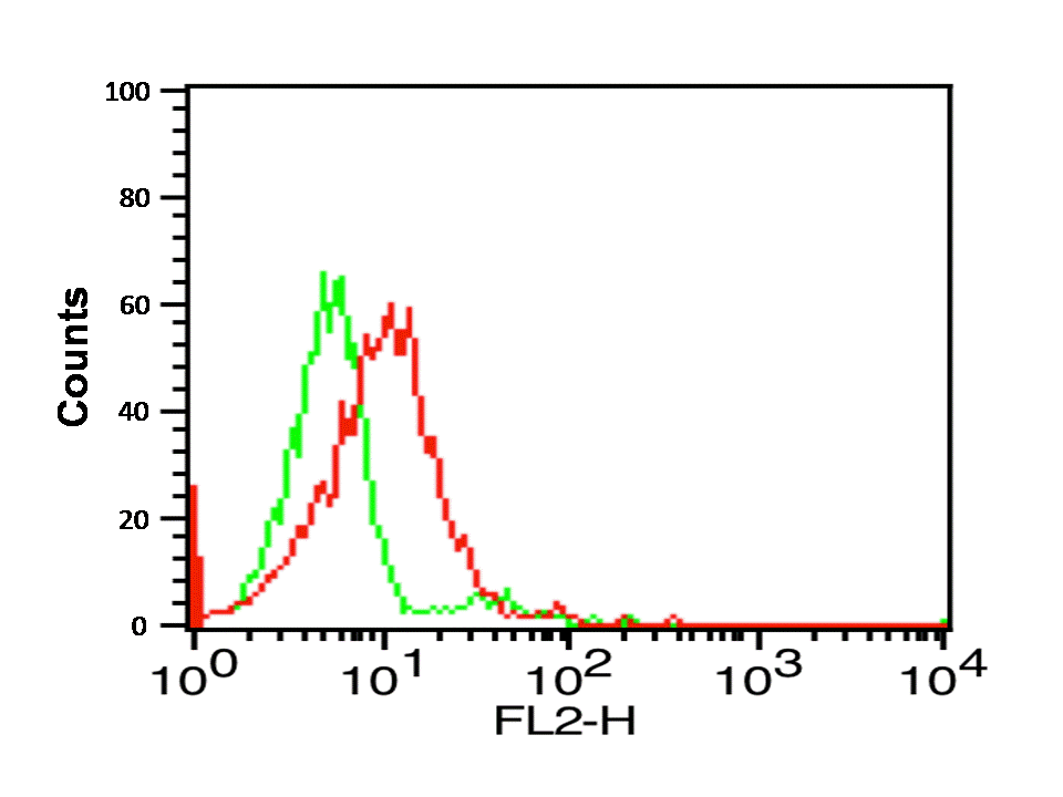 |
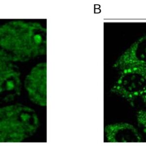
| Flow Cytometry Validation of CX3CR1 in THP-1 Cells Overlay histogram showing THP-1 cells stained with 1398 (red line, 1ug/1x106 cells). 1 h incubation at 4C in 2% FBS/PBS. Followed by secondary antibody 488 goat anti-rabbit IgG (H+L) at 1/500 dilution for 1 h 4C. Isotype control antibody (Green line) was rabbit IgG1 (1ug/1x106 cells) used under the same conditions. Acquisition of >10,000 events was performed. |
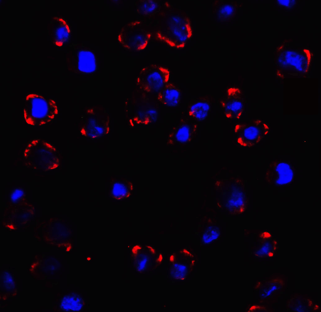 |
| Immunofluorescence Validation of CX3CR1 In K562 Cells Immunofluorescent analysis of 4% paraformaldehyde-fixed K562 cells labeling CX3CR1 with 1398 at 10 µg/mL, followed by goat anti-rabbit IgG secondary antibody at 1/500 dilution (red) and DAPI staining (blue). Image showing membrane staining on K562 cells. |
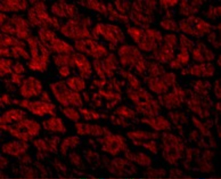 |
| Immunofluorescence Validation of CX3CR1 in Human Heart Immunofluorescent analysis of 4% paraformaldehyde-fixed human heart tissue labeling CX3CR1 with 1398 at 10 µg/mL, followed by goat anti-rabbit IgG secondary antibody at 1/500 dilution (red). |
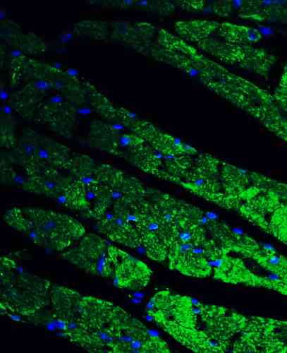 |
| Immunofluorescence Validation of CX3CR1 in Mouse Heart Tissue Immunofluorescent analysis of 4% paraformaldehyde-fixed mouse heart tissue labeling CX3CR1 with 1398 at 20 µg/mL, followed by goat anti-rabbit IgG secondary antibody at 1/500 dilution (green) and DAPI staining (blue). |
Publications
| pmid | title | authors | citation |
|---|---|---|---|
| We haven't added any publications to our database yet. | |||
Protocols
| relevant to this product |
|---|
| Western blot IHC ICC |
Documents
| # | SDS | Certificate | |
|---|---|---|---|
| Please enter your product and batch number here to retrieve product datasheet, SDS, and QC information. | |||
Only logged in customers who have purchased this product may leave a review.

Reviews
There are no reviews yet.