| Weight | 1 lbs |
|---|---|
| Dimensions | 9 × 5 × 2 in |
| host | mouse |
| isotype | IgG2b |
| clonality | monoclonal |
| concentration | 1 mg/mL |
| applications | ICC/IF, WB |
| reactivity | p84 |
| available sizes | 100 µg |


mouse anti-p84 monoclonal antibody (5E9) 9573
$503.00
Antibody summary
- Mouse monoclonal to p84
- Suitable for: WB,ICC/IF,IHC-P,IP,ChIP,IHC
- Isotype: IgG2b
- 100 µg
mouse anti-p84 monoclonal antibody (5E9) 9573
| antibody |
|---|
| Tested applications WB,IHC,IHC,ICC/IF |
| Recommended dilutions Immunoblotting, immunoprecipitation, and Immunofluorescence: use at 0.5-2 ug/mL.In immunoblots, a band of 84 kD is detected. Positive controls: Lysates of Molt 4, HeLa, or Raji cells. |
| Immunogen Fusion protein containing amino acids 15-374 of human p84 expressed in E. coli. |
| Size and concentration 100µg and lot specific |
| Form liquid |
| Storage Instructions This antibody is stable for at least one (1) year at -70°C. Avoid multiple freeze- thaw cycles. |
| Storage buffer PBS, pH 7.4. |
| Purity protein affinity purification |
| Clonality monoclonal |
| Isotype IgG2b |
| Compatible secondaries goat anti-mouse IgG, H&L chain specific, peroxidase conjugated polyclonal antibody 5486 goat anti-mouse IgG, H&L chain specific, biotin conjugated, Conjugate polyclonal antibody 2685 goat anti-mouse IgG, H&L chain specific, FITC conjugated polyclonal antibody 7854 goat anti-mouse IgG, H&L chain specific, peroxidase conjugated polyclonal antibody, crossabsorbed 1706 goat anti-mouse IgG, H&L chain specific, biotin conjugated polyclonal antibody, crossabsorbed 1716 goat anti-mouse IgG, H&L chain specific, FITC conjugated polyclonal antibody, crossabsorbed 1721 |
| Isotype control Mouse monocolonal IgG2b - Isotype Control |
| target relevance |
|---|
| Homo sapiens THOC1 THO complex subunit 1 |
| Protein names THO complex subunit 1 |
| Alternative names Nuclear matrix protein p84, hTREX84 |
| Gene names THOC1 |
| Protein family Belongs to the THOC1 family |
| Function Component of the THO subcomplex of the TREX complex which is thought to couple mRNA transcription, processing and nuclear export, and which specifically associates with spliced mRNA and not with unspliced pre-mRNA (PubMed:15833825, PubMed:15998806, PubMed:17190602). Required for efficient export of polyadenylated RNA (PubMed:23222130). The THOC1-THOC2-THOC3 core complex alone is sufficient to bind export factor NXF1-NXT1 and promote ATPase activity of DDX39B/UAP56 (PubMed:33191911). TREX is recruited to spliced mRNAs by a transcription-independent mechanism, binds to mRNA upstream of the exon-junction complex (EJC) and is recruited in a splicing- and cap-dependent manner to a region near the 5' end of the mRNA where it functions in mRNA export to the cytoplasm via the TAP/NXF1 pathway (PubMed:15833825, PubMed:15998806, PubMed:17190602). Regulates transcriptional elongation of a subset of genes (PubMed:22144908). Involved in genome stability by preventing co-transcriptional R-loop formation (By similarity). May play a role in hair cell formation, hence may be involved in hearing (By similarity) |
| Subcellular location Cytoplasm |
| Structure Component of the THO subcomplex, which is composed of THOC1, THOC2, THOC3, THOC5, THOC6 and THOC7 (PubMed:33191911, PubMed:37020021). The THO subcomplex interacts with DDX39B to form the THO-DDX39B complex which multimerizes into a 28-subunit tetrameric assembly (PubMed:33191911, PubMed:37020021). Component of the transcription/export (TREX) complex at least composed of ALYREF/THOC4, DDX39B, SARNP/CIP29, CHTOP and the THO subcomplex; in the complex interacts with THOC2, THOC5 and THOC7 (PubMed:33191911, PubMed:37020021). TREX seems to have a dynamic structure involving ATP-dependent remodeling (PubMed:23222130, PubMed:37020021). Binds to the hypophosphorylated form of RB1. Interacts with RNA polymerase II. Interacts with LUZP4 |
| Post-translational modification Expression is altered specifically during apoptosis and is accompanied by the appearance of novel forms with smaller apparent molecular mass Polyubiquitinated, leading to proteasomal degradation; probably involves NEDD4 |
| Involvement in disease Deafness, autosomal dominant, 86 A form of non-syndromic, sensorineural hearing loss. Sensorineural hearing loss results from damage to the neural receptors of the inner ear, the nerve pathways to the brain, or the area of the brain that receives sound information. DFNA86 is characterized by progressive, bilateral hearing loss that is most predominant in the high frequencies, begins mildly during the fourth decade and gradually progresses to severe-to-profound deafness in the seventh and eighth decades. Affected subjects have tinnitus, while vestibular dysfunction or other clinical abnormalities are not present. |
| Keywords 3D-structure, Acetylation, Alternative splicing, Apoptosis, Cytoplasm, Deafness, Disease variant, DNA-binding, Isopeptide bond, mRNA processing, mRNA splicing, mRNA transport, Non-syndromic deafness, Nucleus, Phosphoprotein, Proteomics identification, Reference proteome, RNA-binding, Transcription, Transcription regulation, Transport, Ubl conjugation |
| Sequence MSPTPPLFSLPEARTRFTKSTREALNNKNIKPLLSTFSQVPGSENEKKCTLDQAFRGILE EEIINHSSCENVLAIISLAIGGVTEGICTASTPFVLLGDVLDCLPLDQCDTIFTFVEKNV ATWKSNTFYSAGKNYLLRMCNDLLRRLSKSQNTVFCGRIQLFLARLFPLSEKSGLNLQSQ FNLENVTVFNTNEQESTLGQKHTEDREEGMDVEEGEMGDEEAPTTCSIPIDYNLYRKFWS LQDYFRNPVQCYEKISWKTFLKYSEEVLAVFKSYKLDDTQASRKKMEELKTGGEHVYFAK FLTSEKLMDLQLSDSNFRRHILLQYLILFQYLKGQVKFKSSNYVLTDEQSLWIEDTTKSV YQLLSENPPDGERFSKMVEHILNTEENWNSWKNEGCPSFVKERTSDTKPTRIIRKRTAPE DFLGKGPTKKILMGNEELTRLWNLCPDNMEACKSETREHMPTLEEFFEEAIEQADPENMV ENEYKAVNNSNYGWRALRLLARRSPHFFQPTNQQFKSLPEYLENMVIKLAKELPPPSEEI KTGEDEDEEDNDALLKENESPDVRRDKPVTGEQIEVFANKLGEQWKILAPYLEMKDSEIR QIECDSEDMKMRAKQLLVAWQDQEGVHATPENLINALNKSGLSDLAESLTNDNETNS |
| UniProt accession: Q96FV9 |
Data
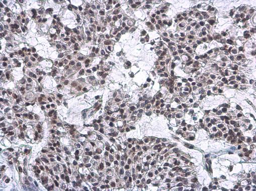 |
| Nuclear Matrix Protein p84 antibody [5E10] detects Nuclear Matrix Protein p84 protein at nucleus by immunohistochemical analysis.Sample: Paraffin-embedded human breast carcinoma.Nuclear Matrix Protein p84 stained by Nuclear Matrix Protein p84 antibody [5E10] (9573) diluted at 1:200.Antigen Retrieval: Citrate buffer, pH 6.0, 15 min |
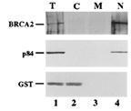 |
| Detection of human p84/N5 in nuclear fraction by anti-p84/N5 5E10 monoclonal antibody (9573) in western blot experiment. Lane 1: total lysate, Lane 2: cytoplasmic fraction, Lane 3: membrane fraction, Lane 4: nuclear fraction. |
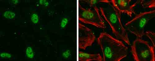 |
| Nuclear Matrix Protein p84 antibody [5E10] detects Nuclear Matrix Protein p84 protein at nucleus by immunofluorescent analysis.Sample: HeLa cells were fixed in 4% paraformaldehyde at RT for 15 min.Green: Nuclear Matrix Protein p84 stained by Nuclear Matrix Protein p84 antibody [5E10] (9573) diluted at 1:500.Red: phalloidin, a cytoskeleton marker, diluted at 1:100. |
 |
| p84 antibody [5E10] immunoprecipitates p84 protein in IP experiments. IP Sample: HepG2 whole cell lysate/extract A : 30 µg whole cell lysate/extract of p84 protein expressing HepG2 cells B : Control with 3 µg of pre-immune mouse IgG C : Immunoprecipitation of p84 by 3 µg of p84 antibody [5E10] (9573) 7.5% SDS-PAGE The immunoprecipitated p84 protein was detected by p84 antibody [5E10] (9573) diluted at 1 : 1000. EasyBlot anti-rabbit IgG (HRP) was used as a secondary reagent. |
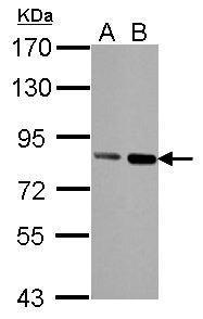 |
| Sample (30 µg of whole cell lysate) A: HeLa B: HeLa nucleus 7.5% SDS PAGE 9573 diluted at 1:1000 The HRP-conjugated anti-mouse IgG antibody was used to detect the primary antibody. |
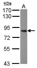 |
| Sample (50 µg of whole cell lysate) A: mouse Cerebellum 7.5% SDS PAGE 9573 diluted at 1:1000 The HRP-conjugated anti-mouse IgG antibody was used to detect the primary antibody. |
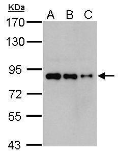 |
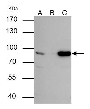
| Sample (whole cell lysate) A: 293T 20ug B: 293T 10ug C: 293T 5ug 7.5% SDS PAGE 9573 diluted at 1:1000 The HRP-conjugated anti-mouse IgG antibody was used to detect the primary antibody. |
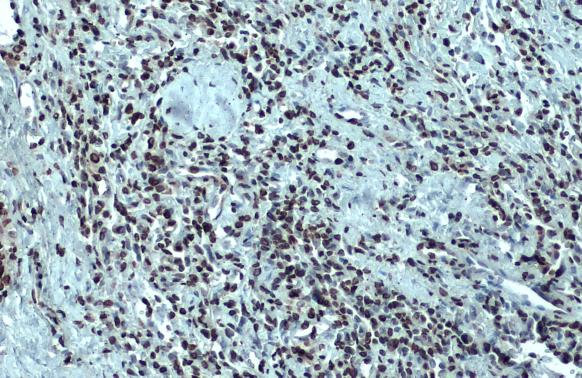 |
| Nuclear Matrix Protein p84 antibody [5E10] detects Nuclear Matrix Protein p84 protein at nucleus by immunohistochemical analysis.Sample: Paraffin-embedded human lung cancer.Nuclear Matrix Protein p84 stained by Nuclear Matrix Protein p84 antibody [5E10] (9573) diluted at 1:100.Antigen Retrieval: Citrate buffer, pH 6.0, 15 min |
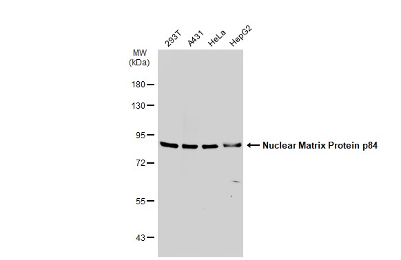 |
| Various whole cell extracts (30 µg) were separated by 7.5% SDS-PAGE, and the membrane was blotted with Nuclear Matrix Protein p84 antibody [5E10] (9573) diluted at 1:500. The HRP-conjugated anti-mouse IgG antibody was used to detect the primary antibody. |
FAQ & Publications
Frequently Asked Questions
What applications has the mouse anti-p84 monoclonal antibody (5E9) been validated for?
This antibody has been tested and is suitable for Western blotting (WB), immunocytochemistry/immunofluorescence (ICC/IF), immunohistochemistry on paraffin-embedded sections (IHC-P), immunoprecipitation (IP), and chromatin immunoprecipitation (ChIP).
How should the mouse anti-p84 antibody be stored to maintain stability?
The antibody should be stored at -70°C and is stable for at least one year under these conditions. It is important to avoid multiple freeze-thaw cycles to preserve antibody integrity.
What is the host species and clonality of the anti-p84 antibody (clone 5E9)?
The antibody is a mouse monoclonal antibody with an IgG2b isotype, derived from a single clone (5E9).
What is the recommended dilution range for immunoblotting and immunofluorescence using this anti-p84 antibody?
For immunoblotting, immunoprecipitation, and immunofluorescence applications, it is recommended to use the antibody at a concentration of 0.5 to 2 µg/mL.
What immunogen was used to generate the mouse anti-p84 monoclonal antibody (5E9)?
The immunogen was a fusion protein containing amino acids 15 to 374 of human p84 expressed in Escherichia coli.
Publications
| pmid | title | authors | citation |
|---|---|---|---|
| 37885910 | A Knock-In Mouse Model of the Gcm2 Variant p.Y392S Develops Normal Parathyroid Glands. | Vaishali I Parekh, Lauren R Brinster, Bin Guan, William F Simonds, Lee S Weinstein, Sunita K Agarwal | J Endocr Soc 7:bvad126 |
| 37790327 | Increased AID Results in Mutations at the CRLF2 Locus Implicated in Latin American ALL Health Disparities. | Nicholas Pannunzio, Valeria Rangel, Jason Sterrenberg, Aya Garawi, Vyanka Mezcord, Melissa Folkerts, Sabrina Caulderon, Jinglong Wang, Eli Soyfer, Oliver Eng, Jennifer Valerin, Sora Tanjasiri, Fabiola Quintero-Rivera, Selma Masri, Marcus Seldin, Richard Frock, Angela Fleischman | Res Sq N/A:N/A |
| 37267101 | Liver and muscle circadian clocks cooperate to support glucose tolerance in mice. | Jacob G Smith, Kevin B Koronowski, Thomas Mortimer, Tomoki Sato, Carolina M Greco, Paul Petrus, Amandine Verlande, Siwei Chen, Muntaha Samad, Ekaterina Deyneka, Lavina Mathur, Ronnie Blazev, Jeffrey Molendijk, Arun Kumar, Oleg Deryagin, Mireia Vaca-Dempere, Valentina Sica, Peng Liu, Valerio Orlando, Benjamin L Parker, Pierre Baldi, Patrick-Simon Welz, Cholsoon Jang, Selma Masri, Salvador Aznar Benitah, Pura Muñoz-Cánoves, Paolo Sassone-Corsi | Cell Rep 42:112588 |
| 36973248 | Time-of-day defines NAD(+) efficacy to treat diet-induced metabolic disease by synchronizing the hepatic clock in mice. | Quetzalcoatl Escalante-Covarrubias, Lucía Mendoza-Viveros, Mirna González-Suárez, Román Sitten-Olea, Laura A Velázquez-Villegas, Fernando Becerril-Pérez, Ignacio Pacheco-Bernal, Erick Carreño-Vázquez, Paola Mass-Sánchez, Marcia Bustamante-Zepeda, Ricardo Orozco-Solís, Lorena Aguilar-Arnal | Nat Commun 14:1685 |
| 36646704 | Expansion of interferon inducible gene pool via USP18 inhibition promotes cancer cell pyroptosis. | Kei-Ichiro Arimoto, Sayuri Miyauchi, Ty D Troutman, Yue Zhang, Mengdan Liu, Samuel A Stoner, Amanda G Davis, Jun-Bao Fan, Yi-Jou Huang, Ming Yan, Christopher K Glass, Dong-Er Zhang | Nat Commun 14:251 |
Published literature highly relevant to the biological target of this product and referencing this antibody or clone are retrieved from the PubMed database provided by the United States National Library of Medicine at the National Institutes of Health.
Protocols
| relevant to this product |
|---|
| Western blot IHC ICC |
Documents
| Batch Number | QC File | SDS |
|---|---|---|
| To view batch-specific Safety Datasheets and Quality Certificates associated with your account, please Log In. | ||
Only logged in customers who have purchased this product may leave a review.


Reviews
There are no reviews yet.