| Weight | 1 lbs |
|---|---|
| Dimensions | 9 × 5 × 2 in |
| host | mouse |
| isotype | IgG2a |
| clonality | monoclonal |
| concentration | concentrate, predilute |
| applications | IHC |
| reactivity | human |
| available size | 0.1 mL, 0.5 mL, 1 mL concentrated, 7 mL prediluted |
mouse anti-Tyrosinase monoclonal antibody (T311) 6398
Price range: $160.00 through $528.00
Antibody summary
- Mouse monoclonal to Tyrosinase
- Suitable for: Immunohistochemistry (formalin-fixed, paraffin-embedded tissues)
- Reacts with: Human
- Isotype:IgG2a
- Control: Melanoma
- Visualization: Cytoplasmic
- 0.1, 0.5, 1.0 mL concentrated, 7 mL prediluted
mouse anti-Tyrosinase monoclonal antibody T311 6398
| target relevance |
|---|
| Protein names Tyrosinase (EC 1.14.18.1) (LB24-AB) (Monophenol monooxygenase) (SK29-AB) (Tumor rejection antigen AB) |
| Gene names TYR,TYR |
| Protein family Tyrosinase family |
| Mass 60393Da |
| Function FUNCTION: This is a copper-containing oxidase that functions in the formation of pigments such as melanins and other polyphenolic compounds. Catalyzes the initial and rate limiting step in the cascade of reactions leading to melanin production from tyrosine (By similarity). In addition to hydroxylating tyrosine to DOPA (3,4-dihydroxyphenylalanine), also catalyzes the oxidation of DOPA to DOPA-quinone, and possibly the oxidation of DHI (5,6-dihydroxyindole) to indole-5,6 quinone (PubMed:28661582). {ECO:0000250|UniProtKB:P11344, ECO:0000269|PubMed:28661582}. |
| Catalytic activity CATALYTIC ACTIVITY: Reaction=2 L-dopa + O2 = 2 L-dopaquinone + 2 H2O; Xref=Rhea:RHEA:34287, ChEBI:CHEBI:15377, ChEBI:CHEBI:15379, ChEBI:CHEBI:57504, ChEBI:CHEBI:57924; EC=1.14.18.1; Evidence={ECO:0000269|PubMed:28661582}; PhysiologicalDirection=left-to-right; Xref=Rhea:RHEA:34288; Evidence={ECO:0000305|PubMed:28661582}; CATALYTIC ACTIVITY: Reaction=L-tyrosine + O2 = L-dopaquinone + H2O; Xref=Rhea:RHEA:18117, ChEBI:CHEBI:15377, ChEBI:CHEBI:15379, ChEBI:CHEBI:57924, ChEBI:CHEBI:58315; EC=1.14.18.1; Evidence={ECO:0000269|PubMed:28661582}; PhysiologicalDirection=left-to-right; Xref=Rhea:RHEA:18118; Evidence={ECO:0000305|PubMed:28661582}; CATALYTIC ACTIVITY: Reaction=2 5,6-dihydroxyindole-2-carboxylate + O2 = 2 indole-5,6-quinone-2-carboxylate + 2 H2O; Xref=Rhea:RHEA:68388, ChEBI:CHEBI:15377, ChEBI:CHEBI:15379, ChEBI:CHEBI:16875, ChEBI:CHEBI:177869; Evidence={ECO:0000269|PubMed:28661582}; PhysiologicalDirection=left-to-right; Xref=Rhea:RHEA:68389; Evidence={ECO:0000305|PubMed:28661582}; |
| Subellular location SUBCELLULAR LOCATION: Melanosome membrane {ECO:0000269|PubMed:12643545, ECO:0000269|PubMed:17081065}; Single-pass type I membrane protein {ECO:0000269|PubMed:12643545, ECO:0000269|PubMed:17081065}. Melanosome {ECO:0000250|UniProtKB:P11344}. Note=Proper trafficking to melanosome is regulated by SGSM2, ANKRD27, RAB9A, RAB32 and RAB38. {ECO:0000250|UniProtKB:P11344}. |
| Structure SUBUNIT: Forms an OPN3-dependent complex with DCT in response to blue light in melanocytes. {ECO:0000269|PubMed:28842328}. |
| Post-translational modification PTM: Glycosylated. {ECO:0000250|UniProtKB:P11344}. |
| Involvement in disease DISEASE: Albinism, oculocutaneous, 1A (OCA1A) [MIM:203100]: An autosomal recessive disorder in which the biosynthesis of melanin pigment is absent in skin, hair, and eyes. It is characterized by complete lack of tyrosinase activity due to production of an inactive enzyme. Patients present with a life-long absence of melanin pigment after birth, and manifest increased sensitivity to ultraviolet radiation with predisposition to skin cancer. Visual anomalies include decreased acuity, nystagmus, strabismus and photophobia. {ECO:0000269|PubMed:10571953, ECO:0000269|PubMed:10671066, ECO:0000269|PubMed:10987646, ECO:0000269|PubMed:11295837, ECO:0000269|PubMed:11858948, ECO:0000269|PubMed:1487241, ECO:0000269|PubMed:15146472, ECO:0000269|PubMed:1642278, ECO:0000269|PubMed:1899321, ECO:0000269|PubMed:1943686, ECO:0000269|PubMed:1970634, ECO:0000269|PubMed:22981120, ECO:0000269|PubMed:2342539, ECO:0000269|PubMed:23504663, ECO:0000269|PubMed:24934919, ECO:0000269|PubMed:7902671, ECO:0000269|PubMed:7955413, ECO:0000269|PubMed:8128955, ECO:0000269|PubMed:8644824, ECO:0000269|PubMed:9259202}. Note=The disease is caused by variants affecting the gene represented in this entry.; DISEASE: Albinism, oculocutaneous, 1B (OCA1B) [MIM:606952]: An autosomal recessive disorder in which the biosynthesis of melanin pigment is reduced in skin, hair, and eyes. It is characterized by partial lack of tyrosinase activity. Patients have white hair at birth that rapidly turns yellow or blond. They manifest the development of minimal-to-moderate amounts of cutaneous and ocular pigment. Some patients may have with white hair in the warmer areas (scalp and axilla) and progressively darker hair in the cooler areas (extremities). This variant phenotype is due to a loss of tyrosinase activity above 35-37 degrees C. {ECO:0000269|PubMed:10987646, ECO:0000269|PubMed:1900309, ECO:0000269|PubMed:1903591, ECO:0000269|PubMed:8128955}. Note=The disease is caused by variants affecting the gene represented in this entry. |
| Target Relevance information above includes information from UniProt accession: P14679 |
| The UniProt Consortium |
Data
 |
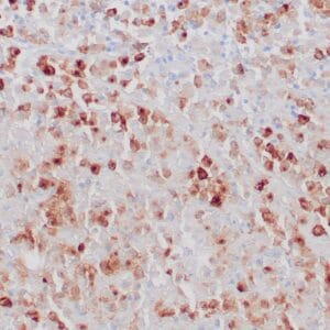
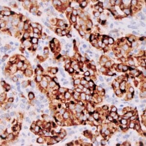
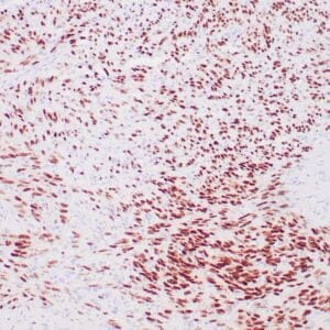
| Human melanoma stained with anti-tyrosinase antibody using peroxidase-conjugate and DAB chromogen. Note the cytoplasmic staining of tumor cells. |
Publications
| pmid | title | authors | citation |
|---|---|---|---|
| We haven't added any publications to our database yet. | |||
Protocols
| relevant to this product |
|---|
| IHC |
Documents
| # | SDS | Certificate | |
|---|---|---|---|
| Please enter your product and batch number here to retrieve product datasheet, SDS, and QC information. | |||
Only logged in customers who have purchased this product may leave a review.


Reviews
There are no reviews yet.