| Weight | 1 lbs |
|---|---|
| Dimensions | 9 × 5 × 2 in |
| host | mouse |
| isotype | IgG1 |
| clonality | monoclonal |
| concentration | 1 mg/mL |
| applications | ICC/IF, WB |
| reactivity | TSG101 |
| available sizes | 100 µL |


mouse anti-TSG101 monoclonal antibody (4A10) 4454
$503.00
Antibody summary
- Mouse monoclonal to TSG101
- Suitable for: WB,ICC/IF,IHC-P,FACS,IP,ELISA,EM,IHC,IHC
- Isotype: IgG1
- 100 µl
mouse anti-TSG101 monoclonal antibody (4A10) 4454
| antibody |
|---|
| Tested applications WB,IHC,IHC,ICC/IF |
| Recommended dilutions ELISA: use at 1-5ug/ml. Immunoblotting: use at a 1:500-1:3,000 dilution. A band of 46kDa is detected. Detection of TSG101 protein in (A) NIH-3T3 cell lysate, (B) JC cell lysate, and (C) BCL-1 cell lysate with #3352 diluted 1:500. Endusers should determine optimal antibody concentra |
| Immunogen Recombinant protein corresponding to aa 167-374 of TSG101 protein |
| Size and concentration 100µL and lot specific |
| Form liquid |
| Storage Instructions This product is stable for at least one (1) year if stored at -20°C. Store product in appropriate aliquots to avoid multiple freeze-thaw cycles. |
| Storage buffer PBS, pH 7.2. |
| Purity affinity purified |
| Clonality monoclonal |
| Isotype IgG1 |
| Compatible secondaries goat anti-mouse IgG, H&L chain specific, peroxidase conjugated polyclonal antibody 5486 goat anti-mouse IgG, H&L chain specific, biotin conjugated, Conjugate polyclonal antibody 2685 goat anti-mouse IgG, H&L chain specific, FITC conjugated polyclonal antibody 7854 goat anti-mouse IgG, H&L chain specific, peroxidase conjugated polyclonal antibody, crossabsorbed 1706 goat anti-mouse IgG, H&L chain specific, biotin conjugated polyclonal antibody, crossabsorbed 1716 goat anti-mouse IgG, H&L chain specific, FITC conjugated polyclonal antibody, crossabsorbed 1721 |
| Isotype control Mouse monocolonal IgG1 - Isotype Control |
| target relevance |
|---|
| Homo sapiens TSG101 Tumor susceptibility gene 101 protein |
| Protein names Tumor susceptibility gene 101 protein |
| Alternative names ESCRT-I complex subunit TSG101 |
| Gene names TSG101 |
| Protein family Belongs to the ubiquitin-conjugating enzyme family. UEV subfamily |
| Function Component of the ESCRT-I complex, a regulator of vesicular trafficking process. Binds to ubiquitinated cargo proteins and is required for the sorting of endocytic ubiquitinated cargos into multivesicular bodies (MVBs). Mediates the association between the ESCRT-0 and ESCRT-I complex. Required for completion of cytokinesis; the function requires CEP55. May be involved in cell growth and differentiation. Acts as a negative growth regulator. Involved in the budding of many viruses through an interaction with viral proteins that contain a late-budding motif P-[ST]-A-P. This interaction is essential for viral particle budding of numerous retroviruses. Required for the exosomal release of SDCBP, CD63 and syndecan (PubMed:22660413). It may also play a role in the extracellular release of microvesicles that differ from the exosomes (PubMed:22315426) |
| Subcellular location Cytoplasm, Early endosome membrane, Late endosome membrane, Cytoplasm, cytoskeleton, microtubule organizing center, centrosome, Midbody, Midbody ring, Nucleus |
| Structure (Microbial infection) Interacts with hepatitis E virus protein ORF3 |
| Post-translational modification Monoubiquitinated at multiple sites by LRSAM1 and by MGRN1. Ubiquitination inactivates it, possibly by regulating its shuttling between an active membrane-bound protein and an inactive soluble form. Ubiquitination by MGRN1 requires the presence of UBE2D1 |
| Keywords 3D-structure, Acetylation, Alternative splicing, Cell cycle, Cell division, Coiled coil, Cytoplasm, Cytoskeleton, Endosome, Growth regulation, Host-virus interaction, Membrane, Nucleus, Phosphoprotein, Protein transport, Proteomics identification, Reference proteome, Transport, Ubl conjugation |
| Sequence MAVSESQLKKMVSKYKYRDLTVRETVNVITLYKDLKPVLDSYVFNDGSSRELMNLTGTIP VPYRGNTYNIPICLWLLDTYPYNPPICFVKPTSSMTIKTGKHVDANGKIYLPYLHEWKHP QSDLLGLIQVMIVVFGDEPPVFSRPISASYPPYQATGPPNTSYMPGMPGGISPYPSGYPP NPSGYPGCPYPPGGPYPATTSSQYPSQPPVTTVGPSRDGTISEDTIRASLISAVSDKLRW RMKEEMDRAQAELNALKRTEEDLKKGHQKLEEMVTRLDQEVAEVDKNIELLKKKDEELSS ALEKMENQSENNDIDEVIIPTAPLYKQILNLYAEENAIEDTIFYLGEALRRGVIDLDVFL KHVRLLSRKQFQLRALMQKARKTAGLSDLY |
| UniProt accession: Q99816 |
Data
 |
| TSG101 antibody [4A10] detects TSG101 protein by western blot analysis. A. 30 µg NIH-3T3 whole cell lysate/extract B. 30 µg JC whole cell lysate/extract C. 30 µg BCL-1 whole cell lysate/extract 10% SDS-PAGE TSG101 antibody [4A10] (4454) dilution: 1:500 The HRP-conjugated anti-mouse IgG antibody was used to detect the primary antibody. |
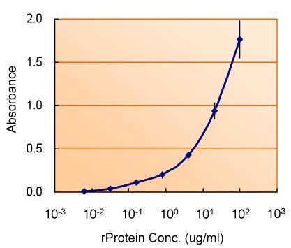 |
| ELISA detection of TSG101 using for capture at a concentration of 5 µg/mL and 4454 for detection at a concentration of 1.5 µg/mL. |
 |
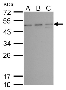
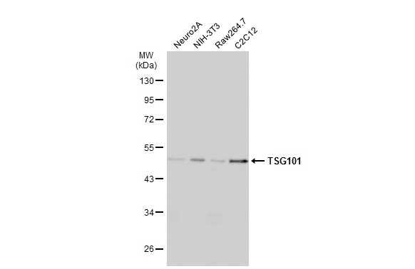
| Various whole cell extracts (30 µg) were separated by 10% SDS-PAGE, and the membrane was blotted with TSG101 antibody [4A10] (4454) diluted at 1:500. The HRP-conjugated anti-mouse IgG antibody was used to detect the primary antibody. |
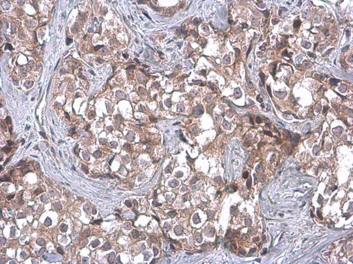 |
| TSG101 antibody [4A10] detects TSG101 protein at cytoplasm by immunohistochemical analysis.Sample: Paraffin-embedded human breast carcinoma.TSG101 stained by TSG101 antibody [4A10] (4454) diluted at 1:100.Antigen Retrieval: Citrate buffer, pH 6.0, 15 min |
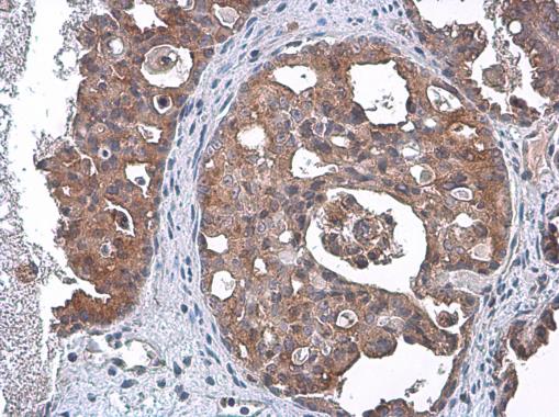 |
| TSG101 antibody [4A10] detects TSG101 protein at cytoplasm by immunohistochemical analysis.Sample: Paraffin-embedded human ovarian cancer.TSG101 stained by TSG101 antibody [4A10] (4454) diluted at 1:100.Antigen Retrieval: Citrate buffer, pH 6.0, 15 min |
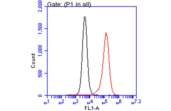 |
| TSG101 antibody [4A10] (4454) detects TSG101 protein by flow cytometry analysis. Sample: THP-1 cell. Black: Unlabelled sample was used as a control. Red: TSG101 antibody [4A10] (4454) dilution: 1:25. Acquisition of 20,000 events were collected using a Dylight 488-conjugated secondary antibody for FACS analysis. |
 |
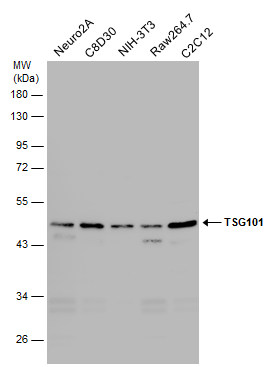
| Various whole cell extracts (30 µg) were separated by 10% SDS-PAGE, and the membrane was blotted with TSG101 antibody [4A10] (4454) diluted at 1:500. The HRP-conjugated anti-mouset IgG antibody was used to detect the primary antibody. |
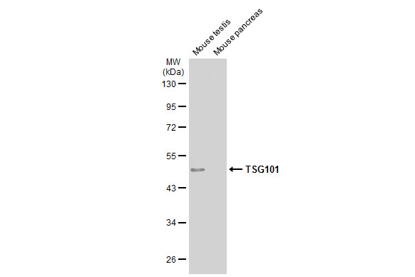 |
| Various tissue extracts (50 µg) were separated by 10% SDS-PAGE, and the membrane was blotted with TSG101 antibody [4A10] (4454) diluted at 1:500. The HRP-conjugated anti-mouse IgG antibody was used to detect the primary antibody. |
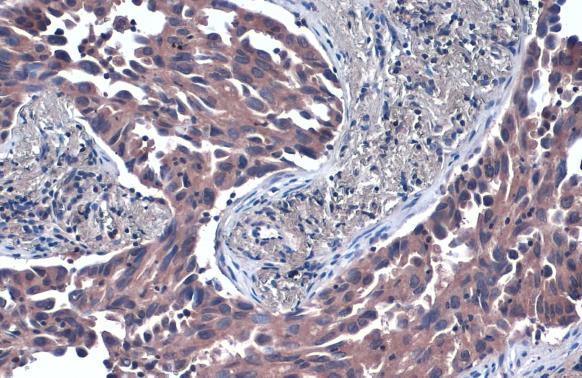 |
| TSG101 antibody [4A10] detects TSG101 protein at cytoplasm by immunohistochemical analysis.Sample: Paraffin-embedded human lung cancer.TSG101 stained by TSG101 antibody [4A10] (4454) diluted at 1:50.Antigen Retrieval: Citrate buffer, pH 6.0, 15 min |
FAQ & Publications
Frequently Asked Questions
What applications is the mouse anti-TSG101 monoclonal antibody (4A10) suitable for?
This antibody is validated for use in Western blot (WB), immunocytochemistry/immunofluorescence (ICC/IF), immunohistochemistry on paraffin-embedded sections (IHC-P), flow cytometry (FACS), immunoprecipitation (IP), ELISA, electron microscopy (EM), and immunohistochemistry (IHC).
How should the mouse anti-TSG101 monoclonal antibody (4A10) be stored to maintain stability?
The antibody should be stored at -20°C in appropriate aliquots to avoid multiple freeze-thaw cycles. Under these conditions, it remains stable for at least one year.
What is the immunogen used to generate the mouse anti-TSG101 monoclonal antibody (4A10)?
The immunogen is a recombinant protein corresponding to amino acids 167-374 of the TSG101 protein.
What is the isotype and clonality of the mouse anti-TSG101 monoclonal antibody (4A10)?
The antibody is a mouse monoclonal of the IgG1 isotype.
What is the recommended dilution for using this antibody in Western blotting and ELISA?
For Western blotting, use a dilution of 1:500 to 1:3,000. For ELISA, use the antibody at a concentration of 1 to 5 µg/mL.
Publications
| pmid | title | authors | citation |
|---|---|---|---|
| 38339765 | Bioengineered small extracellular vesicles deliver multiple SARS-CoV-2 antigenic fragments and drive a broad immunological response. | Hannah K Jackson, Heather M Long, Juan Carlos Yam-Puc, Roberta Palmulli, Tracey A Haigh, Pehuén Pereyra Gerber, Jin S Lee, Nicholas J Matheson, Lesley Young, John Trowsdale, Mathew Lo, Graham S Taylor, James E Thaventhiran, James R Edgar | J Extracell Vesicles 13:e12412 |
| 38339765 | Bioengineered small extracellular vesicles deliver multiple SARS-CoV-2 antigenic fragments and drive a broad immunological response. | Hannah K Jackson, Heather M Long, Juan Carlos Yam-Puc, Roberta Palmulli, Tracey A Haigh, Pehuén Pereyra Gerber, Jin S Lee, Nicholas J Matheson, Lesley Young, John Trowsdale, Mathew Lo, Graham S Taylor, James E Thaventhiran, James R Edgar | J Extracell Vesicles 13:e12412 |
| 38338867 | CD99 Modulates the Proteomic Landscape of Ewing Sarcoma Cells and Related Extracellular Vesicles. | Alessandra De Feo, Marcello Manfredi, Caterina Mancarella, Joaquín J Maqueda, Veronica De Giorgis, Ymera Pignochino, Marika Sciandra, Camilla Cristalli, Massimo Donadelli, Katia Scotlandi | Int J Mol Sci 25:N/A |
| 38338867 | CD99 Modulates the Proteomic Landscape of Ewing Sarcoma Cells and Related Extracellular Vesicles. | Alessandra De Feo, Marcello Manfredi, Caterina Mancarella, Joaquín J Maqueda, Veronica De Giorgis, Ymera Pignochino, Marika Sciandra, Camilla Cristalli, Massimo Donadelli, Katia Scotlandi | Int J Mol Sci 25:N/A |
| 38225453 | Deep proteomic analysis of obstetric antiphospholipid syndrome by DIA-MS of extracellular vesicle enriched fractions. | Wenmin Tian, Dongxue Shi, Yinmei Zhang, Hongli Wang, Haohao Tang, Zhongyu Han, Catherine C L Wong, Liyan Cui, Jiajia Zheng, Yang Chen | Commun Biol 7:99 |
Published literature highly relevant to the biological target of this product and referencing this antibody or clone are retrieved from the PubMed database provided by the United States National Library of Medicine at the National Institutes of Health.
Protocols
| relevant to this product |
|---|
| Western blot IHC ICC |
Documents
| Batch Number | QC File | SDS |
|---|---|---|
| To view batch-specific Safety Datasheets and Quality Certificates associated with your account, please Log In. | ||
Only logged in customers who have purchased this product may leave a review.


Reviews
There are no reviews yet.