| Weight | 1 lbs |
|---|---|
| Dimensions | 9 × 5 × 2 in |
| host | mouse |
| isotype | IgG2a |
| clonality | monoclonal |
| concentration | 1 mg/mL |
| applications | ICC/IF, WB |
| reactivity | Myoferlin |
| available sizes | 100 µg |

mouse anti-Myoferlin monoclonal antibody (7D2) 2343
$520.00
Antibody summary
- Mouse monoclonal to Myoferlin
- Suitable for: WB,ICC
- Isotype: IgG2a
- 100 µg
mouse anti-Myoferlin monoclonal antibody (7D2) 2343
| antibody |
|---|
| Tested applications WB,ICC/IF |
| Recommended dilutions Immunoblotting: use at 1-10ug/mL. A band of ~230kDa is detected. Immunocytochemistry: use at 1- 20ug/mL. These are recommended concentrations. Enduser should determine optimal concentrations for their applications. |
| Immunogen Synthetic peptide derived from the N-terminal domain of the human FER1L3 protein. Accession no. Q9NZM1. |
| Size and concentration 100µg and |
| Form lyophilized |
| Storage Instructions This product is stable for at least one (1) year at -20°C to -70°C. Reconstituted product should be stored in appropriate aliquots to avoid repeated freeze-thaw cycles. |
| Storage buffer Lyophilized, 0.1M Tris, 0.1M glycine, 2% sucrose |
| Purity protein affinity purification |
| Clonality monoclonal |
| Isotype IgG2a |
| Compatible secondaries goat anti-mouse IgG, H&L chain specific, peroxidase conjugated polyclonal antibody 5486 goat anti-mouse IgG, H&L chain specific, biotin conjugated, Conjugate polyclonal antibody 2685 goat anti-mouse IgG, H&L chain specific, FITC conjugated polyclonal antibody 7854 goat anti-mouse IgG, H&L chain specific, peroxidase conjugated polyclonal antibody, crossabsorbed 1706 goat anti-mouse IgG, H&L chain specific, biotin conjugated polyclonal antibody, crossabsorbed 1716 goat anti-mouse IgG, H&L chain specific, FITC conjugated polyclonal antibody, crossabsorbed 1721 |
| Isotype control Mouse monocolonal IgG2a - Isotype Control |
| target relevance |
|---|
| Protein names Myoferlin (Fer-1-like protein 3) |
| Gene names MYOF,MYOF FER1L3 KIAA1207 |
| Protein family Ferlin family |
| Mass 234709Da |
| Function FUNCTION: Calcium/phospholipid-binding protein that plays a role in the plasmalemma repair mechanism of endothelial cells that permits rapid resealing of membranes disrupted by mechanical stress. Involved in endocytic recycling. Implicated in VEGF signal transduction by regulating the levels of the receptor KDR (By similarity). {ECO:0000250}. |
| Subellular location SUBCELLULAR LOCATION: Cell membrane; Single-pass type II membrane protein. Nucleus membrane; Single-pass type II membrane protein. Cytoplasmic vesicle membrane; Single-pass type II membrane protein. Note=Concentrated at the membrane sites of both myoblast-myoblast and myoblast-myotube fusions. Detected at the plasmalemma in endothelial cells lining intact blood vessels (By similarity). Found at nuclear and plasma membranes. Enriched in undifferentiated myoblasts near the plasma membrane in puncate structures. {ECO:0000250}. |
| Tissues TISSUE SPECIFICITY: Expressed in myoblast and endothelial cells (at protein level). Highly expressed in cardiac and skeletal muscles. Also present in lung, and at very low levels in kidney, placenta and brain. {ECO:0000269|PubMed:11959863, ECO:0000269|PubMed:17702744, ECO:0000269|PubMed:18502764}. |
| Structure SUBUNIT: Interacts with DNM2 and KDR. Interacts with EHD1 (By similarity). Interacts with EHD2; the interaction is direct (PubMed:18502764). Interacts with RIPOR2 (PubMed:24687993). {ECO:0000250|UniProtKB:Q69ZN7, ECO:0000269|PubMed:18502764, ECO:0000269|PubMed:24687993}. |
| Domain DOMAIN: The C2 domain 1 associates with lipid membranes in a calcium-dependent manner. |
| Involvement in disease DISEASE: Angioedema, hereditary, 7 (HAE7) [MIM:619366]: A form of angioedema, a disorder characterized by episodic local swelling involving subcutaneous or submucous tissue of the upper respiratory and gastrointestinal tracts, face, extremities, and genitalia. HAE7 is an autosomal dominant form characterized by onset of recurrent swelling of the face, lips, and oral mucosa in the second decade. {ECO:0000269|PubMed:32542751}. Note=The disease may be caused by variants affecting the gene represented in this entry. |
| Target Relevance information above includes information from UniProt accession: Q9NZM1 |
| The UniProt Consortium |
Data
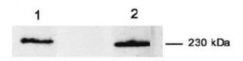 |
| Immunoblotting: use at 1-10ug/ml. A band of ~230kDa is detected. Detection of myoferlin in (1) C2C12 cell lysate and (2) human fibroblast cell lysate with #2343. |
FAQ & Publications
Frequently Asked Questions
What are the recommended applications and dilution ranges for the mouse anti-Myoferlin monoclonal antibody (7D2)?
This antibody is validated for Western Blot (WB) and Immunocytochemistry/Immunofluorescence (ICC/IF). The recommended dilution for immunoblotting is 1-10 µg/mL, where a band of approximately 230 kDa is detected. For immunocytochemistry, use at 1-20 µg/mL. Users should optimize concentrations for their specific experimental conditions.
How should the mouse anti-Myoferlin monoclonal antibody (7D2) be stored to maintain stability?
The lyophilized antibody is stable for at least one year when stored at temperatures between -20°C and -70°C. After reconstitution, it is advisable to aliquot the product and avoid repeated freeze-thaw cycles to preserve antibody integrity.
What is the immunogen used to generate the mouse anti-Myoferlin monoclonal antibody (7D2), and what is the antibody's isotype?
The antibody was raised against a synthetic peptide derived from the N-terminal domain of the human FER1L3 protein (UniProt Accession Q9NZM1). The monoclonal antibody is of the IgG2a isotype, produced in mouse host cells.
Publications
| pmid | title | authors | citation |
|---|---|---|---|
| We haven't added any publications to our database yet. | |||
Published literature highly relevant to the biological target of this product and referencing this antibody or clone are retrieved from the PubMed database provided by the United States National Library of Medicine at the National Institutes of Health.
Protocols
| relevant to this product |
|---|
| Western blot IHC ICC |
Documents
| Batch Number | QC File | SDS |
|---|---|---|
| To view batch-specific Safety Datasheets and Quality Certificates associated with your account, please Log In. | ||
Only logged in customers who have purchased this product may leave a review.


Reviews
There are no reviews yet.