| Weight | 1 lbs |
|---|---|
| Dimensions | 9 × 5 × 2 in |
| host | mouse |
| isotype | IgG2a |
| clonality | monoclonal |
| concentration | 1 mg/mL |
| applications | ICC/IF, WB |
| reactivity | Mitosin |
| available sizes | 100 µg |


mouse anti-Mitosin monoclonal antibody (14C10) 2862
$503.00
Antibody summary
- Mouse monoclonal to Mitosin
- Suitable for: WB,ICC/IF,IHC-P,IHC-Fr,FACS,IP
- Isotype: IgG2a
- 100 µg
mouse anti-Mitosin monoclonal antibody (14C10) 2862
| antibody |
|---|
| Tested applications WB,IHC,IHC,ICC/IF |
| Recommended dilutions Immunoblotting: use at 1-10 ug/mL. Immunoprecipitation: use at 1-10 ug/mL. Immunohistochemistry: use at 10 ug/mL with frozen or paraffin-embedded sections. Positive controls: Human tissue containing rapidly proliferating cells, such as lymphoid germinal centers. |
| Immunogen GST fusion protein expressed in E. coli corresponding to aa 1759-2093 of human mitosin. |
| Size and concentration 100µg and lot specific |
| Form liquid |
| Storage Instructions This antibody is stable for at least one (1) year at -70°C. Avoid multiple freeze-thaw cycles. |
| Storage buffer PBS, pH 7.4 |
| Purity protein affinity purification |
| Clonality monoclonal |
| Isotype IgG2a |
| Compatible secondaries goat anti-mouse IgG, H&L chain specific, peroxidase conjugated polyclonal antibody 5486 goat anti-mouse IgG, H&L chain specific, biotin conjugated, Conjugate polyclonal antibody 2685 goat anti-mouse IgG, H&L chain specific, FITC conjugated polyclonal antibody 7854 goat anti-mouse IgG, H&L chain specific, peroxidase conjugated polyclonal antibody, crossabsorbed 1706 goat anti-mouse IgG, H&L chain specific, biotin conjugated polyclonal antibody, crossabsorbed 1716 goat anti-mouse IgG, H&L chain specific, FITC conjugated polyclonal antibody, crossabsorbed 1721 |
| Isotype control Mouse monocolonal IgG2a - Isotype Control |
| target relevance |
|---|
| Homo sapiens CENPF Centromere protein F |
| Protein names Centromere protein F |
| Alternative names AH antigen, Kinetochore protein CENPF, Mitosin |
| Gene names CENPF |
| Protein family Belongs to the centromere protein F family |
| Function Required for kinetochore function and chromosome segregation in mitosis. Required for kinetochore localization of dynein, LIS1, NDE1 and NDEL1. Regulates recycling of the plasma membrane by acting as a link between recycling vesicles and the microtubule network though its association with STX4 and SNAP25. Acts as a potential inhibitor of pocket protein-mediated cellular processes during development by regulating the activity of RB proteins during cell division and proliferation. May play a regulatory or permissive role in the normal embryonic cardiomyocyte cell cycle and in promoting continued mitosis in transformed, abnormally dividing neonatal cardiomyocytes. Interaction with RB directs embryonic stem cells toward a cardiac lineage. Involved in the regulation of DNA synthesis and hence cell cycle progression, via its C-terminus. Has a potential role regulating skeletal myogenesis and in cell differentiation in embryogenesis. Involved in dendritic cell regulation of T-cell immunity against chlamydia |
| Subcellular location Cytoplasm, perinuclear region, Nucleus matrix, Chromosome, centromere, kinetochore, Cytoplasm, cytoskeleton, spindle |
| Structure Interacts with and STX4 (via C-terminus) (By similarity). Interacts (via N-terminus) with RBL1, RBL2 and SNAP25 (By similarity). Self-associates. Interacts with CENP-E and BUBR1 (via C-terminus). Interacts (via C-terminus) with NDE1, NDEL1 and RB1 |
| Post-translational modification Hyperphosphorylated during mitosis |
| Involvement in disease Stromme syndrome An autosomal recessive congenital disorder characterized by intestinal atresia, ocular anomalies, microcephaly, and renal and cardiac abnormalities in some patients. The disease has features of a ciliopathy, and lethality in early childhood is observed in severe cases. |
| Keywords Acetylation, Cell cycle, Cell division, Centromere, Chromosome, Ciliopathy, Coiled coil, Cytoplasm, Cytoskeleton, Developmental protein, Differentiation, DNA synthesis, Kinetochore, Lipoprotein, Methylation, Mitosis, Myogenesis, Nucleus, Phosphoprotein, Prenylation, Primary ciliary dyskinesia, Proteomics identification, Reference proteome, Repeat |
| Sequence MSWALEEWKEGLPTRALQKIQELEGQLDKLKKEKQQRQFQLDSLEAALQKQKQKVENEKT EGTNLKRENQRLMEICESLEKTKQKISHELQVKESQVNFQEGQLNSGKKQIEKLEQELKR CKSELERSQQAAQSADVSLNPCNTPQKIFTTPLTPSQYYSGSKYEDLKEKYNKEVEERKR LEAEVKALQAKKASQTLPQATMNHRDIARHQASSSVFSWQQEKTPSHLSSNSQRTPIRRD FSASYFSGEQEVTPSRSTLQIGKRDANSSFFDNSSSPHLLDQLKAQNQELRNKINELELR LQGHEKEMKGQVNKFQELQLQLEKAKVELIEKEKVLNKCRDELVRTTAQYDQASTKYTAL EQKLKKLTEDLSCQRQNAESARCSLEQKIKEKEKEFQEELSRQQRSFQTLDQECIQMKAR LTQELQQAKNMHNVLQAELDKLTSVKQQLENNLEEFKQKLCRAEQAFQASQIKENELRRS MEEMKKENNLLKSHSEQKAREVCHLEAELKNIKQCLNQSQNFAEEMKAKNTSQETMLRDL QEKINQQENSLTLEKLKLAVADLEKQRDCSQDLLKKREHHIEQLNDKLSKTEKESKALLS ALELKKKEYEELKEEKTLFSCWKSENEKLLTQMESEKENLQSKINHLETCLKTQQIKSHE YNERVRTLEMDRENLSVEIRNLHNVLDSKSVEVETQKLAYMELQQKAEFSDQKHQKEIEN MCLKTSQLTGQVEDLEHKLQLLSNEIMDKDRCYQDLHAEYESLRDLLKSKDASLVTNEDH QRSLLAFDQQPAMHHSFANIIGEQGSMPSERSECRLEADQSPKNSAILQNRVDSLEFSLE SQKQMNSDLQKQCEELVQIKGEIEENLMKAEQMHQSFVAETSQRISKLQEDTSAHQNVVA ETLSALENKEKELQLLNDKVETEQAEIQELKKSNHLLEDSLKELQLLSETLSLEKKEMSS IISLNKREIEELTQENGTLKEINASLNQEKMNLIQKSESFANYIDEREKSISELSDQYKQ EKLILLQRCEETGNAYEDLSQKYKAAQEKNSKLECLLNECTSLCENRKNELEQLKEAFAK EHQEFLTKLAFAEERNQNLMLELETVQQALRSEMTDNQNNSKSEAGGLKQEIMTLKEEQN KMQKEVNDLLQENEQLMKVMKTKHECQNLESEPIRNSVKERESERNQCNFKPQMDLEVKE ISLDSYNAQLVQLEAMLRNKELKLQESEKEKECLQHELQTIRGDLETSNLQDMQSQEISG LKDCEIDAEEKYISGPHELSTSQNDNAHLQCSLQTTMNKLNELEKICEILQAEKYELVTE LNDSRSECITATRKMAEEVGKLLNEVKILNDDSGLLHGELVEDIPGGEFGEQPNEQHPVS LAPLDESNSYEHLTLSDKEVQMHFAELQEKFLSLQSEHKILHDQHCQMSSKMSELQTYVD SLKAENLVLSTNLRNFQGDLVKEMQLGLEEGLVPSLSSSCVPDSSSLSSLGDSSFYRALL EQTGDMSLLSNLEGAVSANQCSVDEVFCSSLQEENLTRKETPSAPAKGVEELESLCEVYR QSLEKLEEKMESQGIMKNKEIQELEQLLSSERQELDCLRKQYLSENEQWQQKLTSVTLEM ESKLAAEKKQTEQLSLELEVARLQLQGLDLSSRSLLGIDTEDAIQGRNESCDISKEHTSE TTERTPKHDVHQICDKDAQQDLNLDIEKITETGAVKPTGECSGEQSPDTNYEPPGEDKTQ GSSECISELSFSGPNALVPMDFLGNQEDIHNLQLRVKETSNENLRLLHVIEDRDRKVESL LNEMKELDSKLHLQEVQLMTKIEACIELEKIVGELKKENSDLSEKLEYFSCDHQELLQRV ETSEGLNSDLEMHADKSSREDIGDNVAKVNDSWKERFLDVENELSRIRSEKASIEHEALY LEADLEVVQTEKLCLEKDNENKQKVIVCLEEELSVVTSERNQLRGELDTMSKKTTALDQL SEKMKEKTQELESHQSECLHCIQVAEAEVKEKTELLQTLSSDVSELLKDKTHLQEKLQSL EKDSQALSLTKCELENQIAQLNKEKELLVKESESLQARLSESDYEKLNVSKALEAALVEK GEFALRLSSTQEEVHQLRRGIEKLRVRIEADEKKQLHIAEKLKERERENDSLKDKVENLE RELQMSEENQELVILDAENSKAEVETLKTQIEEMARSLKVFELDLVTLRSEKENLTKQIQ EKQGQLSELDKLLSSFKSLLEEKEQAEIQIKEESKTAVEMLQNQLKELNEAVAALCGDQE IMKATEQSLDPPIEEEHQLRNSIEKLRARLEADEKKQLCVLQQLKESEHHADLLKGRVEN LERELEIARTNQEHAALEAENSKGEVETLKAKIEGMTQSLRGLELDVVTIRSEKENLTNE LQKEQERISELEIINSSFENILQEKEQEKVQMKEKSSTAMEMLQTQLKELNERVAALHND QEACKAKEQNLSSQVECLELEKAQLLQGLDEAKNNYIVLQSSVNGLIQEVEDGKQKLEKK DEEISRLKNQIQDQEQLVSKLSQVEGEHQLWKEQNLELRNLTVELEQKIQVLQSKNASLQ DTLEVLQSSYKNLENELELTKMDKMSFVEKVNKMTAKETELQREMHEMAQKTAELQEELS GEKNRLAGELQLLLEEIKSSKDQLKELTLENSELKKSLDCMHKDQVEKEGKVREEIAEYQ LRLHEAEKKHQALLLDTNKQYEVEIQTYREKLTSKEECLSSQKLEIDLLKSSKEELNNSL KATTQILEELKKTKMDNLKYVNQLKKENERAQGKMKLLIKSCKQLEEEKEILQKELSQLQ AAQEKQKTGTVMDTKVDELTTEIKELKETLEEKTKEADEYLDKYCSLLISHEKLEKAKEM LETQVAHLCSQQSKQDSRGSPLLGPVVPGPSPIPSVTEKRLSSGQNKASGKRQRSSGIWE NGRGPTPATPESFSKKSKKAVMSGIHPAEDTEGTEFEPEGLPEVVKKGFADIPTGKTSPY ILRRTTMATRTSPRLAAQKLALSPLSLGKENLAESSKPTAGGSRSQKVKVAQRSPVDSGT ILREPTTKSVPVNNLPERSPTDSPREGLRVKRGRLVPSPKAGLESNGSENCKVQ |
| UniProt accession: P49454 |
Data
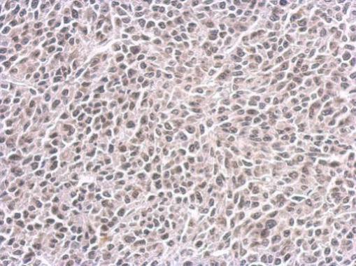 |
| CENPF antibody [14C10 1D8] detects CENPF protein at nucleus on BT483 xenograft by immunohistochemical analysis. Sample: Paraffin-embedded BT483 xenograft. CENPF antibody [14C10 1D8] (2862) dilution: 1:200. Antigen Retrieval: Trilogy™ (EDTA based, pH 8.0) buffer, 15min |
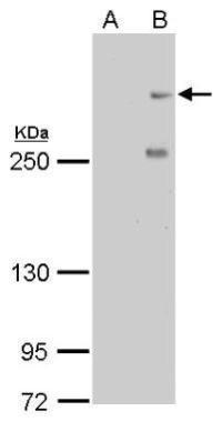 |
| CENPF antibody [14C10 1D8] detects CENPF protein by western blot analysis. A. 30 µg HeLa whole cell lysate/extract (untreated) B. 30 µg HeLa whole cell lysate/extract (100ng/ml Nocodazole treatment for 24hr) 5% SDS-PAGE CENPF antibody [14C10 1D8] (2862) dilution: 1:500 The HRP-conjugated anti-mouse IgG antibody was used to detect the primary antibody. |
 |
| Confocal immunofluorescence staining (Olympus FV10i) of Mitosin. U2OS cells were fixed by 4% PFA and costained with Mitosin 14C10 1D8 AB (Red; cat# 2862; 1:500 Ab dilution) and alpha-tubulin rabbit polyclonal AB, a spindle marker. DAPI (blue), chromosomes. Scale bar, 5 um |
FAQ & Publications
Frequently Asked Questions
What applications has the mouse anti-Mitosin monoclonal antibody (14C10) been validated for?
This antibody has been tested and is suitable for use in Western blotting (WB), immunocytochemistry/immunofluorescence (ICC/IF), immunohistochemistry on both paraffin-embedded and frozen sections (IHC-P, IHC-Fr), flow cytometry (FACS), and immunoprecipitation (IP). Recommended dilutions vary by application, for example, use 1-10 µg/mL for immunoblotting and immunoprecipitation, and 10 µg/mL for immunohistochemistry.
How should the mouse anti-Mitosin monoclonal antibody (14C10) be stored to maintain stability?
The antibody should be stored at -70°C to ensure stability for at least one year. It is important to avoid multiple freeze-thaw cycles to preserve antibody integrity. The antibody is supplied in PBS buffer, pH 7.4, in liquid form at a concentration of 1 mg/mL.
Publications
| pmid | title | authors | citation |
|---|---|---|---|
| We haven't added any publications to our database yet. | |||
Published literature highly relevant to the biological target of this product and referencing this antibody or clone are retrieved from the PubMed database provided by the United States National Library of Medicine at the National Institutes of Health.
Protocols
| relevant to this product |
|---|
| Western blot IHC ICC |
Documents
| Batch Number | QC File | SDS |
|---|---|---|
| To view batch-specific Safety Datasheets and Quality Certificates associated with your account, please Log In. | ||
Only logged in customers who have purchased this product may leave a review.


Reviews
There are no reviews yet.