| Weight | 1 lbs |
|---|---|
| Dimensions | 9 × 5 × 2 in |
| host | mouse |
| isotype | IgG2a |
| clonality | monoclonal |
| concentration | 1 mg/mL |
| applications | ICC/IF, WB |
| reactivity | GPX7 |
| available sizes | 100 µg |


mouse anti-GPX7 monoclonal antibody (2704) 3065
$503.00
Antibody summary
- Mouse monoclonal to GPX7
- Suitable for: WB,IHC-P,IHC
- Isotype: IgG2a
- 100 µg
mouse anti-GPX7 monoclonal antibody (2704) 3065
| antibody |
|---|
| Tested applications WB,IHC,IHC |
| Recommended dilutions Immunoblotting: use at a 1:500-1:2,000 dilution. A band of ~18kDa is detected. Detection of GPX7 in HeLa cell extract. |
| Immunogen Full length human GPX7 |
| Size and concentration 100µg and lot specific |
| Form liquid |
| Storage Instructions This product is stable for at least one (1) year if stored at -20°C. Store product in appropriate aliquots to avoid multiple freeze-thaw cycles. |
| Storage buffer PBS, pH 7.2. |
| Purity protein affinity purification |
| Clonality monoclonal |
| Isotype IgG2a |
| Compatible secondaries goat anti-mouse IgG, H&L chain specific, peroxidase conjugated polyclonal antibody 5486 goat anti-mouse IgG, H&L chain specific, biotin conjugated, Conjugate polyclonal antibody 2685 goat anti-mouse IgG, H&L chain specific, FITC conjugated polyclonal antibody 7854 goat anti-mouse IgG, H&L chain specific, peroxidase conjugated polyclonal antibody, crossabsorbed 1706 goat anti-mouse IgG, H&L chain specific, biotin conjugated polyclonal antibody, crossabsorbed 1716 goat anti-mouse IgG, H&L chain specific, FITC conjugated polyclonal antibody, crossabsorbed 1721 |
| Isotype control Mouse monocolonal IgG2a - Isotype Control |
| target relevance |
|---|
| Protein names Glutathione peroxidase 7 (GPx-7) (GSHPx-7) (EC 1.11.1.9) (CL683) |
| Gene names GPX7,GPX7 GPX6 UNQ469/PRO828 |
| Protein family Glutathione peroxidase family |
| Mass 20996Da |
| Function FUNCTION: It protects esophageal epithelia from hydrogen peroxide-induced oxidative stress. It suppresses acidic bile acid-induced reactive oxygen species (ROS) and protects against oxidative DNA damage and double-strand breaks. {ECO:0000269|PubMed:22157330}. |
| Catalytic activity CATALYTIC ACTIVITY: Reaction=2 glutathione + H2O2 = glutathione disulfide + 2 H2O; Xref=Rhea:RHEA:16833, ChEBI:CHEBI:15377, ChEBI:CHEBI:16240, ChEBI:CHEBI:57925, ChEBI:CHEBI:58297; EC=1.11.1.9; |
| Subellular location SUBCELLULAR LOCATION: Secreted {ECO:0000305}. |
| Tissues TISSUE SPECIFICITY: Expressed in esophageal epithelial cells; expression is up-regulated after exposure to acidic bile acids. {ECO:0000269|PubMed:22157330}. |
| Involvement in disease DISEASE: Barrett esophagus (BE) [MIM:614266]: A condition characterized by a metaplastic change in which normal esophageal squamous epithelium is replaced by a columnar and intestinal-type epithelium. Patients with Barrett esophagus have an increased risk of esophageal adenocarcinoma. The main cause of Barrett esophagus is gastroesophageal reflux. The retrograde movement of acid and bile salts from the stomach into the esophagus causes prolonged injury to the esophageal epithelium and induces chronic esophagitis, which in turn is believed to trigger the pathologic changes. {ECO:0000269|PubMed:22157330}. Note=The disease is caused by variants affecting the gene represented in this entry. The pathologic mechanisms leading to Barrett esophagus involve GPX7 dysfunction that results in higher levels of hydrogen peroxide and ROS-induced oxidative stress and DNA damage in esophageal cells. |
| Target Relevance information above includes information from UniProt accession: Q96SL4 |
| The UniProt Consortium |
Data
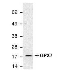 |
| Detection of GPX7 using GPX7 antibody [2704] (3065) in HeLa whole cell extract. |
 |
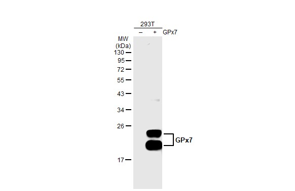 |
| Non-transfected (-) and transfected (-) 293T whole cell extracts (30 µg) were separated by 12% SDS-PAGE, and the membrane was blotted with GPX7 antibody [2704] (3065) diluted at 1:500 The HRP-conjugated anti-mouse IgG antibody was used to detect the primary antibody. |
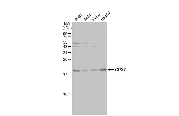 |
| Various whole cell extracts (30 µg) were separated by 15% SDS-PAGE, and the membrane was blotted with GPX7 antibody [2704] (3065) diluted at 1:1000. The HRP-conjugated anti-mouse IgG antibody was used to detect the primary antibody, and the signal was developed with Trident ECL plus-Enhanced. |
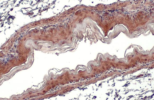 |
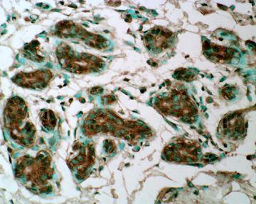
| GPX7 antibody [2704] detects GPX7 protein at cytoplasm by immunohistochemical analysis.Sample: Paraffin-embedded mouse stomach.GPX7 stained by GPX7 antibody [2704] (3065) diluted at 1:100.Antigen Retrieval: Citrate buffer, pH 6.0, 15 min |
FAQ & Publications
Frequently Asked Questions
What applications is the mouse anti-GPX7 monoclonal antibody (2704) validated for?
This antibody is validated for use in Western Blot (WB), Immunohistochemistry on paraffin-embedded sections (IHC-P), and Immunocytochemistry/Immunofluorescence (ICC/IF) applications.
How should the mouse anti-GPX7 monoclonal antibody be stored to maintain stability?
The antibody should be stored at -20°C in appropriate aliquots to avoid multiple freeze-thaw cycles. Under these conditions, it is stable for at least one year.
What is the recommended dilution range for immunoblotting with this GPX7 antibody?
For Western blotting, the antibody is recommended to be used at a dilution of 1:500 to 1:2,000, with a typical detection of a band around 18 kDa in HeLa cell extracts.
What is the host species and isotype of the anti-GPX7 monoclonal antibody?
The antibody is a mouse monoclonal with an IgG2a isotype.
Publications
| pmid | title | authors | citation |
|---|---|---|---|
| We haven't added any publications to our database yet. | |||
Published literature highly relevant to the biological target of this product and referencing this antibody or clone are retrieved from the PubMed database provided by the United States National Library of Medicine at the National Institutes of Health.
Protocols
| relevant to this product |
|---|
| Western blot IHC |
Documents
| Batch Number | QC File | SDS |
|---|---|---|
| To view batch-specific Safety Datasheets and Quality Certificates associated with your account, please Log In. | ||
Only logged in customers who have purchased this product may leave a review.


Reviews
There are no reviews yet.