| Weight | 1 lbs |
|---|---|
| Dimensions | 9 × 5 × 2 in |
| host | mouse |
| isotype | IgG1 |
| clonality | monoclonal |
| concentration | concentrate, predilute |
| applications | IHC |
| reactivity | human |
| available size | 0.1 mL, 0.5 mL, 1 mL concentrated, 7 mL prediluted |

mouse anti-GFAP monoclonal antibody GA-5 6194
Price range: $160.00 through $528.00
Antibody summary
- Mouse monoclonal to GFAP
- Suitable for: Immunohistochemistry (formalin-fixed, paraffin-embedded tissues)
- Reacts with: Human
- Isotype:IgG1
- Control: Brain or astrocytoma
- Visualization: Cytoplasmic
- 0.1, 0.5, 1.0 mL concentrated, 7 mL prediluted
mouse anti-GFAP monoclonal antibody GA-5 6194
| target relevance |
|---|
| Homo sapiens GFAP Glial fibrillary acidic protein |
| Protein names Glial fibrillary acidic protein |
| Gene names GFAP |
| Protein family Belongs to the intermediate filament family |
| Function GFAP, a class-III intermediate filament, is a cell-specific marker that, during the development of the central nervous system, distinguishes astrocytes from other glial cells |
| Subcellular location Cytoplasm |
| Structure Interacts with PSEN1 (via N-terminus) |
| Post-translational modification Phosphorylated by PKN1 |
| Involvement in disease Alexander disease A rare disorder of the central nervous system. The most common form affects infants and young children, and is characterized by progressive failure of central myelination, usually leading to death within the first decade. Infants with Alexander disease develop a leukodystrophy with macrocephaly, seizures, and psychomotor retardation. Patients with juvenile or adult forms typically experience ataxia, bulbar signs and spasticity, and a more slowly progressive course. Histologically, Alexander disease is characterized by Rosenthal fibers, homogeneous eosinophilic inclusions in astrocytes. |
| Keywords 3D-structure, Alternative splicing, Citrullination, Coiled coil, Cytoplasm, Direct protein sequencing, Disease variant, Intermediate filament, Leukodystrophy, Methylation, Phosphoprotein, Proteomics identification, Reference proteome |
| Sequence MERRRITSAARRSYVSSGEMMVGGLAPGRRLGPGTRLSLARMPPPLPTRVDFSLAGALNA GFKETRASERAEMMELNDRFASYIEKVRFLEQQNKALAAELNQLRAKEPTKLADVYQAEL RELRLRLDQLTANSARLEVERDNLAQDLATVRQKLQDETNLRLEAENNLAAYRQEADEAT LARLDLERKIESLEEEIRFLRKIHEEEVRELQEQLARQQVHVELDVAKPDLTAALKEIRT QYEAMASSNMHEAEEWYRSKFADLTDAAARNAELLRQAKHEANDYRRQLQSLTCDLESLR GTNESLERQMREQEERHVREAASYQEALARLEEEGQSLKDEMARHLQEYQDLLNVKLALD IEIATYRKLLEGEENRITIPVQTFSNLQIRETSLDTKSVSEGHLKRNIVVKTVEMRDGEV IKESKQEHKDVM |
| UniProt accession: P14136 |
Data
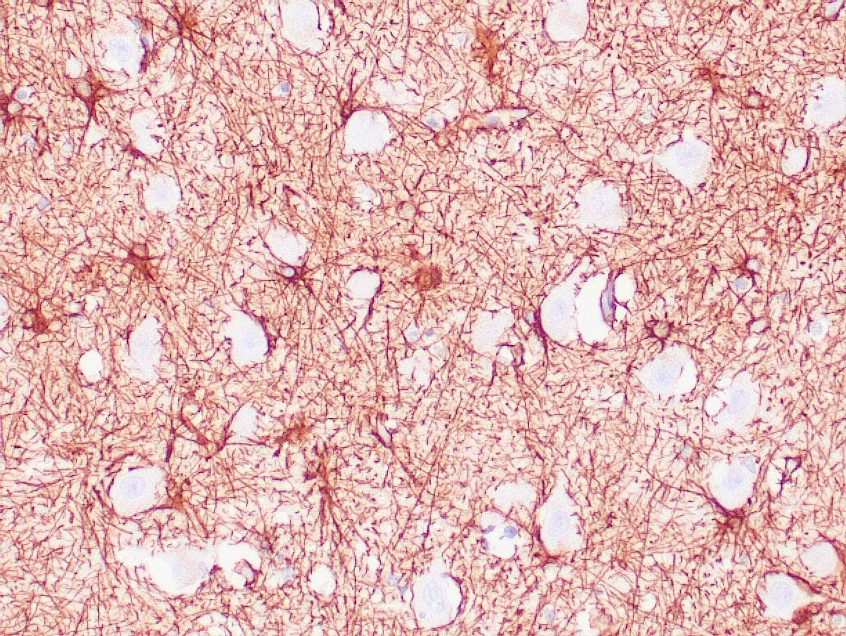 |
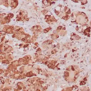
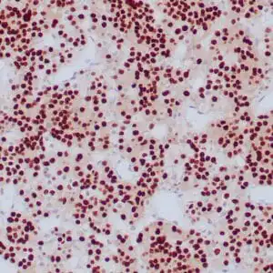
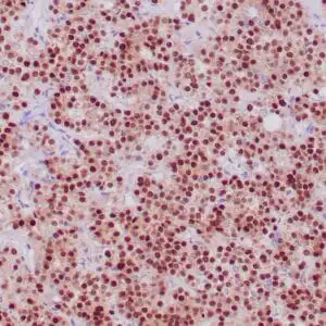
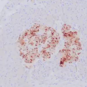
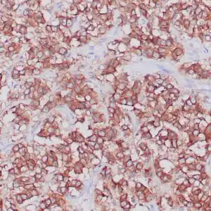
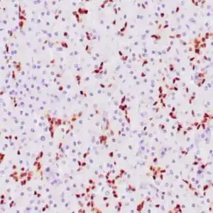
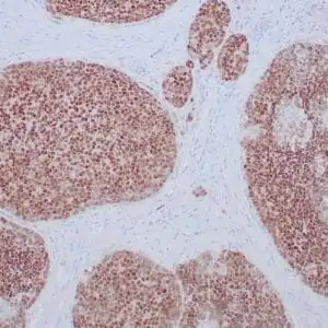
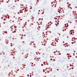
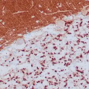
| Human brain stained with anti-GFAP antibody using peroxidase-conjugate and DAB chromogen. Note positive staining of astrocytic processes. |
FAQ & Publications
Frequently Asked Questions
What species does the mouse anti-GFAP monoclonal antibody GA-5 6194 react with?
This antibody reacts specifically with human GFAP (glial fibrillary acidic protein).
What applications is this anti-GFAP antibody suitable for?
It is suitable for immunohistochemistry (IHC), particularly on formalin-fixed, paraffin-embedded tissues.
How should the mouse anti-GFAP monoclonal antibody be stored to maintain its stability?
For short-term storage, keep the antibody at 2-8°C. For long-term storage, it should be stored at -20°C, avoiding freeze-thaw cycles to preserve antibody integrity.
What is the recommended dilution range for using the concentrated form of this GFAP antibody in IHC?
The concentrated antibody is recommended to be diluted between 1:100 and 1:200 for immunohistochemistry applications.
What type of control tissue is recommended when using this antibody for IHC?
Brain tissue or astrocytoma samples are recommended as positive controls for validating staining with this GFAP antibody.
Publications
| pmid | title | authors | citation |
|---|---|---|---|
| We haven't added any publications to our database yet. | |||
Published literature highly relevant to the biological target of this product and referencing this antibody or clone are retrieved from the PubMed database provided by the United States National Library of Medicine at the National Institutes of Health.
Protocols
| relevant to this product |
|---|
| IHC |
Documents
| Batch Number | QC File | SDS |
|---|---|---|
| To view batch-specific Safety Datasheets and Quality Certificates associated with your account, please Log In. | ||
Only logged in customers who have purchased this product may leave a review.


Reviews
There are no reviews yet.