| Weight | 1 lbs |
|---|---|
| Dimensions | 9 × 5 × 2 in |
| host | mouse |
| isotype | IgG1 |
| clonality | monoclonal |
| concentration | 1 mg/mL |
| applications | ICC/IF, WB |
| reactivity | Estrogen Receptorα |
| available sizes | 100 µg |


mouse anti-Estrogen Receptor α monoclonal antibody (1F3) 3616
$503.00
Antibody summary
- Mouse monoclonal to Estrogen Receptor α
- Suitable for: WB,IHC-Fr,FACS,IP
- Isotype: IgG1
- 100 µg
mouse anti-Estrogen Receptor α monoclonal antibody (1F3) 3616
| antibody |
|---|
| Tested applications WB,IHC,IHC,IP |
| Recommended dilutions Immunoblotting: use at 1-5ug/mL. Immunohistochemistry: frozen sections, use at 1-10ug/mL.FACS: use at 1-10ug/mL. Immunoprecipitation: use at 1-5ug/mL. Positive controls: MCF-7 cells and recombinant protein. These are recommended concentrations. Endusers shoul |
| Immunogen 6-His fusion protein containing the region encoding aa 1-190 of human estrogen receptor-a (ER-a) expressed in E. coli. |
| Size and concentration 100µg and 1 mg/mL |
| Form liquid |
| Storage Instructions This antibody is stable for at least one (1) year at -20°C. Avoid multiple freeze-thaw cycles. |
| Storage buffer PBS, pH 7.4, 1mg/ml. |
| Purity protein affinity purification |
| Clonality monoclonal |
| Isotype IgG1 |
| Compatible secondaries goat anti-mouse IgG, H&L chain specific, peroxidase conjugated polyclonal antibody 5486 goat anti-mouse IgG, H&L chain specific, biotin conjugated, Conjugate polyclonal antibody 2685 goat anti-mouse IgG, H&L chain specific, FITC conjugated polyclonal antibody 7854 goat anti-mouse IgG, H&L chain specific, peroxidase conjugated polyclonal antibody, crossabsorbed 1706 goat anti-mouse IgG, H&L chain specific, biotin conjugated polyclonal antibody, crossabsorbed 1716 goat anti-mouse IgG, H&L chain specific, FITC conjugated polyclonal antibody, crossabsorbed 1721 |
| Isotype control Mouse monocolonal IgG1 - Isotype Control |
| target relevance |
|---|
| Homo sapiens ESR1 Estrogen receptor |
| Protein names Estrogen receptor |
| Alternative names ER-alpha, Estradiol receptor, Nuclear receptor subfamily 3 group A member 1 |
| Gene names ESR1 |
| Protein family Belongs to the nuclear hormone receptor family. NR3 subfamily |
| Function Nuclear hormone receptor. The steroid hormones and their receptors are involved in the regulation of eukaryotic gene expression and affect cellular proliferation and differentiation in target tissues. Ligand-dependent nuclear transactivation involves either direct homodimer binding to a palindromic estrogen response element (ERE) sequence or association with other DNA-binding transcription factors, such as AP-1/c-Jun, c-Fos, ATF-2, Sp1 and Sp3, to mediate ERE-independent signaling. Ligand binding induces a conformational change allowing subsequent or combinatorial association with multiprotein coactivator complexes through LXXLL motifs of their respective components. Mutual transrepression occurs between the estrogen receptor (ER) and NF-kappa-B in a cell-type specific manner. Decreases NF-kappa-B DNA-binding activity and inhibits NF-kappa-B-mediated transcription from the IL6 promoter and displace RELA/p65 and associated coregulators from the promoter. Recruited to the NF-kappa-B response element of the CCL2 and IL8 promoters and can displace CREBBP. Present with NF-kappa-B components RELA/p65 and NFKB1/p50 on ERE sequences. Can also act synergistically with NF-kappa-B to activate transcription involving respective recruitment adjacent response elements; the function involves CREBBP. Can activate the transcriptional activity of TFF1. Also mediates membrane-initiated estrogen signaling involving various kinase cascades. Essential for MTA1-mediated transcriptional regulation of BRCA1 and BCAS3 (PubMed:17922032). Maintains neuronal survival in response to ischemic reperfusion injury when in the presence of circulating estradiol (17-beta-estradiol/E2) (By similarity) |
| Subcellular location Nucleus, Golgi apparatus, Cell membrane |
| Structure Probably homodimerizes or heterodimerizes with isoform 1 and ESR2 |
| Post-translational modification Phosphorylated by cyclin A/CDK2 and CK1. Phosphorylation probably enhances transcriptional activity. Self-association induces phosphorylation. Dephosphorylation at Ser-118 by PPP5C inhibits its transactivation activity. Phosphorylated by LMTK3 in vitro Glycosylated; contains N-acetylglucosamine, probably O-linked Ubiquitinated; regulated by LATS1 via DCAF1 it leads to ESR1 proteasomal degradation (PubMed:21602804, PubMed:28068668). Deubiquitinated by OTUB1 (PubMed:19383985). Ubiquitinated by STUB1/CHIP; in the CA1 hippocampal region following loss of endogenous circulating estradiol (17-beta-estradiol/E2) (By similarity). Ubiquitinated by UBR5, leading to its degradation: UBR5 specifically recognizes and binds ligand-bound ESR1 when it is not associated with coactivators (NCOAs) (PubMed:37478846). In presence of NCOAs, the UBR5-degron is not accessible, preventing its ubiquitination and degradation (PubMed:37478846) Dimethylated by PRMT1 at Arg-260. The methylation may favor cytoplasmic localization (PubMed:18657504, PubMed:24498420). Demethylated by JMJD6 at Arg-260 (PubMed:24498420) Palmitoylated (isoform 3). Not biotinylated (isoform 3) Palmitoylated by ZDHHC7 and ZDHHC21. Palmitoylation is required for plasma membrane targeting and for rapid intracellular signaling via ERK and AKT kinases and cAMP generation, but not for signaling mediated by the nuclear hormone receptor |
| Involvement in disease Estrogen resistance A disorder characterized by partial or complete resistance to estrogens, in the presence of elevated estrogen serum levels. Clinical features include absence of the pubertal growth spurt, delayed bone maturation, unfused epiphyses, reduced bone mineral density, osteoporosis, continued growth into adulthood and very tall adult stature. Glucose intolerance, hyperinsulinemia and lipid abnormalities may also be present. |
| Keywords 3D-structure, Activator, Alternative promoter usage, Alternative splicing, Cell membrane, Cytoplasm, Direct protein sequencing, Disease variant, DNA-binding, Glycoprotein, Golgi apparatus, Lipid-binding, Lipoprotein, Membrane, Metal-binding, Methylation, Nucleus, Osteoporosis, Palmitate, Phosphoprotein, Proteomics identification, Receptor, Reference proteome, Steroid-binding, Transcription, Transcription regulation, Transmembrane, Ubl conjugation, Zinc, Zinc-finger |
| Sequence MTMTLHTKASGMALLHQIQGNELEPLNRPQLKIPLERPLGEVYLDSSKPAVYNYPEGAAY EFNAAAAANAQVYGQTGLPYGPGSEAAAFGSNGLGGFPPLNSVSPSPLMLLHPPPQLSPF LQPHGQQVPYYLENEPSGYTVREAGPPAFYRPNSDNRRQGGRERLASTNDKGSMAMESAK ETRYCAVCNDYASGYHYGVWSCEGCKAFFKRSIQGHNDYMCPATNQCTIDKNRRKSCQAC RLRKCYEVGMMKGGIRKDRRGGRMLKHKRQRDDGEGRGEVGSAGDMRAANLWPSPLMIKR SKKNSLALSLTADQMVSALLDAEPPILYSEYDPTRPFSEASMMGLLTNLADRELVHMINW AKRVPGFVDLTLHDQVHLLECAWLEILMIGLVWRSMEHPGKLLFAPNLLLDRNQGKCVEG MVEIFDMLLATSSRFRMMNLQGEEFVCLKSIILLNSGVYTFLSSTLKSLEEKDHIHRVLD KITDTLIHLMAKAGLTLQQQHQRLAQLLLILSHIRHMSNKGMEHLYSMKCKNVVPLYDLL LEMLDAHRLHAPTSRGGASVEETDQSHLATAGSTSSHSLQKYYITGEAEGFPATV |
| UniProt accession: P03372 |
Data
 |
| Whole cell extract (60 µg) was separated by 7.5% SDS-PAGE, and the membrane was blotted with Estrogen Receptor alpha antibody (3616) diluted at 1:500. The HRP-conjugated anti-mouse IgG antibody was used to detect the primary antibody, and the signal was developed with Trident femto Western HRP Substrate. |
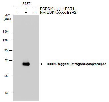 |
| Non-transfected (-) and transfected (-) 293T whole cell extracts (30 µg) were separated by 7.5% SDS-PAGE, and the membrane was blotted with Estrogen Receptor alpha antibody [1F3] (3616) diluted at 1:5000. The HRP-conjugated anti-mouse IgG antibody was used to detect the primary antibody. |
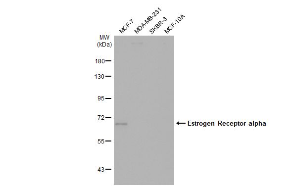 |
| Various whole cell extracts (30 µg) were separated by 7.5% SDS-PAGE, and the membrane was blotted with Estrogen Receptor alpha antibody [1F3] (3616) diluted at 1:1000. The HRP-conjugated anti-mouse IgG antibody was used to detect the primary antibody. |
FAQ & Publications
Frequently Asked Questions
What are the recommended applications and dilutions for the mouse anti-Estrogen Receptor α monoclonal antibody (1F3)?
This antibody is suitable for Western Blot (WB), Immunohistochemistry on frozen sections (IHC-Fr), Flow Cytometry (FACS), and Immunoprecipitation (IP). Recommended dilutions are 1-5 µg/mL for immunoblotting and immunoprecipitation, and 1-10 µg/mL for immunohistochemistry and FACS.
How should the mouse anti-Estrogen Receptor α monoclonal antibody (1F3) be stored to maintain its stability?
The antibody should be stored at -20°C and is stable for at least one year under these conditions. It is advised to avoid multiple freeze-thaw cycles to preserve antibody integrity.
Publications
| pmid | title | authors | citation |
|---|---|---|---|
| We haven't added any publications to our database yet. | |||
Published literature highly relevant to the biological target of this product and referencing this antibody or clone are retrieved from the PubMed database provided by the United States National Library of Medicine at the National Institutes of Health.
Protocols
| relevant to this product |
|---|
| Western blot IHC |
Documents
| Batch Number | QC File | SDS |
|---|---|---|
| To view batch-specific Safety Datasheets and Quality Certificates associated with your account, please Log In. | ||
Only logged in customers who have purchased this product may leave a review.


Reviews
There are no reviews yet.