| Weight | 1 lbs |
|---|---|
| Dimensions | 9 × 5 × 2 in |
| host | mouse |
| isotype | IgG2b |
| clonality | monoclonal |
| concentration | concentrate, predilute |
| applications | IHC |
| reactivity | human |
| available size | 0.1 mL, 0.5 mL, 1 mL concentrated, 7 mL prediluted |
mouse anti-EP-CAM monoclonal antibody (ZM131) 6170
Price range: $160.00 through $528.00
Antibody summary
- Mouse monoclonal to EP-CAM
- Suitable for: Immunohistochemistry (formalin-fixed, paraffin-embedded tissues)
- Reacts with: Human
- Isotype:IgG2b
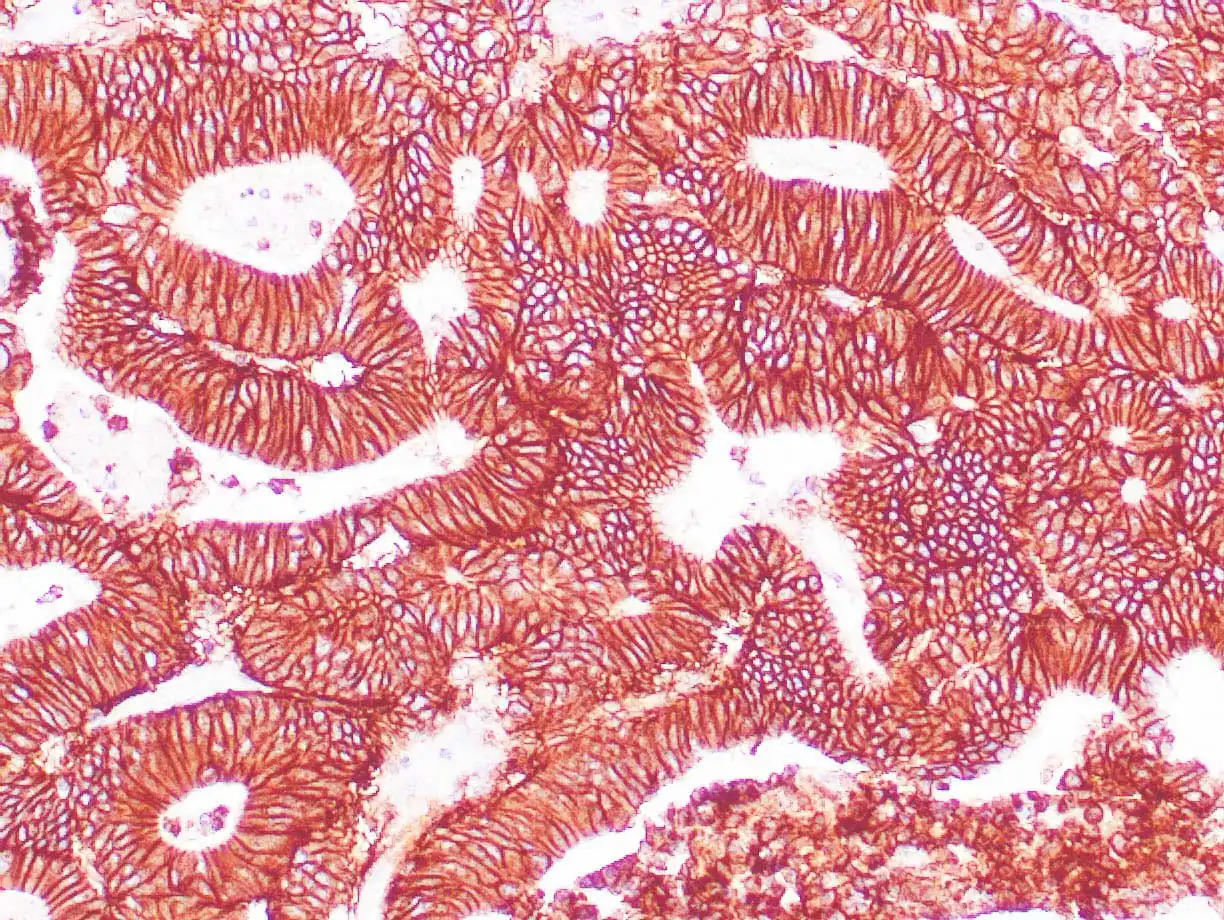
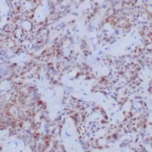
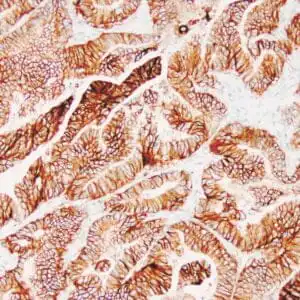
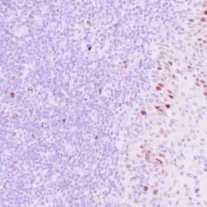
- Control: Colon carcinoma
- Visualization: Cytoplasmic
- 0.1, 0.5, 1.0 mL concentrated, 7 mL prediluted
mouse anti-EP-CAM monoclonal antibody ZM131 6170
| target relevance |
|---|
| Homo sapiens EPCAM Epithelial cell adhesion molecule |
| Protein names Epithelial cell adhesion molecule |
| Alternative names Adenocarcinoma-associated antigen, Cell surface glycoprotein Trop-1, Epithelial cell surface antigen, Epithelial glycoprotein, Epithelial glycoprotein 314, KS 1/4 antigen, KSA, Major gastrointestinal tumor-associated protein GA733-2, Tumor-associated calcium signal transducer 1 |
| Gene names EPCAM |
| Protein family Belongs to the EPCAM family |
| Function May act as a physical homophilic interaction molecule between intestinal epithelial cells (IECs) and intraepithelial lymphocytes (IELs) at the mucosal epithelium for providing immunological barrier as a first line of defense against mucosal infection. Plays a role in embryonic stem cells proliferation and differentiation. Up-regulates the expression of FABP5, MYC and cyclins A and E |
| Subcellular location Lateral cell membrane, Cell junction, tight junction |
| Structure Monomer. Interacts with phosphorylated CLDN7 |
| Post-translational modification Hyperglycosylated in carcinoma tissue as compared with autologous normal epithelia. Glycosylation at Asn-198 is crucial for protein stability |
| Involvement in disease Diarrhea 5, with tufting enteropathy, congenital An intractable diarrhea of infancy characterized by villous atrophy and absence of inflammation, with intestinal epithelial cell dysplasia manifesting as focal epithelial tufts in the duodenum and jejunum. Lynch syndrome 8 A form of Lynch syndrome, an autosomal dominant disease associated with marked increase in cancer susceptibility. It is characterized by a familial predisposition to early-onset colorectal carcinoma (CRC) and extra-colonic tumors of the gastrointestinal, urological and female reproductive tracts. Lynch syndrome is reported to be the most common form of inherited colorectal cancer in the Western world. Clinically, it is often divided into two subgroups. Type I is characterized by hereditary predisposition to colorectal cancer, a young age of onset, and carcinoma observed in the proximal colon. Type II is characterized by increased risk for cancers in certain tissues such as the uterus, ovary, breast, stomach, small intestine, skin, and larynx in addition to the colon. Diagnosis of classical Lynch syndrome is based on the Amsterdam criteria: 3 or more relatives affected by colorectal cancer, one a first degree relative of the other two; 2 or more generation affected; 1 or more colorectal cancers presenting before 50 years of age; exclusion of hereditary polyposis syndromes. The term 'suspected Lynch syndrome' or 'incomplete Lynch syndrome' can be used to describe families who do not or only partially fulfill the Amsterdam criteria, but in whom a genetic basis for colon cancer is strongly suspected. |
| Keywords 3D-structure, Cell junction, Cell membrane, Direct protein sequencing, Disease variant, Disulfide bond, Glycoprotein, Hereditary nonpolyposis colorectal cancer, Membrane, Proteomics identification, Reference proteome, Repeat, Signal, Tight junction, Transmembrane, Transmembrane helix, Tumor antigen |
| Sequence MAPPQVLAFGLLLAAATATFAAAQEECVCENYKLAVNCFVNNNRQCQCTSVGAQNTVICS KLAAKCLVMKAEMNGSKLGRRAKPEGALQNNDGLYDPDCDESGLFKAKQCNGTSMCWCVN TAGVRRTDKDTEITCSERVRTYWIIIELKHKAREKPYDSKSLRTALQKEITTRYQLDPKF ITSILYENNVITIDLVQNSSQKTQNDVDIADVAYYFEKDVKGESLFHSKKMDLTVNGEQL DLDPGQTLIYYVDEKAPEFSMQGLKAGVIAVIVVVVIAVVAGIVVLVISRKKRMAKYEKA EIKEMGEMHRELNA |
| UniProt accession: P16422 |
Data
 |
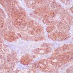
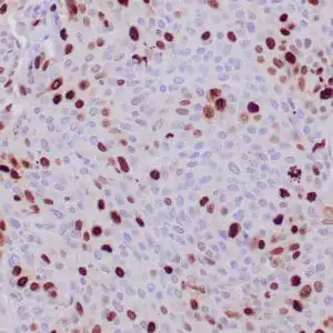
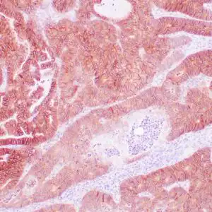
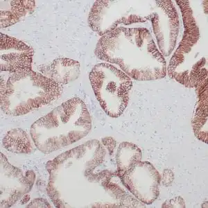
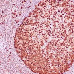
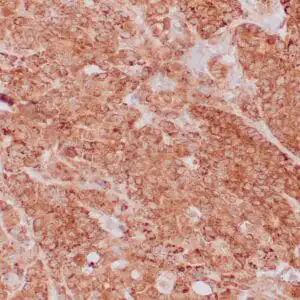
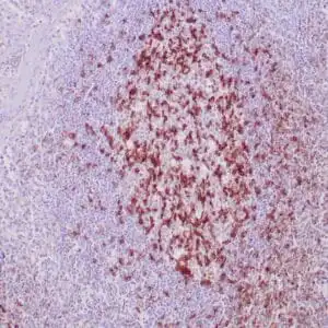
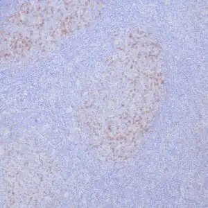
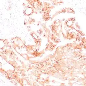
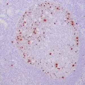
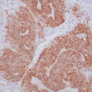
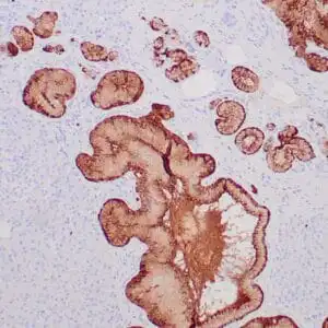
| Human colon adenocarcinoma stained with EP-CAM antibody using peroxidase-conjugate and DAB chromogen. Note the membranous staining of tumor cells. |
FAQ & Publications
Frequently Asked Questions
What is the recommended dilution for using the mouse anti-EP-CAM monoclonal antibody (ZM131) in immunohistochemistry?
For immunohistochemistry applications, the recommended dilution for the concentrated mouse anti-EP-CAM monoclonal antibody (ZM131) is between 1:100 and 1:200.
How should the mouse anti-EP-CAM monoclonal antibody (ZM131) be stored to maintain its stability?
The antibody should be stored at 2-8°C for short-term use and at -20°C for long-term storage. It is important to avoid freeze/thaw cycles to preserve antibody integrity.
Publications
| pmid | title | authors | citation |
|---|---|---|---|
| We haven't added any publications to our database yet. | |||
Published literature highly relevant to the biological target of this product and referencing this antibody or clone are retrieved from the PubMed database provided by the United States National Library of Medicine at the National Institutes of Health.
Protocols
| relevant to this product |
|---|
| IHC |
Documents
| Batch Number | QC File | SDS |
|---|---|---|
| To view batch-specific Safety Datasheets and Quality Certificates associated with your account, please Log In. | ||
Only logged in customers who have purchased this product may leave a review.
Reviews
There are no reviews yet.