| Weight | 1 lbs |
|---|---|
| Dimensions | 9 × 5 × 2 in |
| host | mouse |
| isotype | IgG1 |
| clonality | monoclonal |
| concentration | 1 mg/mL |
| applications | ICC/IF, WB |
| reactivity | E2F-1 |
| available sizes | 100 µg |


mouse anti-E2F-1 monoclonal antibody (6B5) 3078
$503.00
Antibody summary
- Mouse monoclonal to E2F-1
- Suitable for: WB,IP
- Isotype: IgG1
- 100 µg
mouse anti-E2F-1 monoclonal antibody (6B5) 3078
| antibody |
|---|
| Tested applications WB,IP |
| Recommended dilutions Immunoblotting: use at 1-5 ug/mL. Immunoprecipitation: use at 1-5 ug/mL. Positive controls: E2F-1 transfected Soas2 cells and recombinant fusion protein. |
| Immunogen GST fusion protein expressed in E. coli corresponding to aa 162-476 of human E2F-1. |
| Size and concentration 100µg and lot specific |
| Form liquid |
| Storage Instructions This antibody is stable for at least one (1) year at -70°C. Avoid multiple freeze- thaw cycles. |
| Storage buffer PBS, pH 7.4 |
| Purity protein affinity purification |
| Clonality monoclonal |
| Isotype IgG1 |
| Compatible secondaries goat anti-mouse IgG, H&L chain specific, peroxidase conjugated polyclonal antibody 5486 goat anti-mouse IgG, H&L chain specific, biotin conjugated, Conjugate polyclonal antibody 2685 goat anti-mouse IgG, H&L chain specific, FITC conjugated polyclonal antibody 7854 goat anti-mouse IgG, H&L chain specific, peroxidase conjugated polyclonal antibody, crossabsorbed 1706 goat anti-mouse IgG, H&L chain specific, biotin conjugated polyclonal antibody, crossabsorbed 1716 goat anti-mouse IgG, H&L chain specific, FITC conjugated polyclonal antibody, crossabsorbed 1721 |
| Isotype control Mouse monocolonal IgG1 - Isotype Control |
| target relevance |
|---|
| Homo sapiens E2F1 Transcription factor E2F1 |
| Protein names Transcription factor E2F1 |
| Alternative names PBR3, Retinoblastoma-associated protein 1, Retinoblastoma-binding protein 3, pRB-binding protein E2F-1 |
| Gene names E2F1 |
| Protein family Belongs to the E2F/DP family |
| Function Transcription activator that binds DNA cooperatively with DP proteins through the E2 recognition site, 5'-TTTC[CG]CGC-3' found in the promoter region of a number of genes whose products are involved in cell cycle regulation or in DNA replication (PubMed:10675335, PubMed:12717439, PubMed:17050006, PubMed:17704056, PubMed:18625225, PubMed:28992046). The DRTF1/E2F complex functions in the control of cell-cycle progression from G1 to S phase (PubMed:10675335, PubMed:12717439, PubMed:17704056). E2F1 binds preferentially RB1 in a cell-cycle dependent manner (PubMed:10675335, PubMed:12717439, PubMed:17704056). It can mediate both cell proliferation and TP53/p53-dependent apoptosis (PubMed:8170954). Blocks adipocyte differentiation by binding to specific promoters repressing CEBPA binding to its target gene promoters (PubMed:20176812). Directly activates transcription of PEG10 (PubMed:17050006, PubMed:18625225, PubMed:28992046). Positively regulates transcription of RRP1B (PubMed:20040599) |
| Subcellular location Nucleus |
| Structure (Microbial infection) Interacts with human cytomegalovirus/HHV-5 protein UL123 |
| Post-translational modification Phosphorylated by CDK2 and cyclin A-CDK2 in the S-phase (PubMed:12717439, PubMed:7838523). Phosphorylation at Ser-364 by CHEK2 stabilizes E2F1 upon DNA damage and regulates its effect on transcription and apoptosis (PubMed:12717439). Phosphorylation at Ser-403 by GSK3B promotes interaction with USP11, leading to its deubiquitination and stabilization (PubMed:28992046) Ubiquitinated via 'Lys-63'-linked ubiquitin, leading to its degradation (PubMed:28992046). Deubiquitinated by USP11 following phosphorylation by GSK3B, promoting its stability (PubMed:28992046) Acetylation stimulates DNA-binding. Enhanced under stress conditions such as DNA damage and inhibited by retinoblastoma protein RB1. Regulated by KAP1/TRIM28 which recruits HDAC1 to E2F1 resulting in deacetylation. Acetylated by P/CAF/KAT2B Methylation at Lys-185 by SETD7 promotes E2F1 ubiquitin-dependent proteasomal degradation |
| Keywords 3D-structure, Acetylation, Activator, Apoptosis, Cell cycle, DNA-binding, Host-virus interaction, Methylation, Nucleus, Phosphoprotein, Proteomics identification, Reference proteome, Transcription, Transcription regulation, Ubl conjugation |
| Sequence MALAGAPAGGPCAPALEALLGAGALRLLDSSQIVIISAAQDASAPPAPTGPAAPAAGPCD PDLLLFATPQAPRPTPSAPRPALGRPPVKRRLDLETDHQYLAESSGPARGRGRHPGKGVK SPGEKSRYETSLNLTTKRFLELLSHSADGVVDLNWAAEVLKVQKRRIYDITNVLEGIQLI AKKSKNHIQWLGSHTTVGVGGRLEGLTQDLRQLQESEQQLDHLMNICTTQLRLLSEDTDS QRLAYVTCQDLRSIADPAEQMVMVIKAPPETQLQAVDSSENFQISLKSKQGPIDVFLCPE ETVGGISPGKTPSQEVTSEEENRATDSATIVSPPPSSPPSSLTTDPSQSLLSLEQEPLLS RMGSLRAPVDEDRLSPLVAADSLLEHVREDFSGLLPEEFISLSPPHEALDYHFGLEEGEG IRDLFDCDFGDLTPLDF |
| UniProt accession: Q01094 |
Data
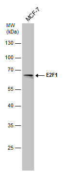 |
| Whole cell extract (30 µg) was separated by 10% SDS-PAGE, and the membrane was blotted with E2F1 antibody [6B5] (3078) diluted at 1:500. The signal was developed with Trident ECL plus-Enhanced. |
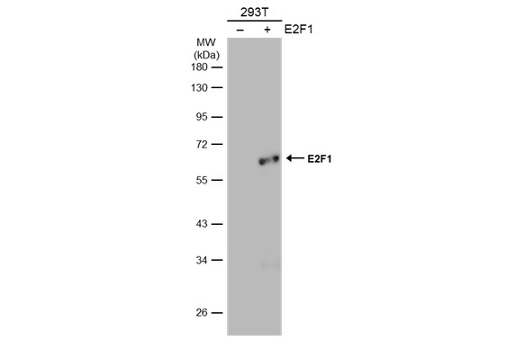 |
| Non-transfected (-) and transfected (-) 293T whole cell extracts (30 µg) were separated by 10% SDS-PAGE, and the membrane was blotted with E2F1 antibody [6B5] (3078) diluted at 1:500. The HRP-conjugated anti-mouse IgG antibody was used to detect the primary antibody. |
FAQ & Publications
Frequently Asked Questions
What are the recommended applications and dilution ranges for the mouse anti-E2F-1 monoclonal antibody (6B5)?
This antibody is suitable for Western blotting (WB) and immunoprecipitation (IP). The recommended dilution range for both immunoblotting and immunoprecipitation is 1-5 µg/mL.
How should the mouse anti-E2F-1 monoclonal antibody (6B5) be stored to maintain its stability?
The antibody should be stored at -70°C and is stable for at least one year under these conditions. It is important to avoid multiple freeze-thaw cycles to preserve antibody integrity.
Publications
| pmid | title | authors | citation |
|---|---|---|---|
| We haven't added any publications to our database yet. | |||
Published literature highly relevant to the biological target of this product and referencing this antibody or clone are retrieved from the PubMed database provided by the United States National Library of Medicine at the National Institutes of Health.
Protocols
| relevant to this product |
|---|
| Western blot |
Documents
| Batch Number | QC File | SDS |
|---|---|---|
| To view batch-specific Safety Datasheets and Quality Certificates associated with your account, please Log In. | ||
Only logged in customers who have purchased this product may leave a review.


Reviews
There are no reviews yet.