| Weight | 1 lbs |
|---|---|
| Dimensions | 9 × 5 × 2 in |
| host | mouse |
| isotype | IgG1 |
| clonality | monoclonal |
| concentration | 1 mg/mL |
| applications | ICC/IF, WB |
| reactivity | DNA Ligase III |
| available sizes | 100 µg |


mouse anti-DNA Ligase III monoclonal antibody (1F3) 4859
$503.00
Antibody summary
- Mouse monoclonal to DNA Ligase III
- Suitable for: WB,ICC/IF,IP,Blocking,PLA
- Isotype: IgG1
- 100 µg
mouse anti-DNA Ligase III monoclonal antibody (1F3) 4859
| antibody |
|---|
| Tested applications WB,ICC/IF,IP |
| Recommended dilutions Immunoblotting: use at 0.5-2ug/mL. Predicted molecular weight ~100kDa. These are recommended concentrations. Endusers should determine optimal concentrations for their applications. |
| Immunogen Full-length recombinant human DNA Ligase III-b. |
| Size and concentration 100µg and 1 mg/mL |
| Form liquid |
| Storage Instructions This antibody is stable for at least one year at -20°C. Store in appropriate aliquots to avoid multiple freeze-thaw cycles. |
| Storage buffer Hybridoma culture supernatant |
| Purity protein affinity purification |
| Clonality monoclonal |
| Isotype IgG1 |
| Compatible secondaries goat anti-mouse IgG, H&L chain specific, peroxidase conjugated polyclonal antibody 5486 goat anti-mouse IgG, H&L chain specific, biotin conjugated, Conjugate polyclonal antibody 2685 goat anti-mouse IgG, H&L chain specific, FITC conjugated polyclonal antibody 7854 goat anti-mouse IgG, H&L chain specific, peroxidase conjugated polyclonal antibody, crossabsorbed 1706 goat anti-mouse IgG, H&L chain specific, biotin conjugated polyclonal antibody, crossabsorbed 1716 goat anti-mouse IgG, H&L chain specific, FITC conjugated polyclonal antibody, crossabsorbed 1721 |
| Isotype control Mouse monocolonal IgG1 - Isotype Control |
| target relevance |
|---|
| Homo sapiens LIG3 DNA ligase 3 |
| Protein names DNA ligase 3 |
| Alternative names DNA ligase III, Polydeoxyribonucleotide synthase [ATP] 3 |
| Gene names LIG3 |
| Protein family Belongs to the ATP-dependent DNA ligase family |
| Function Isoform 3 functions as a heterodimer with DNA-repair protein XRCC1 in the nucleus and can correct defective DNA strand-break repair and sister chromatid exchange following treatment with ionizing radiation and alkylating agents. Isoform 1 is targeted to mitochondria, where it functions as a DNA ligase in mitochondrial base-excision DNA repair (PubMed:10207110, PubMed:24674627) |
| Catalytic activity ATP + (deoxyribonucleotide)n-3'-hydroxyl + 5'-phospho-(deoxyribonucleotide)m = (deoxyribonucleotide)n+m + AMP + diphosphate. |
| Subcellular location Nucleus |
| Structure Isoform 3 interacts (via BRCT domain) with the nuclear DNA-repair protein XRCC1. Interacts with POLG (PubMed:33855352). Interacts with POLB (PubMed:19336415) |
| Involvement in disease Mitochondrial DNA depletion syndrome 20, MNGIE type An autosomal recessive mitochondrial disorder characterized by severe gut dysmotility, muscle weakness and atrophy, neurological abnormalities including epilepsy, migraine, stroke-like episodes, learning difficulties or cognitive decline, and neurogenic bladder. Brain imaging usually shows diffuse leukoencephalopathy and may show cerebellar atrophy. Disease onset can range from infancy to the teenage years. |
| Keywords 3D-structure, Alternative initiation, Alternative splicing, ATP-binding, Cell cycle, Cell division, Disease variant, DNA damage, DNA recombination, DNA repair, DNA replication, Ligase, Magnesium, Metal-binding, Mitochondrion, Nucleotide-binding, Nucleus, Phosphoprotein, Primary mitochondrial disease, Proteomics identification, Reference proteome, Transit peptide, Zinc, Zinc-finger |
| Sequence MSLAFKIFFPQTLRALSRKELCLFRKHHWRDVRQFSQWSETDLLHGHPLFLRRKPVLSFQ GSHLRSRATYLVFLPGLHVGLCSGPCEMAEQRFCVDYAKRGTAGCKKCKEKIVKGVCRIG KVVPNPFSESGGDMKEWYHIKCMFEKLERARATTKKIEDLTELEGWEELEDNEKEQITQH IADLSSKAAGTPKKKAVVQAKLTTTGQVTSPVKGASFVTSTNPRKFSGFSAKPNNSGEAP SSPTPKRSLSSSKCDPRHKDCLLREFRKLCAMVADNPSYNTKTQIIQDFLRKGSAGDGFH GDVYLTVKLLLPGVIKTVYNLNDKQIVKLFSRIFNCNPDDMARDLEQGDVSETIRVFFEQ SKSFPPAAKSLLTIQEVDEFLLRLSKLTKEDEQQQALQDIASRCTANDLKCIIRLIKHDL KMNSGAKHVLDALDPNAYEAFKASRNLQDVVERVLHNAQEVEKEPGQRRALSVQASLMTP VQPMLAEACKSVEYAMKKCPNGMFSEIKYDGERVQVHKNGDHFSYFSRSLKPVLPHKVAH FKDYIPQAFPGGHSMILDSEVLLIDNKTGKPLPFGTLGVHKKAAFQDANVCLFVFDCIYF NDVSLMDRPLCERRKFLHDNMVEIPNRIMFSEMKRVTKALDLADMITRVIQEGLEGLVLK DVKGTYEPGKRHWLKVKKDYLNEGAMADTADLVVLGAFYGQGSKGGMMSIFLMGCYDPGS QKWCTVTKCAGGHDDATLARLQNELDMVKISKDPSKIPSWLKVNKIYYPDFIVPDPKKAA VWEITGAEFSKSEAHTADGISIRFPRCTRIRDDKDWKSATNLPQLKELYQLSKEKADFTV VAGDEGSSTTGGSSEENKGPSGSAVSRKAPSKPSASTKKAEGKLSNSNSKDGNMQTAKPS AMKVGEKLATKSSPVKVGEKRKAADETLCQTKVLLDIFTGVRLYLPPSTPDFSRLRRYFV AFDGDLVQEFDMTSATHVLGSRDKNPAAQQVSPEWIWACIRKRRLVAPC |
| UniProt accession: P49916 |
Data
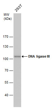 |
| DNA ligase III antibody detects DNA ligase III protein by western blot analysis. Whole cell extracts (30 µg) was separated by 7.5% SDS-PAGE, and the membrane was blotted with DNA ligase III antibody (4859) diluted by 1:500. The HRP-conjugated anti-mouse IgG antibody was used to detect the primary antibody. |
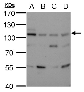 |
| DNA ligase III detects DNA ligase III antibody [1F3] protein by western blot analysis. A. 30 µg 293T whole cell lysate/extract B. 30 µg A431 whole cell lysate/extract C. 30 µg HeLa whole cell lysate/extract D. 30 µg HepG2 whole cell lysate/extract 7.5% SDS-PAGE DNA ligase III (4859) dilution: 1:500 The HRP-conjugated anti-mouse IgG antibody was used to detect the primary antibody. |
FAQ & Publications
Frequently Asked Questions
What applications is the mouse anti-DNA Ligase III monoclonal antibody (1F3) validated for?
This antibody is suitable and tested for Western blotting (WB), immunocytochemistry/immunofluorescence (ICC/IF), and immunoprecipitation (IP). It can also be used in blocking and proximity ligation assays (PLA).
How should the mouse anti-DNA Ligase III monoclonal antibody be stored to maintain stability?
The antibody should be stored at -20°C in appropriate aliquots to avoid multiple freeze-thaw cycles. Under these conditions, it remains stable for at least one year.
What is the recommended dilution range for using this antibody in immunoblotting?
For immunoblotting applications, the recommended dilution range is 0.5 to 2 µg/mL. Users should optimize the concentration for their specific experimental conditions.
Publications
| pmid | title | authors | citation |
|---|---|---|---|
| We haven't added any publications to our database yet. | |||
Published literature highly relevant to the biological target of this product and referencing this antibody or clone are retrieved from the PubMed database provided by the United States National Library of Medicine at the National Institutes of Health.
Protocols
| relevant to this product |
|---|
| Western blot IHC ICC |
Documents
| Batch Number | QC File | SDS |
|---|---|---|
| To view batch-specific Safety Datasheets and Quality Certificates associated with your account, please Log In. | ||
Only logged in customers who have purchased this product may leave a review.


Reviews
There are no reviews yet.