| Weight | 1 lbs |
|---|---|
| Dimensions | 9 × 5 × 2 in |
| host | mouse |
| isotype | IgG1 |
| clonality | monoclonal |
| concentration | 1 mg/mL |
| applications | ICC/IF, WB |
| reactivity | CD46/Membrane Cofactor Protein |
| available sizes | 100 µg |

mouse anti-CD46/Membrane Cofactor Protein monoclonal antibody (3F1) 7171
$520.00
Antibody summary
- Mouse monoclonal to CD46/Membrane Cofactor Protein
- Suitable for: WB,IHC,ELISA
- Isotype: IgG1
- 100 µg
mouse anti-CD46/Membrane Cofactor Protein monoclonal antibody (3F1) 7171
| antibody |
|---|
| Tested applications WB,IHC,IHC,ELISA |
| Recommended dilutions Immunohistochemistry: use at 1- 10ug/ml. Immunoblotting: use at 0.5-1ug/ml. A band of ~44kDa is detected. Additional bands due to glycosylation of CD46 may be detected.ELISA: use at 0.1-1ug/ml with human CD46 on the solid phase. These are recommended concentrations. |
| Immunogen Recombinant human CD46. |
| Size and concentration 100µg and |
| Form lyophilized |
| Storage Instructions This product is stable for at least one (1) year at -20°C to -70°C. Reconstituted product should be stored in appropriate aliquots to avoid repeated freeze-thaw cycles. |
| Storage buffer Lyophilized in PBS. |
| Purity protein affinity purification |
| Clonality monoclonal |
| Isotype IgG1 |
| Compatible secondaries goat anti-mouse IgG, H&L chain specific, peroxidase conjugated polyclonal antibody 5486 goat anti-mouse IgG, H&L chain specific, biotin conjugated, Conjugate polyclonal antibody 2685 goat anti-mouse IgG, H&L chain specific, FITC conjugated polyclonal antibody 7854 goat anti-mouse IgG, H&L chain specific, peroxidase conjugated polyclonal antibody, crossabsorbed 1706 goat anti-mouse IgG, H&L chain specific, biotin conjugated polyclonal antibody, crossabsorbed 1716 goat anti-mouse IgG, H&L chain specific, FITC conjugated polyclonal antibody, crossabsorbed 1721 |
| Isotype control Mouse monocolonal IgG1 - Isotype Control |
| target relevance |
|---|
| Homo sapiens CD46 Membrane cofactor protein |
| Protein names Membrane cofactor protein |
| Alternative names TLX, Trophoblast leukocyte common antigen |
| Gene names CD46 |
| Function Acts as a cofactor for complement factor I, a serine protease which protects autologous cells against complement-mediated injury by cleaving C3b and C4b deposited on host tissue. May be involved in the fusion of the spermatozoa with the oocyte during fertilization. Also acts as a costimulatory factor for T-cells which induces the differentiation of CD4+ into T-regulatory 1 cells. T-regulatory 1 cells suppress immune responses by secreting interleukin-10, and therefore are thought to prevent autoimmunity |
| Subcellular location Cytoplasmic vesicle, secretory vesicle, acrosome inner membrane |
| Structure (Microbial infection) Binds to Streptococcus pyogenes M protein and to type IV pili from Neisseria (PubMed:11260136, PubMed:11971006, PubMed:7708671, PubMed:9379894) |
| Post-translational modification N-glycosylated on Asn-83; Asn-114 and Asn-273 in most tissues, but probably less N-glycosylated in testis. N-glycosylation on Asn-114 and Asn-273 is required for cytoprotective function. N-glycosylation on Asn-114 is required for Measles virus binding. N-glycosylation on Asn-273 is required for Neisseria binding. N-glycosylation is not required for human adenovirus binding Extensively O-glycosylated in the Ser/Thr-rich domain. O-glycosylation is required for Neisseria binding but not for Measles virus or human adenovirus binding In epithelial cells, isoforms B/D/F/H/J/L/3 are phosphorylated by YES1 in response to infection by Neisseria gonorrhoeae; which promotes infectivity. In T-cells, these isoforms may be phosphorylated by LCK |
| Involvement in disease Hemolytic uremic syndrome, atypical, 2 An atypical form of hemolytic uremic syndrome. It is a complex genetic disease characterized by microangiopathic hemolytic anemia, thrombocytopenia, renal failure and absence of episodes of enterocolitis and diarrhea. In contrast to typical hemolytic uremic syndrome, atypical forms have a poorer prognosis, with higher death rates and frequent progression to end-stage renal disease. |
| Keywords 3D-structure, Alternative splicing, Complement pathway, Cytoplasmic vesicle, Direct protein sequencing, Disease variant, Disulfide bond, Fertilization, Glycoprotein, Hemolytic uremic syndrome, Host cell receptor for virus entry, Host-virus interaction, Immunity, Innate immunity, Membrane, Phosphoprotein, Proteomics identification, Receptor, Reference proteome, Repeat, Signal, Sushi, Transmembrane, Transmembrane helix |
| Sequence MEPPGRRECPFPSWRFPGLLLAAMVLLLYSFSDACEEPPTFEAMELIGKPKPYYEIGERV DYKCKKGYFYIPPLATHTICDRNHTWLPVSDDACYRETCPYIRDPLNGQAVPANGTYEFG YQMHFICNEGYYLIGEEILYCELKGSVAIWSGKPPICEKVLCTPPPKIKNGKHTFSEVEV FEYLDAVTYSCDPAPGPDPFSLIGESTIYCGDNSVWSRAAPECKVVKCRFPVVENGKQIS GFGKKFYYKATVMFECDKGFYLDGSDTIVCDSNSTWDPPVPKCLKVLPPSSTKPPALSHS VSTSSTTKSPASSASGPRPTYKPPVSNYPGYPKPEEGILDSLDVWVIAVIVIAIVVGVAV ICVVPYRYLQRRKKKGTYLTDETHREVKFTSL |
| UniProt accession: P15529 |
Data
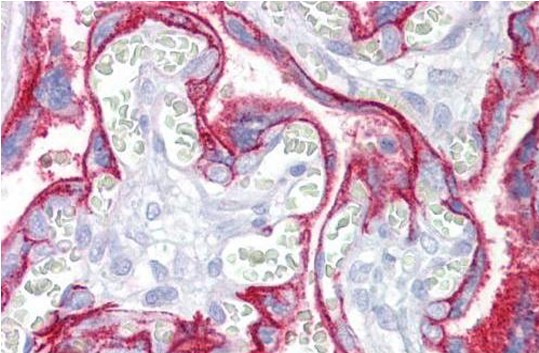 |
| Immunohistochemistry: use at 110ug/ml. Paraffin-embedded human placenta stained with #7171. |
FAQ & Publications
Frequently Asked Questions
What applications is the mouse anti-CD46 monoclonal antibody (3F1) suitable for?
This antibody is suitable for Western Blot (WB), Immunohistochemistry (IHC), and ELISA applications.
How should the mouse anti-CD46 antibody be stored to ensure stability?
The lyophilized antibody is stable for at least one year when stored at -20°C to -70°C. After reconstitution, aliquot the antibody to avoid repeated freeze-thaw cycles.
What is the recommended dilution for using this antibody in immunohistochemistry?
For immunohistochemistry, use the antibody at a concentration between 1 to 10 µg/mL.
What is the host species and isotype of this monoclonal antibody?
The antibody is a mouse monoclonal of the IgG1 isotype.
Publications
| pmid | title | authors | citation |
|---|---|---|---|
| We haven't added any publications to our database yet. | |||
Published literature highly relevant to the biological target of this product and referencing this antibody or clone are retrieved from the PubMed database provided by the United States National Library of Medicine at the National Institutes of Health.
Protocols
| relevant to this product |
|---|
| Western blot IHC |
Documents
| Batch Number | QC File | SDS |
|---|---|---|
| To view batch-specific Safety Datasheets and Quality Certificates associated with your account, please Log In. | ||
Only logged in customers who have purchased this product may leave a review.


Reviews
There are no reviews yet.