| Weight | 1 lbs |
|---|---|
| Dimensions | 9 × 5 × 2 in |
| host | mouse |
| isotype | IgG2b/κ |
| clonality | monoclonal |
| concentration | concentrate, predilute |
| applications | IHC |
| reactivity | human |
| available size | 0.1 mL, 0.5 mL, 1 mL concentrated, 7 mL prediluted |

mouse anti-C3d monoclonal antibody (ZM369) 6041
Price range: $160.00 through $528.00
Antibody summary
- Mouse monoclonal to C3d
- Suitable for: Immunohistochemistry (formalin-fixed, paraffin-embedded tissues)
- Reacts with: Human
- Isotype:IgG2b/κ
- Control: Acute rejected kidney transplant
- Visualization: Membrane or cytoplasmic
- 0.1, 0.5, 1.0 mL concentrated, 7 mL prediluted
mouse anti-C3d monoclonal antibody ZM369 6041
| target relevance |
|---|
| Homo sapiens C3 Complement C3 |
| Protein names Complement C3 |
| Alternative names C3 and PZP-like alpha-2-macroglobulin domain-containing protein 1 |
| Gene names C3 |
| Function Precursor of non-enzymatic components of the classical, alternative, lectin and GZMK complement pathways, which consist in a cascade of proteins that leads to phagocytosis and breakdown of pathogens and signaling that strengthens the adaptive immune system |
| Subcellular location Secreted |
| Structure (Microbial infection) Interacts with Staphylococcus aureus protein Fib |
| Post-translational modification C3 precursor is first processed by the removal of 4 Arg residues, forming two chains, beta and alpha, linked by a disulfide bond (PubMed:7240134). During activation of the complement systems, the alpha chain is cleaved into C3a and C3b by the C3 convertase: C3b stays linked to the beta chain, while C3a is released in the plasma (PubMed:6611150, PubMed:6906228). The alpha chain is cleaved by the serine protease complement C2b component of the C3 convertase to generate C3a and C3b following activation by the classical, lectin and GZMK complement systems (PubMed:39914456, PubMed:39814882, PubMed:6611150, PubMed:6906228). The alpha chain is cleaved by CFB component of the C3 convertase to generate C3a and C3b following activation by the alternative complement system (PubMed:28264884, PubMed:31507604, PubMed:3638964, PubMed:6919543, PubMed:9748277) C3a is further processed by carboxypeptidases to release the C-terminal arginine residue generating the acylation stimulating protein (ASP) (PubMed:4098172, PubMed:6968751). Levels of ASP are increased in adipocytes in the postprandial period and by insulin and dietary chylomicrons (PubMed:15833747) Complement C3b is rapidly split in two positions by factor I (CFI) and a cofactor (CFH) to form iC3b (inactivated C3b) and C3f which is released (PubMed:17320177, PubMed:28671664, PubMed:7360115). CFI and CFH catalyze proteolytic degradation of already-deposited complement C3b (PubMed:28671664). Then iC3b is slowly cleaved (possibly by CFI) to form C3c (beta chain + alpha' chain fragment 1 + alpha' chain fragment 2), C3dg and C3f (PubMed:16177781, PubMed:17051150, PubMed:17684013). Other proteases produce other fragments such as C3d or C3g (PubMed:7539791) Upon activation, the internal thioester bond reacts with carbohydrate antigens on the target surface to form amide or ester bonds, leading to covalent association with the surface of pathogens Complement C3b interacts with complement C4b via a thioester linkage Phosphorylated by FAM20C in the extracellular medium (Microbial infection) C3 is cleaved by Staphylococcus aureus aureolysin; this cleavage renders C3a and C3b inactive. C3b is rapidly degraded by host factors CFH and CFI preventing its deposition on the bacterial surface while C3a is further inactivated by aureolysin (Microbial infection) Complement C3 beta chain is cleaved and inactivated by S.pyogenes SpeB (Microbial infection) Cleaved by N.meningitidis NalP between Leu-744 and Gly-745, generating a slightly shorter C3 alpha form and a slightly longer C3 beta form. The C3b-like fragment is degraded in the presence of the complement regulators CFH and CFI, preventing its deposition on the bacterial surface |
| Involvement in disease Complement component 3 deficiency A rare defect of the complement classical pathway. Patients develop recurrent, severe, pyogenic infections because of ineffective opsonization of pathogens. Some patients may also develop autoimmune disorders, such as arthralgia and vasculitic rashes, lupus-like syndrome and membranoproliferative glomerulonephritis. Macular degeneration, age-related, 9 A form of age-related macular degeneration, a multifactorial eye disease and the most common cause of irreversible vision loss in the developed world. In most patients, the disease is manifest as ophthalmoscopically visible yellowish accumulations of protein and lipid that lie beneath the retinal pigment epithelium and within an elastin-containing structure known as Bruch membrane. Hemolytic uremic syndrome, atypical, 5 An atypical form of hemolytic uremic syndrome. It is a complex genetic disease characterized by microangiopathic hemolytic anemia, thrombocytopenia, renal failure and absence of episodes of enterocolitis and diarrhea. In contrast to typical hemolytic uremic syndrome, atypical forms have a poorer prognosis, with higher death rates and frequent progression to end-stage renal disease. |
| Keywords 3D-structure, Age-related macular degeneration, Cleavage on pair of basic residues, Complement alternate pathway, Complement pathway, Direct protein sequencing, Disease variant, Disulfide bond, Fatty acid metabolism, Glycoprotein, Hemolytic uremic syndrome, Immunity, Inflammatory response, Innate immunity, Lipid metabolism, Phosphoprotein, Proteomics identification, Reference proteome, Secreted, Signal, Thioester bond |
| Sequence MGPTSGPSLLLLLLTHLPLALGSPMYSIITPNILRLESEETMVLEAHDAQGDVPVTVTVH DFPGKKLVLSSEKTVLTPATNHMGNVTFTIPANREFKSEKGRNKFVTVQATFGTQVVEKV VLVSLQSGYLFIQTDKTIYTPGSTVLYRIFTVNHKLLPVGRTVMVNIENPEGIPVKQDSL SSQNQLGVLPLSWDIPELVNMGQWKIRAYYENSPQQVFSTEFEVKEYVLPSFEVIVEPTE KFYYIYNEKGLEVTITARFLYGKKVEGTAFVIFGIQDGEQRISLPESLKRIPIEDGSGEV VLSRKVLLDGVQNPRAEDLVGKSLYVSATVILHSGSDMVQAERSGIPIVTSPYQIHFTKT PKYFKPGMPFDLMVFVTNPDGSPAYRVPVAVQGEDTVQSLTQGDGVAKLSINTHPSQKPL SITVRTKKQELSEAEQATRTMQALPYSTVGNSNNYLHLSVLRTELRPGETLNVNFLLRMD RAHEAKIRYYTYLIMNKGRLLKAGRQVREPGQDLVVLPLSITTDFIPSFRLVAYYTLIGA SGQREVVADSVWVDVKDSCVGSLVVKSGQSEDRQPVPGQQMTLKIEGDHGARVVLVAVDK GVFVLNKKNKLTQSKIWDVVEKADIGCTPGSGKDYAGVFSDAGLTFTSSSGQQTAQRAEL QCPQPAARRRRSVQLTEKRMDKVGKYPKELRKCCEDGMRENPMRFSCQRRTRFISLGEAC KKVFLDCCNYITELRRQHARASHLGLARSNLDEDIIAEENIVSRSEFPESWLWNVEDLKE PPKNGISTKLMNIFLKDSITTWEILAVSMSDKKGICVADPFEVTVMQDFFIDLRLPYSVV RNEQVEIRAVLYNYRQNQELKVRVELLHNPAFCSLATTKRRHQQTVTIPPKSSLSVPYVI VPLKTGLQEVEVKAAVYHHFISDGVRKSLKVVPEGIRMNKTVAVRTLDPERLGREGVQKE DIPPADLSDQVPDTESETRILLQGTPVAQMTEDAVDAERLKHLIVTPSGCGEQNMIGMTP TVIAVHYLDETEQWEKFGLEKRQGALELIKKGYTQQLAFRQPSSAFAAFVKRAPSTWLTA YVVKVFSLAVNLIAIDSQVLCGAVKWLILEKQKPDGVFQEDAPVIHQEMIGGLRNNNEKD MALTAFVLISLQEAKDICEEQVNSLPGSITKAGDFLEANYMNLQRSYTVAIAGYALAQMG RLKGPLLNKFLTTAKDKNRWEDPGKQLYNVEATSYALLALLQLKDFDFVPPVVRWLNEQR YYGGGYGSTQATFMVFQALAQYQKDAPDHQELNLDVSLQLPSRSSKITHRIHWESASLLR SEETKENEGFTVTAEGKGQGTLSVVTMYHAKAKDQLTCNKFDLKVTIKPAPETEKRPQDA KNTMILEICTRYRGDQDATMSILDISMMTGFAPDTDDLKQLANGVDRYISKYELDKAFSD RNTLIIYLDKVSHSEDDCLAFKVHQYFNVELIQPGAVKVYAYYNLEESCTRFYHPEKEDG KLNKLCRDELCRCAEENCFIQKSDDKVTLEERLDKACEPGVDYVYKTRLVKVQLSNDFDE YIMAIEQTIKSGSDEVQVGQQRTFISPIKCREALKLEEKKHYLMWGLSSDFWGEKPNLSY IIGKDTWVEHWPEEDECQDEENQKQCQDLGAFTESMVVFGCPN |
| UniProt accession: P01024 |
Data
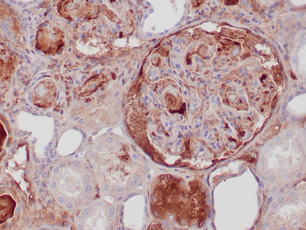 |
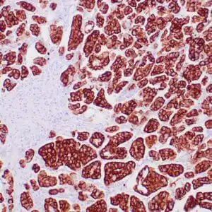
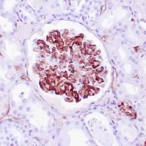
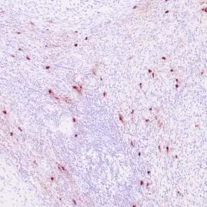
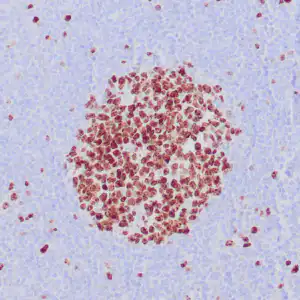
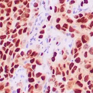
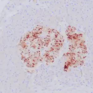
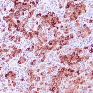
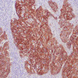
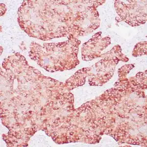
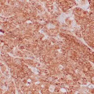
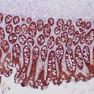
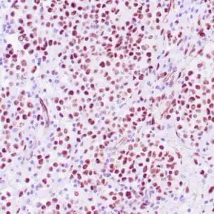
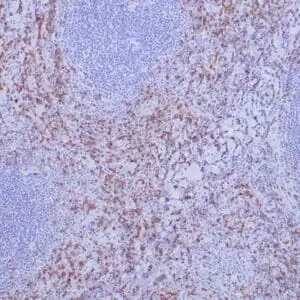
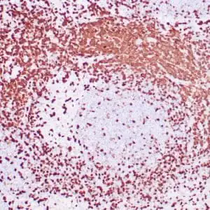
| Human transplanted kidney stained with anti-C3d antibody using peroxidase-conjugate and DAB chromogen. Note basement membrane staining of renal tubules and glomeruli. |
FAQ & Publications
Frequently Asked Questions
What species does the mouse anti-C3d monoclonal antibody (ZM369) specifically react with?
This monoclonal antibody specifically reacts with human species.
How should the mouse anti-C3d monoclonal antibody be stored to maintain its stability?
For short-term storage, keep the antibody at 2-8°C. For long-term storage, it should be kept at -20°C while avoiding freeze/thaw cycles.
Publications
| pmid | title | authors | citation |
|---|---|---|---|
| We haven't added any publications to our database yet. | |||
Published literature highly relevant to the biological target of this product and referencing this antibody or clone are retrieved from the PubMed database provided by the United States National Library of Medicine at the National Institutes of Health.
Protocols
| relevant to this product |
|---|
| IHC |
Documents
| Batch Number | QC File | SDS |
|---|---|---|
| To view batch-specific Safety Datasheets and Quality Certificates associated with your account, please Log In. | ||
Only logged in customers who have purchased this product may leave a review.

Reviews
There are no reviews yet.