| Weight | 1 lbs |
|---|---|
| Dimensions | 9 × 5 × 2 in |
| host | mouse |
| isotype | IgG1 |
| clonality | monoclonal |
| concentration | 1 mg/mL |
| applications | ICC/IF, WB |
| reactivity | ATM |
| available sizes | 100 µg |


mouse anti-ATM monoclonal antibody (2C1) 2952
$503.00
Antibody summary
- Mouse monoclonal to ATM
- Suitable for: WB,ICC/IF,IHC-P,FACS,IP,ELISA,ChIP,IHC
- Isotype: IgG1
- 100 µg
mouse anti-ATM monoclonal antibody (2C1) 2952
| antibody |
|---|
| Tested applications WB,IHC,IHC,ICC/IF |
| Recommended dilutions Immunoblotting: use at 1-10 ug/mL. Immunoprecipitation: use at 1-10 ug/mL. Positive controls: Raji or Akata cells. |
| Immunogen GST fusion protein expressed in E. coli corresponding to aa 2577- 3056 of full-length ATM. |
| Size and concentration 100µg and lot specific |
| Form liquid |
| Storage Instructions This antibody is stable for at least one (1) year at -70°C. Avoid multiple freeze-thaw cycles. |
| Storage buffer PBS, pH 7.4 |
| Purity protein affinity purification |
| Clonality monoclonal |
| Isotype IgG1 |
| Compatible secondaries goat anti-mouse IgG, H&L chain specific, peroxidase conjugated polyclonal antibody 5486 goat anti-mouse IgG, H&L chain specific, biotin conjugated, Conjugate polyclonal antibody 2685 goat anti-mouse IgG, H&L chain specific, FITC conjugated polyclonal antibody 7854 goat anti-mouse IgG, H&L chain specific, peroxidase conjugated polyclonal antibody, crossabsorbed 1706 goat anti-mouse IgG, H&L chain specific, biotin conjugated polyclonal antibody, crossabsorbed 1716 goat anti-mouse IgG, H&L chain specific, FITC conjugated polyclonal antibody, crossabsorbed 1721 |
| Isotype control Mouse monocolonal IgG1 - Isotype Control |
| target relevance |
|---|
| Homo sapiens ATM Serine-protein kinase ATM |
| Protein names Serine-protein kinase ATM |
| Alternative names Ataxia telangiectasia mutated |
| Gene names ATM |
| Protein family Belongs to the PI3/PI4-kinase family. ATM subfamily |
| Function Serine/threonine protein kinase which activates checkpoint signaling upon double strand breaks (DSBs), apoptosis and genotoxic stresses such as ionizing ultraviolet A light (UVA), thereby acting as a DNA damage sensor (PubMed:10550055, PubMed:10839545, PubMed:10910365, PubMed:12556884, PubMed:14871926, PubMed:15064416, PubMed:15448695, PubMed:15456891, PubMed:15790808, PubMed:15916964, PubMed:17923702, PubMed:21757780, PubMed:24534091, PubMed:35076389, PubMed:9733514). Recognizes the substrate consensus sequence [ST]-Q (PubMed:10550055, PubMed:10839545, PubMed:10910365, PubMed:12556884, PubMed:14871926, PubMed:15448695, PubMed:15456891, PubMed:15916964, PubMed:17923702, PubMed:24534091, PubMed:9733514). Phosphorylates 'Ser-139' of histone variant H2AX at double strand breaks (DSBs), thereby regulating DNA damage response mechanism (By similarity). Also plays a role in pre-B cell allelic exclusion, a process leading to expression of a single immunoglobulin heavy chain allele to enforce clonality and monospecific recognition by the B-cell antigen receptor (BCR) expressed on individual B-lymphocytes. After the introduction of DNA breaks by the RAG complex on one immunoglobulin allele, acts by mediating a repositioning of the second allele to pericentromeric heterochromatin, preventing accessibility to the RAG complex and recombination of the second allele. Also involved in signal transduction and cell cycle control. May function as a tumor suppressor. Necessary for activation of ABL1 and SAPK. Phosphorylates DYRK2, CHEK2, p53/TP53, FBXW7, FANCD2, NFKBIA, BRCA1, CREBBP/CBP, RBBP8/CTIP, FBXO46, MRE11, nibrin (NBN), RAD50, RAD17, PELI1, TERF1, UFL1, RAD9, UBQLN4 and DCLRE1C (PubMed:10550055, PubMed:10766245, PubMed:10802669, PubMed:10839545, PubMed:10910365, PubMed:10973490, PubMed:11375976, PubMed:12086603, PubMed:15456891, PubMed:19965871, PubMed:21757780, PubMed:24534091, PubMed:26240375, PubMed:26774286, PubMed:30171069, PubMed:30612738, PubMed:30886146, PubMed:30952868, PubMed:38128537, PubMed:9733515, PubMed:9843217). May play a role in vesicle and/or protein transport. Could play a role in T-cell development, gonad and neurological function. Plays a role in replication-dependent histone mRNA degradation. Binds DNA ends. Phosphorylation of DYRK2 in nucleus in response to genotoxic stress prevents its MDM2-mediated ubiquitination and subsequent proteasome degradation (PubMed:19965871). Phosphorylates ATF2 which stimulates its function in DNA damage response (PubMed:15916964). Phosphorylates ERCC6 which is essential for its chromatin remodeling activity at DNA double-strand breaks (PubMed:29203878). Phosphorylates TTC5/STRAP at 'Ser-203' in the cytoplasm in response to DNA damage, which promotes TTC5/STRAP nuclear localization (PubMed:15448695). Also involved in pexophagy by mediating phosphorylation of PEX5: translocated to peroxisomes in response to reactive oxygen species (ROS), and catalyzes phosphorylation of PEX5, promoting PEX5 ubiquitination and induction of pexophagy (PubMed:26344566) |
| Catalytic activity L-seryl-[protein] + ATP = O-phospho-L-seryl-[protein] + ADP + H(+) L-threonyl-[protein] + ATP = O-phospho-L-threonyl-[protein] + ADP + H(+) |
| Subcellular location Nucleus, Cytoplasmic vesicle, Cytoplasm, cytoskeleton, microtubule organizing center, centrosome, Peroxisome matrix |
| Structure Homodimer (PubMed:12556884, PubMed:15790808, PubMed:28508083). Dimers or tetramers in inactive state (PubMed:12556884, PubMed:15790808, PubMed:28508083). On DNA damage, autophosphorylation dissociates ATM into monomers rendering them catalytically active (PubMed:12556884, PubMed:28508083). Binds p53/TP53, ABL1, BRCA1 and TERF1 (PubMed:15790808, PubMed:9168117, PubMed:9843217). Interacts with NBN (via FxF/Y motif) (PubMed:35076389). Part of the BRCA1-associated genome surveillance complex (BASC), which contains BRCA1, MSH2, MSH6, MLH1, ATM, BLM, PMS2 and the RAD50-MRE11-NBN protein complex (PubMed:10783165). This association could be a dynamic process changing throughout the cell cycle and within subnuclear domains (PubMed:10783165). Interacts with RAD17; DNA damage promotes the association (PubMed:11418864). Interacts with EEF1E1; the interaction, induced on DNA damage, up-regulates TP53 (PubMed:15680327). Interacts with KAT8, NABP2, ATMIN and CEP164 (PubMed:15923642, PubMed:17525732, PubMed:18283122, PubMed:18449195). Interacts with AP2B1 and AP3B2; the interaction occurs in cytoplasmic vesicles (By similarity). Interacts with TELO2 and TTI1 (PubMed:20427287, PubMed:20801936, PubMed:20810650). Interacts with DDX1 (PubMed:18710941). Interacts with BRAT1 (PubMed:22977523). Interacts with CYREN (via XLF motif) (By similarity). Interacts (via microbody targeting signal) with PEX5; promoting translocation to peroxisomes in response to reactive oxygen species (ROS) (PubMed:26344566) |
| Post-translational modification Phosphorylated by NUAK1/ARK5 (PubMed:12409306). Autophosphorylation on Ser-367, Ser-1893, Ser-1981 correlates with DNA damage-mediated activation of the kinase (PubMed:12556884, PubMed:15790808, PubMed:16141325, PubMed:16858402, PubMed:21144835, PubMed:27664052). During the late stages of DNA damage response, dephosphorylated following deacetylation by SIRT7, leading to ATM deactivation (PubMed:30944854) Acetylation, on DNA damage, is required for activation of the kinase activity, dimer-monomer transition, and subsequent autophosphorylation on Ser-1981 (PubMed:12556884, PubMed:16141325, PubMed:16858402, PubMed:17923702, PubMed:21144835). Acetylated in vitro by KAT5/TIP60 (PubMed:16141325). Deacetylated by SIRT7 during the late stages of DNA damage response, promoting ATM dephosphorylation and subsequent deactivation (PubMed:30944854) |
| Involvement in disease Ataxia telangiectasia A rare recessive disorder characterized by progressive cerebellar ataxia, dilation of the blood vessels in the conjunctiva and eyeballs, immunodeficiency, growth retardation and sexual immaturity. Patients have a strong predisposition to cancer; about 30% of patients develop tumors, particularly lymphomas and leukemias. Cells from affected individuals are highly sensitive to damage by ionizing radiation and resistant to inhibition of DNA synthesis following irradiation. |
| Keywords 3D-structure, Acetylation, ATP-binding, Cell cycle, Cytoplasm, Cytoplasmic vesicle, Cytoskeleton, Disease variant, DNA damage, DNA-binding, Kinase, Neurodegeneration, Nucleotide-binding, Nucleus, Peroxisome, Phosphoprotein, Proteomics identification, Reference proteome, Serine/threonine-protein kinase, Transferase, Tumor suppressor |
| Sequence MSLVLNDLLICCRQLEHDRATERKKEVEKFKRLIRDPETIKHLDRHSDSKQGKYLNWDAV FRFLQKYIQKETECLRIAKPNVSASTQASRQKKMQEISSLVKYFIKCANRRAPRLKCQEL LNYIMDTVKDSSNGAIYGADCSNILLKDILSVRKYWCEISQQQWLELFSVYFRLYLKPSQ DVHRVLVARIIHAVTKGCCSQTDGLNSKFLDFFSKAIQCARQEKSSSGLNHILAALTIFL KTLAVNFRIRVCELGDEILPTLLYIWTQHRLNDSLKEVIIELFQLQIYIHHPKGAKTQEK GAYESTKWRSILYNLYDLLVNEISHIGSRGKYSSGFRNIAVKENLIELMADICHQVFNED TRSLEISQSYTTTQRESSDYSVPCKRKKIELGWEVIKDHLQKSQNDFDLVPWLQIATQLI SKYPASLPNCELSPLLMILSQLLPQQRHGERTPYVLRCLTEVALCQDKRSNLESSQKSDL LKLWNKIWCITFRGISSEQIQAENFGLLGAIIQGSLVEVDREFWKLFTGSACRPSCPAVC CLTLALTTSIVPGTVKMGIEQNMCEVNRSFSLKESIMKWLLFYQLEGDLENSTEVPPILH SNFPHLVLEKILVSLTMKNCKAAMNFFQSVPECEHHQKDKEELSFSEVEELFLQTTFDKM DFLTIVRECGIEKHQSSIGFSVHQNLKESLDRCLLGLSEQLLNNYSSEITNSETLVRCSR LLVGVLGCYCYMGVIAEEEAYKSELFQKAKSLMQCAGESITLFKNKTNEEFRIGSLRNMM QLCTRCLSNCTKKSPNKIASGFFLRLLTSKLMNDIADICKSLASFIKKPFDRGEVESMED DTNGNLMEVEDQSSMNLFNDYPDSSVSDANEPGESQSTIGAINPLAEEYLSKQDLLFLDM LKFLCLCVTTAQTNTVSFRAADIRRKLLMLIDSSTLEPTKSLHLHMYLMLLKELPGEEYP LPMEDVLELLKPLSNVCSLYRRDQDVCKTILNHVLHVVKNLGQSNMDSENTRDAQGQFLT VIGAFWHLTKERKYIFSVRMALVNCLKTLLEADPYSKWAILNVMGKDFPVNEVFTQFLAD NHHQVRMLAAESINRLFQDTKGDSSRLLKALPLKLQQTAFENAYLKAQEGMREMSHSAEN PETLDEIYNRKSVLLTLIAVVLSCSPICEKQALFALCKSVKENGLEPHLVKKVLEKVSET FGYRRLEDFMASHLDYLVLEWLNLQDTEYNLSSFPFILLNYTNIEDFYRSCYKVLIPHLV IRSHFDEVKSIANQIQEDWKSLLTDCFPKILVNILPYFAYEGTRDSGMAQQRETATKVYD MLKSENLLGKQIDHLFISNLPEIVVELLMTLHEPANSSASQSTDLCDFSGDLDPAPNPPH FPSHVIKATFAYISNCHKTKLKSILEILSKSPDSYQKILLAICEQAAETNNVYKKHRILK IYHLFVSLLLKDIKSGLGGAWAFVLRDVIYTLIHYINQRPSCIMDVSLRSFSLCCDLLSQ VCQTAVTYCKDALENHLHVIVGTLIPLVYEQVEVQKQVLDLLKYLVIDNKDNENLYITIK LLDPFPDHVVFKDLRITQQKIKYSRGPFSLLEEINHFLSVSVYDALPLTRLEGLKDLRRQ LELHKDQMVDIMRASQDNPQDGIMVKLVVNLLQLSKMAINHTGEKEVLEAVGSCLGEVGP IDFSTIAIQHSKDASYTKALKLFEDKELQWTFIMLTYLNNTLVEDCVKVRSAAVTCLKNI LATKTGHSFWEIYKMTTDPMLAYLQPFRTSRKKFLEVPRFDKENPFEGLDDINLWIPLSE NHDIWIKTLTCAFLDSGGTKCEILQLLKPMCEVKTDFCQTVLPYLIHDILLQDTNESWRN LLSTHVQGFFTSCLRHFSQTSRSTTPANLDSESEHFFRCCLDKKSQRTMLAVVDYMRRQK RPSSGTIFNDAFWLDLNYLEVAKVAQSCAAHFTALLYAEIYADKKSMDDQEKRSLAFEEG SQSTTISSLSEKSKEETGISLQDLLLEIYRSIGEPDSLYGCGGGKMLQPITRLRTYEHEA MWGKALVTYDLETAIPSSTRQAGIIQALQNLGLCHILSVYLKGLDYENKDWCPELEELHY QAAWRNMQWDHCTSVSKEVEGTSYHESLYNALQSLRDREFSTFYESLKYARVKEVEEMCK RSLESVYSLYPTLSRLQAIGELESIGELFSRSVTHRQLSEVYIKWQKHSQLLKDSDFSFQ EPIMALRTVILEILMEKEMDNSQRECIKDILTKHLVELSILARTFKNTQLPERAIFQIKQ YNSVSCGVSEWQLEEAQVFWAKKEQSLALSILKQMIKKLDASCAANNPSLKLTYTECLRV CGNWLAETCLENPAVIMQTYLEKAVEVAGNYDGESSDELRNGKMKAFLSLARFSDTQYQR IENYMKSSEFENKQALLKRAKEEVGLLREHKIQTNRYTVKVQRELELDELALRALKEDRK RFLCKAVENYINCLLSGEEHDMWVFRLCSLWLENSGVSEVNGMMKRDGMKIPTYKFLPLM YQLAARMGTKMMGGLGFHEVLNNLISRISMDHPHHTLFIILALANANRDEFLTKPEVARR SRITKNVPKQSSQLDEDRTEAANRIICTIRSRRPQMVRSVEALCDAYIILANLDATQWKT QRKGINIPADQPITKLKNLEDVVVPTMEIKVDHTGEYGNLVTIQSFKAEFRLAGGVNLPK IIDCVGSDGKERRQLVKGRDDLRQDAVMQQVFQMCNTLLQRNTETRKRKLTICTYKVVPL SQRSGVLEWCTGTVPIGEFLVNNEDGAHKRYRPNDFSAFQCQKKMMEVQKKSFEEKYEVF MDVCQNFQPVFRYFCMEKFLDPAIWFEKRLAYTRSVATSSIVGYILGLGDRHVQNILINE QSAELVHIDLGVAFEQGKILPTPETVPFRLTRDIVDGMGITGVEGVFRRCCEKTMEVMRN SQETLLTIVEVLLYDPLFDWTMNPLKALYLQQRPEDETELHPTLNADDQECKRNLSDIDQ SFNKVAERVLMRLQEKLKGVEEGTVLSVGGQVNLLIQQAIDPKNLSRLFPGWKAWV |
| UniProt accession: Q13315 |
Data
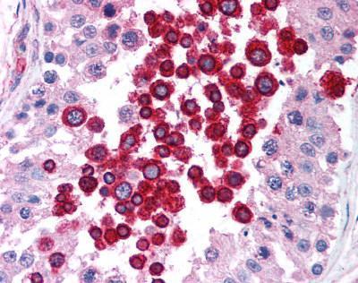 |
| Human Testis (formalin-fixed, paraffin-embedded) stained with ATM antibody at 5 µg/mL followed by biotinylated anti-mouse IgG secondary antibody, alkaline phosphatase-streptavidin and chromogen. |
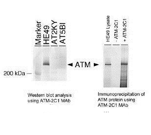 |
| Detection of human ATM protein using anti-ATM 2C1 monoclonal antibody (2952) by western blot or immunoprecipitation. |
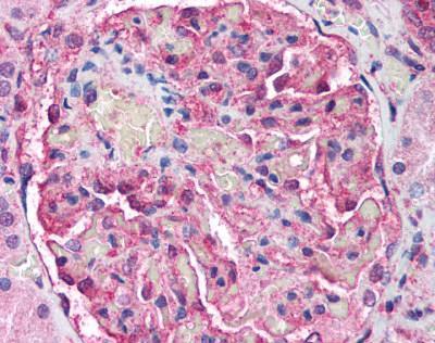 |
| Human Kidney (formalin-fixed, paraffin-embedded) stained with ATM antibody at 5 µg/mL followed by biotinylated anti-mouse IgG secondary antibody, alkaline phosphatase-streptavidin and chromogen. |
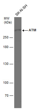 |
| Whole cell extract (30 µg) was separated by 5% SDS-PAGE, and the membrane was blotted with ATM antibody [2C1] (2952) diluted at 1:1000. |
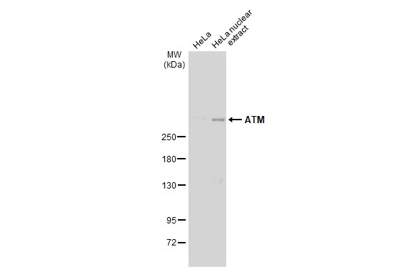 |
| HeLa whole cell extract and nuclear extracts (30 µg) were separated by 5% SDS-PAGE, and the membrane was blotted with ATM antibody [2C1] (2952) diluted at 1:500. The HRP-conjugated anti-mouse IgG antibody was used to detect the primary antibody. |
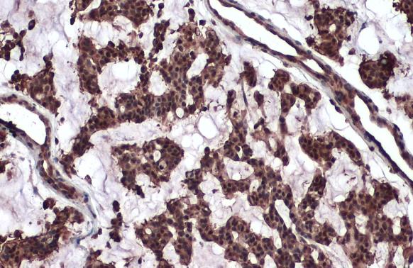 |
| ATM antibody [2C1] detects ATM protein at nucleus by immunohistochemical analysis.Sample: Paraffin-embedded human breast carcinoma.ATM stained by ATM antibody [2C1] (2952) diluted at 1:100.Antigen Retrieval: Citrate buffer, pH 6.0, 15 min |
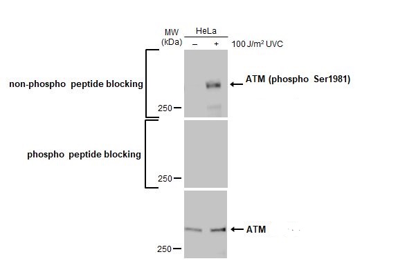 |
| Untreated (-) and treated (-) 293T whole cell extracts (60 µg) were separated by 5% SDS-PAGE, and the membrane was blotted with ATM antibody [2C1] (2952) diluted at 1:1000. The HRP-conjugated anti-mouse IgG antibody was used to detect the primary antibody, and the signal was developed with Trident ECL plus-Enhanced. |
FAQ & Publications
Frequently Asked Questions
What applications has the mouse anti-ATM monoclonal antibody (2C1) been validated for?
This antibody has been tested and validated for use in Western Blotting (WB), Immunohistochemistry on paraffin-embedded sections (IHC-P), Immunocytochemistry/Immunofluorescence (ICC/IF), Fluorescence-activated cell sorting (FACS), Immunoprecipitation (IP), Enzyme-linked immunosorbent assay (ELISA), and Chromatin Immunoprecipitation (ChIP).
How should the mouse anti-ATM monoclonal antibody (2C1) be stored to maintain stability?
The antibody should be stored at -70°C and is stable for at least one year under these conditions. It is important to avoid multiple freeze-thaw cycles to preserve antibody integrity.
Publications
| pmid | title | authors | citation |
|---|---|---|---|
| 37503842 | The HDAC6-RNF168 axis regulates H2A/H2A.X ubiquitination to enable double-strand break repair. | Lingyu Qiu, Wenchao Xu, Xiaopeng Lu, Feng Chen, Yongcan Chen, Yuan Tian, Qian Zhu, Xiangyu Liu, Yongqing Wang, Xin-Hai Pei, Xingzhi Xu, Jun Zhang, Wei-Guo Zhu | Nucleic Acids Res 51:9166-9182 |
| 36603026 | Ataxia Telangiectasia Mutated and MSH2 Control Blunt DNA End Joining in Ig Class Switch Recombination. | Emily Sible, Mary Attaway, Giuseppe Fiorica, Genesis Michel, Jayanta Chaudhuri, Bao Q Vuong | J Immunol 210:369-376 |
| 36200829 | APE1 assembles biomolecular condensates to promote the ATR-Chk1 DNA damage response in nucleolus. | Jia Li, Haichao Zhao, Anne McMahon, Shan Yan | Nucleic Acids Res 50:10503-10525 |
| 36185355 | RPRM negatively regulates ATM levels through its nuclear translocation on irradiation mediated by CDK4/6 and IPO11. | Yarui Zhang, Guomin Ou, Zhujing Ye, Zhou Zhou, Qianlin Cao, Mengting Li, Jingdong Wang, Jianping Cao, Hongying Yang | iScience 25:105115 |
| 36139416 | Inhibition of BRD4 Promotes Pexophagy by Increasing ROS and ATM Activation. | Yong Hwan Kim, Doo Sin Jo, Na Yeon Park, Ji-Eun Bae, Joon Bum Kim, Ha Jung Lee, So Hyun Kim, Seong Hyun Kim, Sunwoo Lee, Mikyung Son, Kyuhee Park, Kwiwan Jeong, Eunbyul Yeom, Dong-Hyung Cho | Cells 11:N/A |
Published literature highly relevant to the biological target of this product and referencing this antibody or clone are retrieved from the PubMed database provided by the United States National Library of Medicine at the National Institutes of Health.
Protocols
| relevant to this product |
|---|
| Western blot IHC ICC |
Documents
| Batch Number | QC File | SDS |
|---|---|---|
| To view batch-specific Safety Datasheets and Quality Certificates associated with your account, please Log In. | ||
Only logged in customers who have purchased this product may leave a review.


Reviews
There are no reviews yet.