| Weight | 1 lbs |
|---|---|
| Dimensions | 9 × 5 × 2 in |
| host | mouse |
| isotype | IgG1 |
| clonality | monoclonal |
| concentration | concentrate, predilute |
| applications | IHC |
| reactivity | human |
| available size | 0.1 mL, 0.5 mL, 1 mL concentrated, 7 mL prediluted |

mouse anti-Melan-A (MART-1) monoclonal antibody (A103) 6251
Price range: $160.00 through $528.00
Antibody summary
- Mouse monoclonal to Melan-A (MART-1)
- Suitable for: Immunohistochemistry (formalin-fixed, paraffin-embedded tissues)
- Reacts with: Human
- Isotype:IgG1
- Control: Skin or melanoma
- Visualization: Cytoplasmic
- 0.1, 0.5, 1.0 mL concentrated, 7 mL prediluted
mouse anti-Melan-A (MART-1) monoclonal antibody A103 6251
| target relevance |
|---|
| Protein names Melanoma antigen recognized by T-cells 1 (MART-1) (Antigen LB39-AA) (Antigen SK29-AA) (Protein Melan-A) |
| Gene names MLANA,MLANA MART1 |
| Mass 13157Da |
| Function FUNCTION: Involved in melanosome biogenesis by ensuring the stability of GPR143. Plays a vital role in the expression, stability, trafficking, and processing of melanocyte protein PMEL, which is critical to the formation of stage II melanosomes. {ECO:0000269|PubMed:15695812, ECO:0000269|PubMed:19717472}. |
| Subellular location SUBCELLULAR LOCATION: Endoplasmic reticulum membrane; Single-pass type III membrane protein. Golgi apparatus. Golgi apparatus, trans-Golgi network membrane. Melanosome. Note=Also found in small vesicles and tubules dispersed over the entire cytoplasm. A small fraction of the protein is inserted into the membrane in an inverted orientation. Inversion of membrane topology results in the relocalization of the protein from a predominant Golgi/post-Golgi area to the endoplasmic reticulum. Melanoma cells expressing the protein with an inverted membrane topology are more effectively recognized by specific cytolytic T-lymphocytes than those expressing the protein in its native membrane orientation. |
| Tissues TISSUE SPECIFICITY: Expression is restricted to melanoma and melanocyte cell lines and retina. |
| Structure SUBUNIT: Interacts with PMEL. Interacts with GPR143. {ECO:0000269|PubMed:15695812, ECO:0000269|PubMed:19717472, ECO:0000269|PubMed:20417641}. |
| Post-translational modification PTM: Acylated. {ECO:0000269|PubMed:12191019}. |
| Target Relevance information above includes information from UniProt accession: Q16655 |
| The UniProt Consortium |
Data
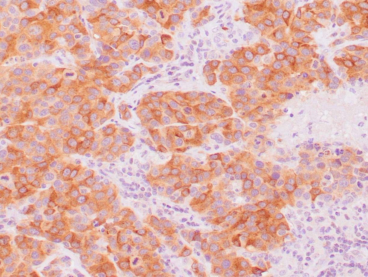 |
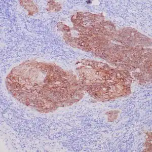
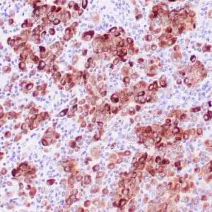
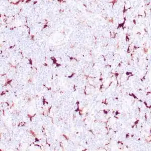
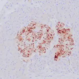
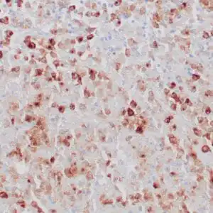
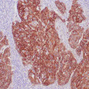
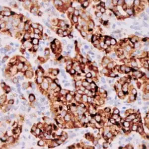
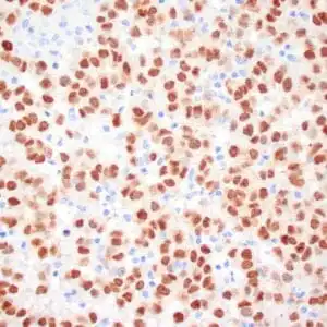
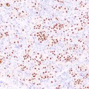
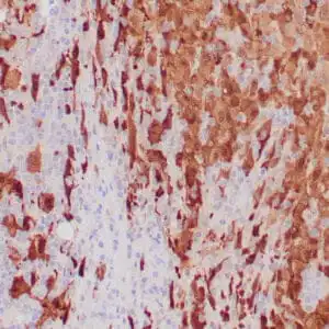
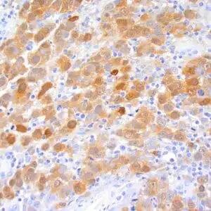
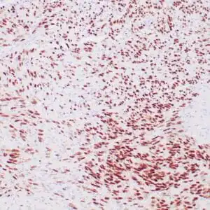
| Human melanoma stained with anti-Melan-A antibody using peroxidase-conjugate and DAB chromogen. Note the cytoplasmic staining of melanoma cells. |
FAQ & Publications
Frequently Asked Questions
What is the recommended dilution range for using the mouse anti-Melan-A (MART-1) monoclonal antibody (A103) in immunohistochemistry?
For immunohistochemistry applications, the concentrated mouse anti-Melan-A (MART-1) monoclonal antibody (A103) is recommended to be diluted between 1:50 and 1:100.
How should the mouse anti-Melan-A (MART-1) monoclonal antibody be stored to maintain its stability?
The antibody should be stored at 2-8°C for short term use, and for longer term storage, it should be kept at -20°C. It is important to avoid freeze/thaw cycles to preserve antibody integrity.
Publications
| pmid | title | authors | citation |
|---|---|---|---|
| We haven't added any publications to our database yet. | |||
Published literature highly relevant to the biological target of this product and referencing this antibody or clone are retrieved from the PubMed database provided by the United States National Library of Medicine at the National Institutes of Health.
Protocols
| relevant to this product |
|---|
| IHC |
Documents
| Batch Number | QC File | SDS |
|---|---|---|
| To view batch-specific Safety Datasheets and Quality Certificates associated with your account, please Log In. | ||
Only logged in customers who have purchased this product may leave a review.
Reviews
There are no reviews yet.