| Weight | 1 lbs |
|---|---|
| Dimensions | 9 × 5 × 2 in |
| host | mouse |
| isotype | IgG |
| clonality | monoclonal |
| concentration | concentrate, predilute |
| applications | IHC |
| reactivity | human |
| available size | 0.1 mL, 0.5 mL, 1 mL concentrated, 7 mL prediluted |

rabbit anti-Villin monoclonal antibody (ZR155) 6402
Price range: $160.00 through $528.00
Antibody summary
- Rabbit monoclonal to Villin
- Suitable for: Immunohistochemistry (formalin-fixed, paraffin-embedded tissues)
- Reacts with: Human
- Isotype:IgG
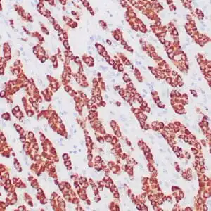
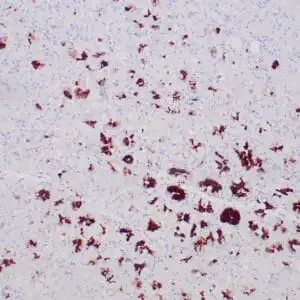
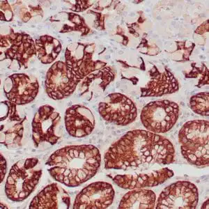
- Control: Colon carcinoma, small intestine
- Visualization: Cytoplasmic
- 0.1, 0.5, 1.0 mL concentrated, 7 mL prediluted
rabbit anti-Villin monoclonal antibody ZR155 6402
| target relevance |
|---|
| Homo sapiens VIL1 Villin-1 |
| Protein names Villin-1 |
| Gene names VIL1 |
| Protein family Belongs to the villin/gelsolin family |
| Function Epithelial cell-specific Ca(2+)-regulated actin-modifying protein that modulates the reorganization of microvillar actin filaments. Plays a role in the actin nucleation, actin filament bundle assembly, actin filament capping and severing. Binds phosphatidylinositol 4,5-bisphosphate (PIP2) and lysophosphatidic acid (LPA); binds LPA with higher affinity than PIP2. Binding to LPA increases its phosphorylation by SRC and inhibits all actin-modifying activities. Binding to PIP2 inhibits actin-capping and -severing activities but enhances actin-bundling activity. Regulates the intestinal epithelial cell morphology, cell invasion, cell migration and apoptosis. Protects against apoptosis induced by dextran sodium sulfate (DSS) in the gastrointestinal epithelium. Appears to regulate cell death by maintaining mitochondrial integrity. Enhances hepatocyte growth factor (HGF)-induced epithelial cell motility, chemotaxis and wound repair. Upon S.flexneri cell infection, its actin-severing activity enhances actin-based motility of the bacteria and plays a role during the dissemination |
| Subcellular location Cytoplasm, cytoskeleton, Cell projection, lamellipodium, Cell projection, ruffle, Cell projection, microvillus, Cell projection, filopodium tip, Cell projection, filopodium |
| Structure Monomer. Homodimer; homodimerization is necessary for actin-bundling. Associates with F-actin; phosphorylation at tyrosine residues decreases the association with F-actin. Interacts (phosphorylated at C-terminus tyrosine phosphorylation sites) with PLCG1 (via the SH2 domains). Interacts (phosphorylated form) with PLCG1; the interaction is enhanced by hepatocyte growth factor (HGF) (By similarity) |
| Post-translational modification Tyrosine phosphorylation is induced by epidermal growth factor (EGF) and stimulates cell migration (By similarity). Phosphorylated on tyrosine residues by SRC. The unphosphorylated form increases the initial rate of actin-nucleating activity, whereas the tyrosine-phosphorylated form inhibits actin-nucleating activity, enhances actin-bundling activity and enhances actin-severing activity by reducing high Ca(2+) requirements. The tyrosine-phosphorylated form does not regulate actin-capping activity. Tyrosine phosphorylation is essential for cell migration: tyrosine phosphorylation sites in the N-terminus half regulate actin reorganization and cell morphology, whereas tyrosine phosphorylation sites in the C-terminus half regulate cell migration via interaction with PLCG1 |
| Keywords 3D-structure, Actin capping, Actin-binding, Alternative splicing, Apoptosis, Calcium, Cell projection, Cytoplasm, Cytoskeleton, Direct protein sequencing, Phosphoprotein, Proteomics identification, Reference proteome, Repeat |
| Sequence MTKLSAQVKGSLNITTPGLQIWRIEAMQMVPVPSSTFGSFFDGDCYIILAIHKTASSLSY DIHYWIGQDSSLDEQGAAAIYTTQMDDFLKGRAVQHREVQGNESEAFRGYFKQGLVIRKG GVASGMKHVETNSYDVQRLLHVKGKRNVVAGEVEMSWKSFNRGDVFLLDLGKLIIQWNGP ESTRMERLRGMTLAKEIRDQERGGRTYVGVVDGENELASPKLMEVMNHVLGKRRELKAAV PDTVVEPALKAALKLYHVSDSEGNLVVREVATRPLTQDLLSHEDCYILDQGGLKIYVWKG KKANEQEKKGAMSHALNFIKAKQYPPSTQVEVQNDGAESAVFQQLFQKWTASNRTSGLGK THTVGSVAKVEQVKFDATSMHVKPQVAAQQKMVDDGSGEVQVWRIENLELVPVDSKWLGH FYGGDCYLLLYTYLIGEKQHYLLYVWQGSQASQDEITASAYQAVILDQKYNGEPVQIRVP MGKEPPHLMSIFKGRMVVYQGGTSRTNNLETGPSTRLFQVQGTGANNTKAFEVPARANFL NSNDVFVLKTQSCCYLWCGKGCSGDEREMAKMVADTISRTEKQVVVEGQEPANFWMALGG KAPYANTKRLQEENLVITPRLFECSNKTGRFLATEIPDFNQDDLEEDDVFLLDVWDQVFF WIGKHANEEEKKAAATTAQEYLKTHPSGRDPETPIIVVKQGHEPPTFTGWFLAWDPFKWS NTKSYEDLKAELGNSRDWSQITAEVTSPKVDVFNANSNLSSGPLPIFPLEQLVNKPVEEL PEGVDPSRKEEHLSIEDFTQAFGMTPAAFSALPRWKQQNLKKEKGLF |
| UniProt accession: P09327 |
Data
 |
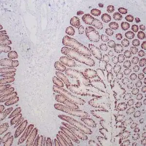
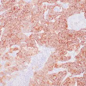
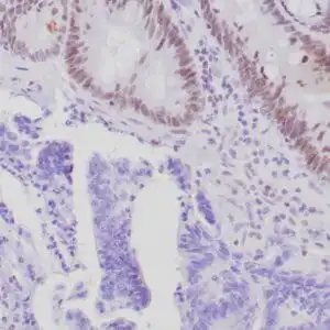
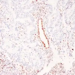
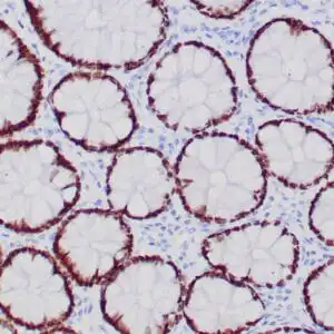
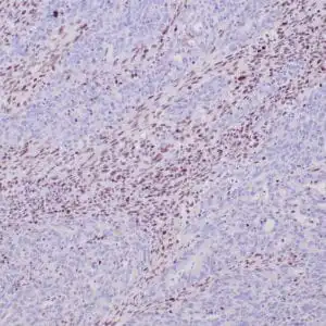
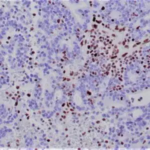
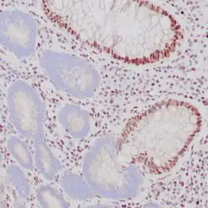
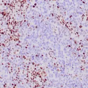
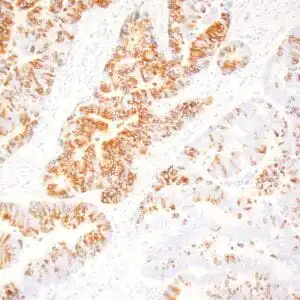
| Formalin-fixed, paraffin-embedded human colon stained with anti-villin antibody using peroxidase-conjugate and DAB chromogen. Note brush border and cytoplasmic staining of epithelial cells |
FAQ & Publications
Frequently Asked Questions
What species does the rabbit anti-Villin monoclonal antibody (ZR155) specifically react with?
This antibody specifically reacts with human Villin protein, making it suitable for studies involving human tissue samples.
What are the recommended storage conditions for the rabbit anti-Villin monoclonal antibody (ZR155) to maintain its stability?
For short-term storage, keep the antibody at 2-8°C. For longer-term preservation, store it at -20°C and avoid repeated freeze/thaw cycles to maintain antibody integrity.
Publications
| pmid | title | authors | citation |
|---|---|---|---|
| We haven't added any publications to our database yet. | |||
Published literature highly relevant to the biological target of this product and referencing this antibody or clone are retrieved from the PubMed database provided by the United States National Library of Medicine at the National Institutes of Health.
Protocols
| relevant to this product |
|---|
| IHC |
Documents
| Batch Number | QC File | SDS |
|---|---|---|
| To view batch-specific Safety Datasheets and Quality Certificates associated with your account, please Log In. | ||
Only logged in customers who have purchased this product may leave a review.

Reviews
There are no reviews yet.