Showing all 5 results

Actin antibodies
Actin is widely used as a reliable loading control in Western blotting (WB) due to its consistent expression levels across different cell types and conditions. Its stable and ubiquitous presence in cells ensures that variations in protein loading are accurately normalized, making actin ideal for comparative analysis in WB experiments.
Beyond WB, actin antibodies offered by your website are also available for use in immunofluorescence (IF) and immunohistochemistry (IHC). These antibodies can detect actin structures in a variety of tissues and cell types, helping visualize cytoskeletal dynamics. Actin is a key component of the cytoskeleton, playing an essential role in maintaining cell shape, facilitating cell movement, and organizing intracellular structures. It forms dynamic structures such as filaments and microfilaments, which are crucial for many cellular processes, including division and signaling.
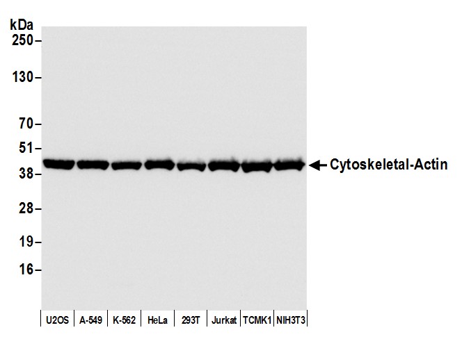
WB with antibody [9038] : Detection of human beta Actin by western blot. Samples: Whole cell lysate (10 �g) from U2OS, A-549, K-562, HeLa, HEK293T, Jurkat, MOLT-4, and Hep-G2 cells prepared using lysis buffer. Antibody: Affinity purified rabbit anti-beta Actin antibody used for WB at 0.04 �g/ml. Detection: Chemiluminescence with an exposure time of 10 seconds.
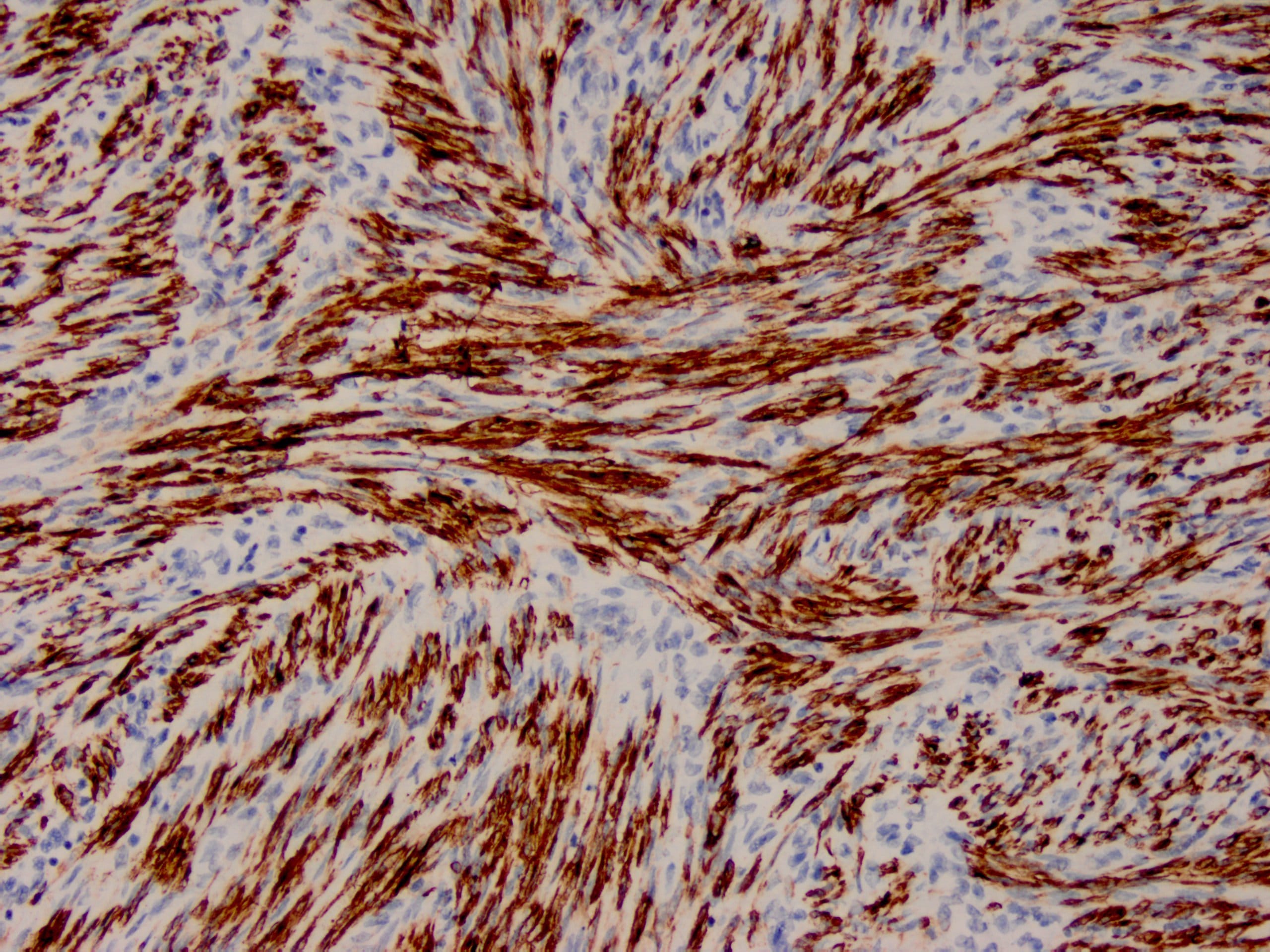
IHC with antibody [6012] : Human uterus stained with anti-SMA antibody using peroxidase-conjugate and DAB chromogen. Note the cytoplasmic staining of smooth muscle.

WB with antibody [5132] : 10µg/lane of mouse brain tissue lysates. 42kDa band is Anti-β-Actin (5132) at 1:1000 dilution (1µg/mL);


